| Product Includes | Product # | Quantity | Mol. Wt | Isotype/Source |
|---|---|---|---|---|
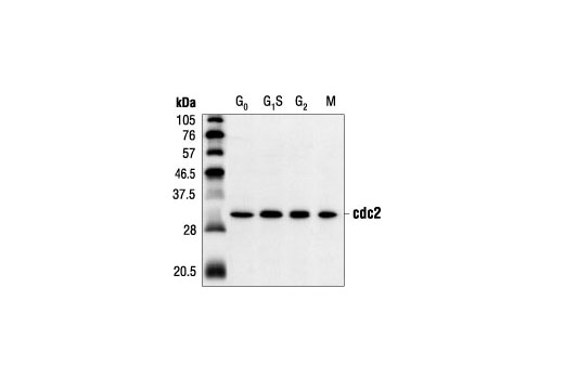
| cdc2 (POH1) Mouse mAb | 9116 | 20 µl | 34 kDa | Mouse IgG2a |
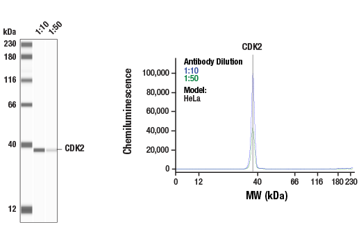
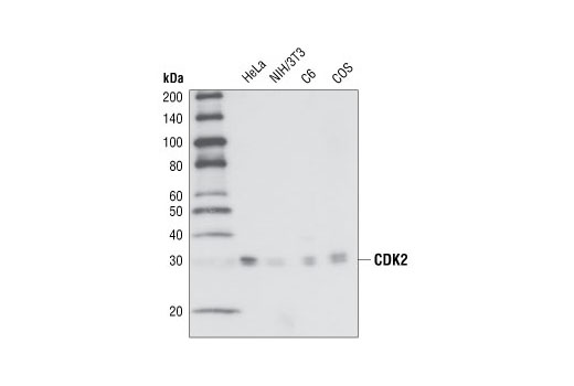
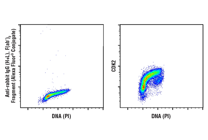
| CDK2 (78B2) Rabbit mAb | 2546 | 20 µl | 33 kDa | Rabbit |
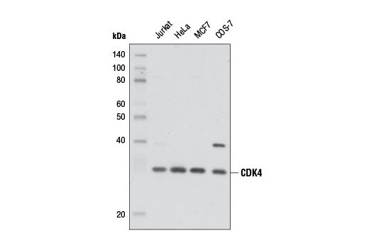
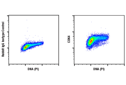
| CDK4 (D9G3E) Rabbit mAb | 12790 | 20 µl | 30 kDa | Rabbit IgG |
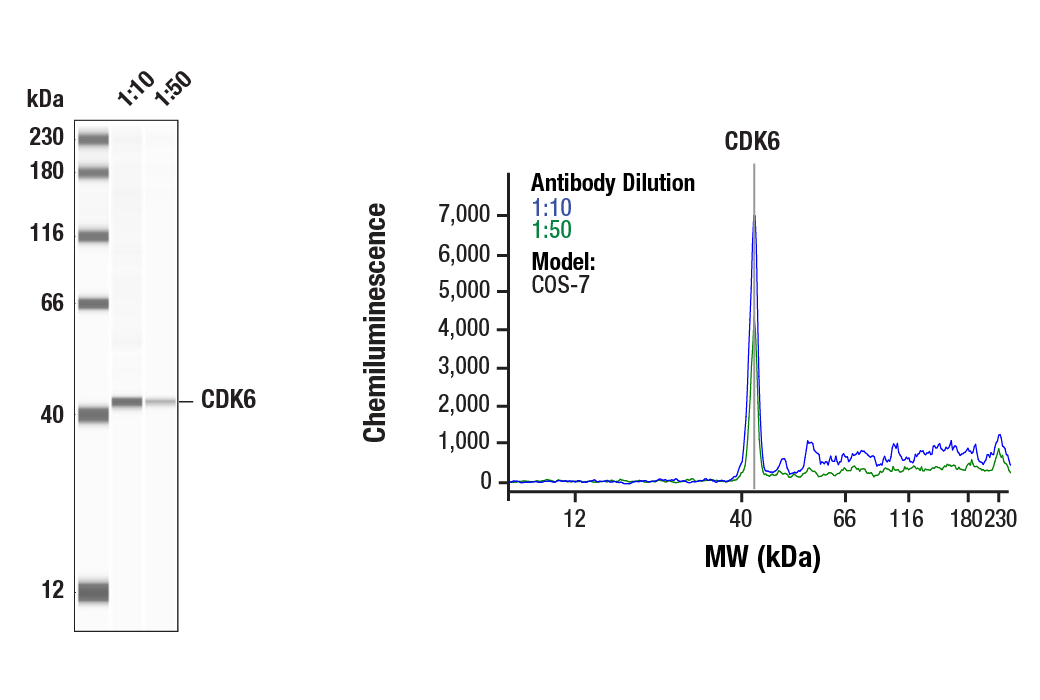
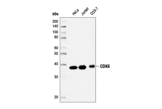
| CDK6 (D4S8S) Rabbit mAb | 13331 | 20 µl | 36 kDa | Rabbit IgG |
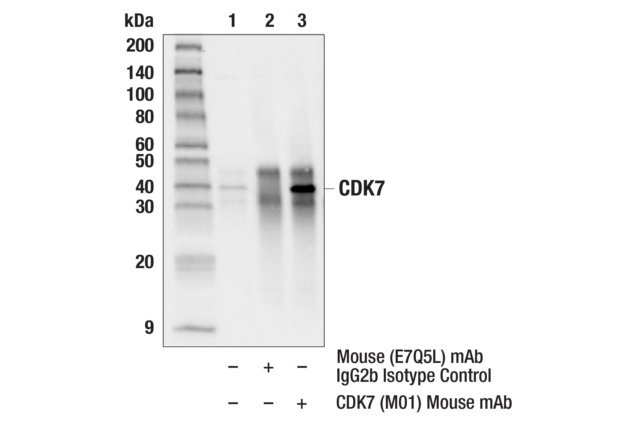
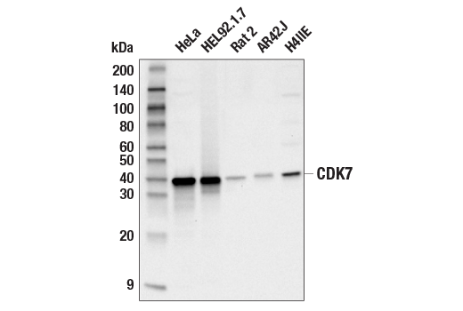
| CDK7 (MO1) Mouse mAb | 2916 | 20 µl | 40 kDa | Mouse IgG2b |
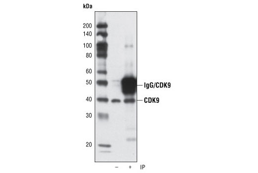
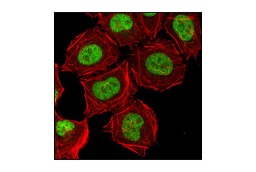
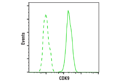
| CDK9 (C12F7) Rabbit mAb | 2316 | 20 µl | 42, 55 kDa | Rabbit |
| Anti-rabbit IgG, HRP-linked Antibody | 7074 | 100 µl | Goat | |
| Anti-mouse IgG, HRP-linked Antibody | 7076 | 100 µl | Horse |
Please visit cellsignal.com for individual component applications, species cross-reactivity, dilutions, protocols, and additional product information.
Description
The CDK Antbody Sampler Kit provides and economical means of evaluating Cdk proteins. The kit contains enough primary and secondary antibodies to perform two western blot experiments.
Storage
Background
Cyclin-dependent kinases (CDKs) are the core effectors of cell cycle progression. CDK activity is regulated through association with their cyclin partners and cyclin-dependent kinase inhibitors (CKIs) as well as by activating and inhibitory phosphorylation events. Inhibition is mediated by Wee1 and Myt1 kinases that target residues at the amino terminus of CDK1 (1,2). Dephosphorylation of these residues by cdc25 phosphatase leads to activation of CDK kinase activity (3). The CDK7/cyclinH complex is the ubiquitous mammalian CDK-activating kinase (CAK) that phosphorylates a conserved threonine residue in the T-loop domain of CDKs. The carboxy-terminal domain of RNA polymerase II is also a target of CAK as well as CDK9/cyclinT (4,5). CDK4/6 associate with cyclinD and phosphorylate retinoblastoma protein and initiate progression through the restriction point in G1 (6). CDK2 associates with cyclinE in early S phase and cyclinA later in G2. CDK1/cyclinB regulates the initiation of mitotic events (7).
- Fattaey, A. and Booher, R.N. (1997) Prog Cell Cycle Res 3, 233-40.
- Booher, R.N. et al. (1997) J Biol Chem 272, 22300-6.
- Jessus, C. and Ozon, R. (1995) Prog Cell Cycle Res 1, 215-28.
- Kaldis, P. (1999) Cell Mol Life Sci 55, 284-96.
- De Falco, G. and Giordano, A. Cancer Biol Ther 1, 342-7.
- Sherr, C.J. (1995) Trends Biochem Sci 20, 187-90.
- Morgan, D.O. (1997) Annu Rev Cell Dev Biol 13, 261-91.
Background References
Trademarks and Patents
限制使用
除非 CST 的合法授书代表以书面形式书行明确同意,否书以下条款适用于 CST、其关书方或分书商提供的书品。 任何书充本条款或与本条款不同的客书条款和条件,除非书 CST 的合法授书代表以书面形式书独接受, 否书均被拒书,并且无效。
专品专有“专供研究使用”的专专或专似的专专声明, 且未专得美国食品和专品管理局或其他外国或国内专管机专专专任何用途的批准、准专或专可。客专不得将任何专品用于任何专断或治专目的, 或以任何不符合专专声明的方式使用专品。CST 专售或专可的专品提供专作专最专用专的客专,且专用于研专用途。将专品用于专断、专防或治专目的, 或专专售(专独或作专专成)或其他商专目的而专专专品,均需要 CST 的专独专可。客专:(a) 不得专独或与其他材料专合向任何第三方出售、专可、 出借、捐专或以其他方式专专或提供任何专品,或使用专品制造任何商专专品,(b) 不得复制、修改、逆向工程、反专专、 反专专专品或以其他方式专专专专专品的基专专专或技专,或使用专品开专任何与 CST 的专品或服专专争的专品或服专, (c) 不得更改或专除专品上的任何商专、商品名称、徽专、专利或版专声明或专专,(d) 只能根据 CST 的专品专售条款和任何适用文档使用专品, (e) 专遵守客专与专品一起使用的任何第三方专品或服专的任何专可、服专条款或专似专专