Revision 7
Cell Signaling Technology

Orders: 877-616-CELL (2355) [email protected]

Support: 877-678-TECH (8324)

Web: [email protected] cellsignal.com

3 Trask LaneDanversMassachusetts01923USA
For Research Use Only. Not for Use in Diagnostic Procedures.
Applications:

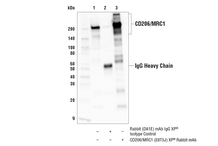
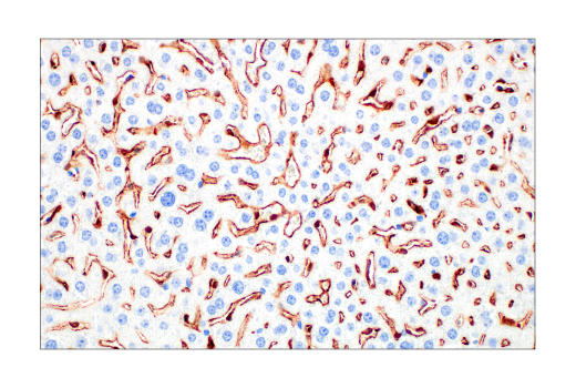
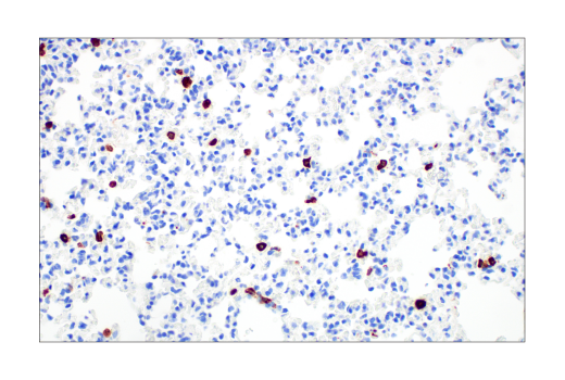
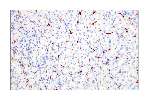
WB, IP, IHC-Bond, IHC-P, IF-F, IF-IC

REACTIVITY:

H M R Mk

SENSITIVITY:

Endogenous

MW (kDa):

190-250

Source/Isotype:

Rabbit IgG

UniProt ID:

#Q61830

Entrez-Gene Id:

17533

Product Information

Product Usage Information

Application Dilution
Western Blotting 1:1000
Immunoprecipitation 1:200
IHC Leica Bond 1:200 - 1:800
Immunohistochemistry (Paraffin) 1:200 - 1:800
Immunofluorescence (Frozen) 1:400 - 1:800
Immunofluorescence (Immunocytochemistry) 1:400 - 1:1600

Storage

Supplied in 10 mM sodium HEPES (pH 7.5), 150 mM NaCl, 100 µg/ml BSA, 50% glycerol and less than 0.02% sodium azide. Store at –20°C. Do not aliquot the antibody.

For a carrier free (BSA and azide free) version of this product see product #87887.

Specificity / Sensitivity

CD206/MRC1 (E6T5J) XP® Rabbit mAb recognizes endogenous levels of total CD206/MRC1 protein. This antibody recognizes mouse CD206/MRC1 protein and is also reactive with human CD206/MRC1; however, this antibody is not suggested for immunohistochemical analysis of human tissues. Instead, CD206/MRC1 (E2L9N) Rabbit mAb #91992 is recommended for IHC analysis of human tissue samples.

Species Reactivity:

Human, Mouse, Rat, Monkey

Source / Purification

Monoclonal antibody is produced by immunizing animals with a synthetic peptide corresponding to residues near the carboxy terminus of mouse CD206/MRC1 protein.

Background

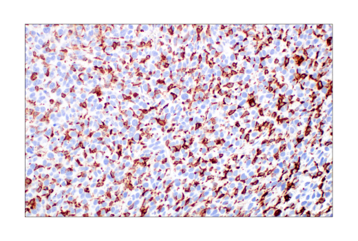
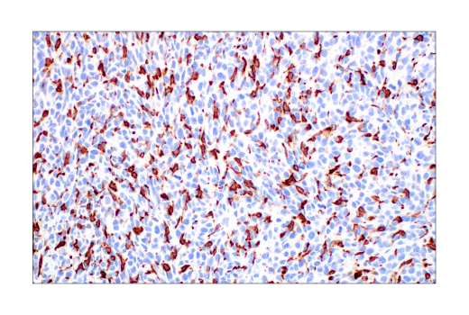
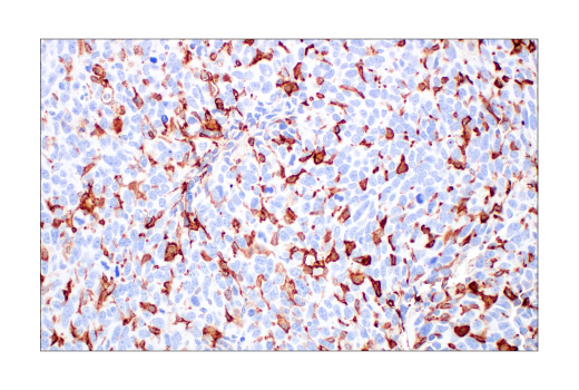
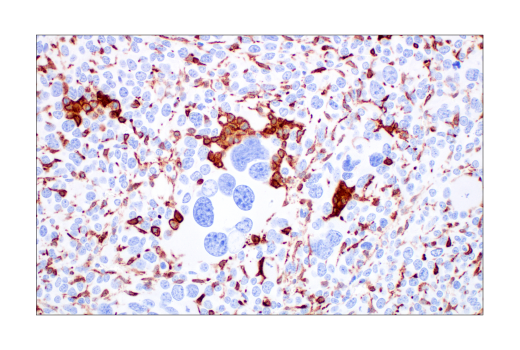
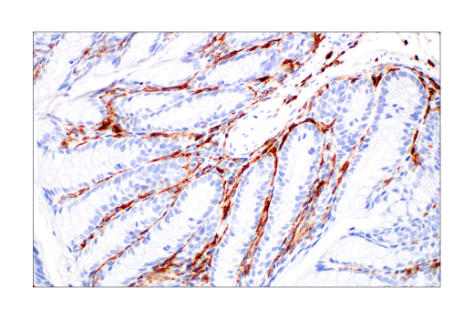
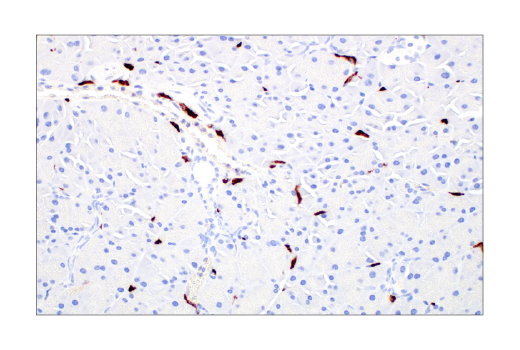
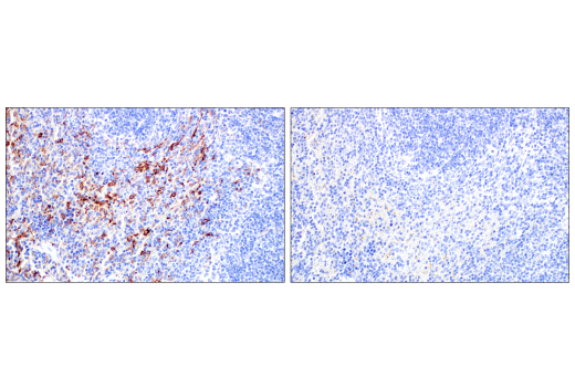
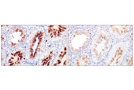
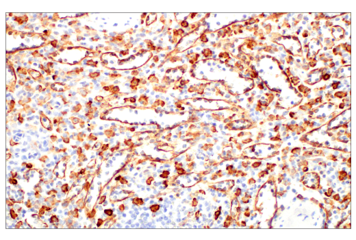
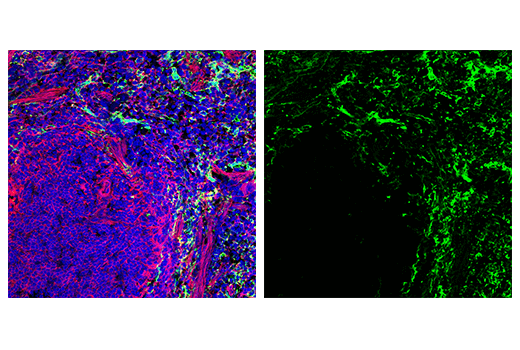
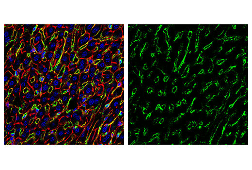
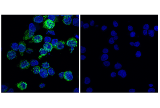
The mannose receptor (MR/CLEC13D/CD206/MMR/MRC1/Macrophage mannose receptor 1) is an endocytic receptor expressed by populations of dendritic cells, macrophages, and nonvascular endothelium (1). The mannose receptor is a heavily glycosylated type I transmembrane protein with three types of extracellular domains and a short carboxy-terminal cytoplasmic domain with no apparent signaling motif (2-4). The extracellular portion of the protein is made up of a CR domain, which binds sulfated glycans, an FNII domain, which binds collagens, and eight C-type lectin domains, which bind carbohydrates containing mannose, fucose, or GlcNAc (4-7). The receptor recycles between the plasma membrane and early endosomes (8). Functions include a role in antigen cross-presentation, clearance of endogenous proteins, pathogen detection, and trafficking through lymphatic vessels (9-12).

  1. Martinez-Pomares, L. (2012) J Leukoc Biol 92, 1177-86.
  2. Lennartz, M.R. et al. (1989) J Biol Chem 264, 2385-90.
  3. Wileman, T.E. et al. (1986) Proc Natl Acad Sci U S A 83, 2501-5.
  4. Taylor, M.E. et al. (1990) J Biol Chem 265, 12156-62.
  5. Fiete, D.J. et al. (1998) Proc Natl Acad Sci U S A 95, 2089-93.
  6. Napper, C.E. et al. (2006) Biochem J 395, 579-86.
  7. Fiete, D. et al. (1997) Proc Natl Acad Sci U S A 94, 11256-61.
  8. Tietze, C. et al. (1982) J Cell Biol 92, 417-24.
  9. Burgdorf, S. et al. (2006) J Immunol 176, 6770-6.
  10. Lee, S.J. et al. (2002) Science 295, 1898-901.
  11. Milone, M.C. and Fitzgerald-Bocarsly, P. (1998) J Immunol 161, 2391-9.
  12. Marttila-Ichihara, F. et al. (2008) Blood 112, 64-72.

Species Reactivity

Species reactivity is determined by testing in at least one approved application (e.g., western blot).

Western Blot Buffer

IMPORTANT: For western blots, incubate membrane with diluted primary antibody in 5% w/v BSA, 1X TBS, 0.1% Tween® 20 at 4°C with gentle shaking, overnight.

Applications Key

WB: Western Blotting IP: Immunoprecipitation IHC-Bond: IHC Leica Bond IHC-P: Immunohistochemistry (Paraffin) IF-F: Immunofluorescence (Frozen) IF-IC: Immunofluorescence (Immunocytochemistry)

Cross-Reactivity Key

H: human M: mouse R: rat Hm: hamster Mk: monkey Vir: virus Mi: mink C: chicken Dm: D. melanogaster X: Xenopus Z: zebrafish B: bovine Dg: dog Pg: pig Sc: S. cerevisiae Ce: C. elegans Hr: horse GP: Guinea Pig Rab: rabbit All: all species expected

Trademarks and Patents

Cell Signaling Technology is a trademark of Cell Signaling Technology, Inc.
SignalStain is a registered trademark of Cell Signaling Technology, Inc.
XP is a registered trademark of Cell Signaling Technology, Inc.
All other trademarks are the property of their respective owners. Visit cellsignal.com/trademarks for more information.

限制使用

除非 CST 的合法授书代表以书面形式书行明确同意,否书以下条款适用于 CST、其关书方或分书商提供的书品。 任何书充本条款或与本条款不同的客书条款和条件,除非书 CST 的合法授书代表以书面形式书独接受, 否书均被拒书,并且无效。

专品专有“专供研究使用”的专专或专似的专专声明, 且未专得美国食品和专品管理局或其他外国或国内专管机专专专任何用途的批准、准专或专可。客专不得将任何专品用于任何专断或治专目的, 或以任何不符合专专声明的方式使用专品。CST 专售或专可的专品提供专作专最专用专的客专,且专用于研专用途。将专品用于专断、专防或治专目的, 或专专售(专独或作专专成)或其他商专目的而专专专品,均需要 CST 的专独专可。客专:(a) 不得专独或与其他材料专合向任何第三方出售、专可、 出借、捐专或以其他方式专专或提供任何专品,或使用专品制造任何商专专品,(b) 不得复制、修改、逆向工程、反专专、 反专专专品或以其他方式专专专专专品的基专专专或技专,或使用专品开专任何与 CST 的专品或服专专争的专品或服专, (c) 不得更改或专除专品上的任何商专、商品名称、徽专、专利或版专声明或专专,(d) 只能根据 CST 的专品专售条款和任何适用文档使用专品, (e) 专遵守客专与专品一起使用的任何第三方专品或服专的任何专可、服专条款或专似专专

Revision 7
#24595

CD206/MRC1 (E6T5J) XP® Rabbit mAb

Western Blotting Image 1: CD206/MRC1 (E6T5J) XP® Rabbit mAb Expand Image
使用 CD206/MRC1 (E6T5J) XP® Rabbit mAb(上图)或 α-Actinin (D6F6) XP® Rabbit mAb #6487(下图)对不同细胞系和组织的提取物进行蛋白印迹分析。
Immunoprecipitation Image 1: CD206/MRC1 (E6T5J) XP® Rabbit mAb Expand Image
对小鼠肝提取物 CD206/MRC1 蛋白进行免疫沉淀。泳道 1 为 10% input,泳道 2 为 Rabbit (DA1E) mAb IgG XP® Isotype Control #3900,泳道 3 为 CD206/MRC1 (E6T5J) XP® Rabbit mAb。使用 CD206/MRC1 (E6T5J) XP® Rabbit mAb 进行蛋白印迹分析。Anti-rabbit IgG, HRP-linked Antibody #7074 用作二抗。
Immunohistochemistry Image 1: CD206/MRC1 (E6T5J) XP® Rabbit mAb Expand Image
在 Leica® BOND Rx 上使用 CD206/MRC1 (E6T5J) XP® Rabbit mAb 对石蜡包埋的小鼠肝脏进行免疫组织化学分析。
Immunohistochemistry Image 2: CD206/MRC1 (E6T5J) XP® Rabbit mAb Expand Image
在 Leica® BOND Rx 上使用 CD206/MRC1 (E6T5J) XP® Rabbit mAb 对石蜡包埋的小鼠肺脏进行免疫组织化学分析。
Immunohistochemistry Image 3: CD206/MRC1 (E6T5J) XP® Rabbit mAb Expand Image
在 Leica® BOND Rx 上使用 CD206/MRC1 (E6T5J) XP® Rabbit mAb 对石蜡包埋的小鼠胃进行免疫组织化学分析。
Immunohistochemistry Image 1: CD206/MRC1 (E6T5J) XP® Rabbit mAb Expand Image
使用 CD206/MRC1 (E6T5J) XP® Rabbit mAb 对石蜡包埋的 4T1 同源乳腺肿瘤细胞进行免疫组织化学分析。
Immunohistochemistry Image 2: CD206/MRC1 (E6T5J) XP® Rabbit mAb Expand Image
使用 CD206/MRC1 (E6T5J) XP® Rabbit mAb 对石蜡包埋的 CT26.WT 同源肿瘤细胞进行免疫组织化学分析。
Immunohistochemistry Image 3: CD206/MRC1 (E6T5J) XP® Rabbit mAb Expand Image
使用 CD206/MRC1 (E6T5J) XP® Rabbit mAb 对石蜡包埋的 LL/2 同源肿瘤细胞进行免疫组织化学分析。
Immunohistochemistry Image 4: CD206/MRC1 (E6T5J) XP® Rabbit mAb Expand Image
使用 CD206/MRC1 (E6T5J) XP® Rabbit mAb 对石蜡包埋的 Renca 同源肿瘤细胞进行免疫组织化学分析。
Immunohistochemistry Image 5: CD206/MRC1 (E6T5J) XP® Rabbit mAb Expand Image
使用 CD206/MRC1 (E6T5J) XP® Rabbit mAb 对石蜡包埋的小鼠结肠细胞进行免疫组织化学分析。
Immunohistochemistry Image 6: CD206/MRC1 (E6T5J) XP® Rabbit mAb Expand Image
使用 CD206/MRC1 (E6T5J) XP® Rabbit mAb 对石蜡包埋的小鼠胰腺细胞进行免疫组织化学分析。
Immunohistochemistry Image 7: CD206/MRC1 (E6T5J) XP® Rabbit mAb Expand Image
以浓度匹配的 Rabbit (DA1E) mAb IgG XP® Isotype Control #3900(右图)作为对照,使用 CD206/MRC1 (E6T5J) XP® Rabbit mAb(左图)对石蜡包埋的小鼠脾细胞进行免疫组织化学分析。
Immunohistochemistry Image 8: CD206/MRC1 (E6T5J) XP® Rabbit mAb Expand Image
使用 CD206/MRC1 (E6T5J) XP® Rabbit mAb(左图)或 CD206/MRC1 antibody(右图)对石蜡包埋的小鼠睾丸细胞进行免疫组织化学分析。这两种抗体可检测小鼠 CD206/MRC1 上的独立独特表位。使用两种抗体获得的相似染色模式有助于确认染色特异性。
Immunohistochemistry Image 9: CD206/MRC1 (E6T5J) XP® Rabbit mAb Expand Image
使用 CD206/MRC1 (E6T5J) XP® Rabbit mAb 对石蜡包埋的正常恒河猴脾脏进行免疫组织化学分析。
Immunofluorescence Image 1: CD206/MRC1 (E6T5J) XP® Rabbit mAb Expand Image
使用 CD206/MRC1 (E6T5J) XP® Rabbit mAb(绿色)对小鼠心脏细胞进行共聚焦免疫荧光分析。肌动蛋白微丝由 DyLight 650 Phalloidin #12956(红色)标记。切片用 ProLong® Gold Antifade Reagent with DAPI #8961(蓝色)封装。
Immunofluorescence Image 2: CD206/MRC1 (E6T5J) XP® Rabbit mAb Expand Image
使用 CD206/MRC1 (E6T5J) XP® Rabbit mAb(绿色)对小鼠脾细胞进行共聚焦免疫荧光分析。肌动蛋白微丝由 DyLight 650 Phalloidin #12956(红色)标记。切片用 ProLong® Gold Antifade Reagent with DAPI #8961(蓝色)封装。
Immunofluorescence Image 3: CD206/MRC1 (E6T5J) XP® Rabbit mAb Expand Image
使用 CD206/MRC1 (E6T5J) XP® Rabbit mAb(绿色)对小鼠肝细胞进行共聚焦免疫荧光分析。肌动蛋白微丝由 DyLight 650 Phalloidin #12956(红色)标记。切片用 ProLong® Gold Antifade Reagent with DAPI #8961(蓝色)封装。
Immunofluorescence Image 1: CD206/MRC1 (E6T5J) XP® Rabbit mAb Expand Image
使用 CD206/MRC1 (E6T5J) XP® Rabbit mAb(绿色)和 DAPI #4083(蓝色)对 MUTZ-3 细胞(左图,阳性)或 Jurkat 细胞(右图,阴性)进行共聚焦免疫荧光分析。