| Product Includes | Product # | Quantity | Mol. Wt | Isotype/Source |
|---|---|---|---|---|
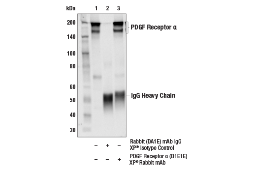
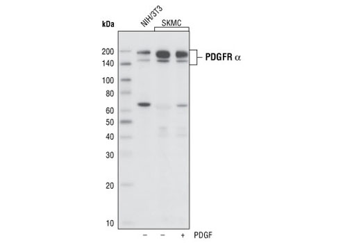
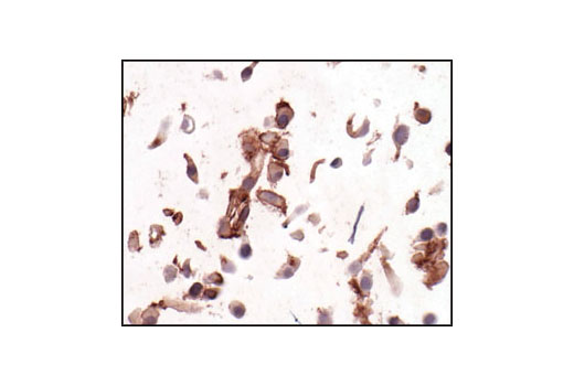
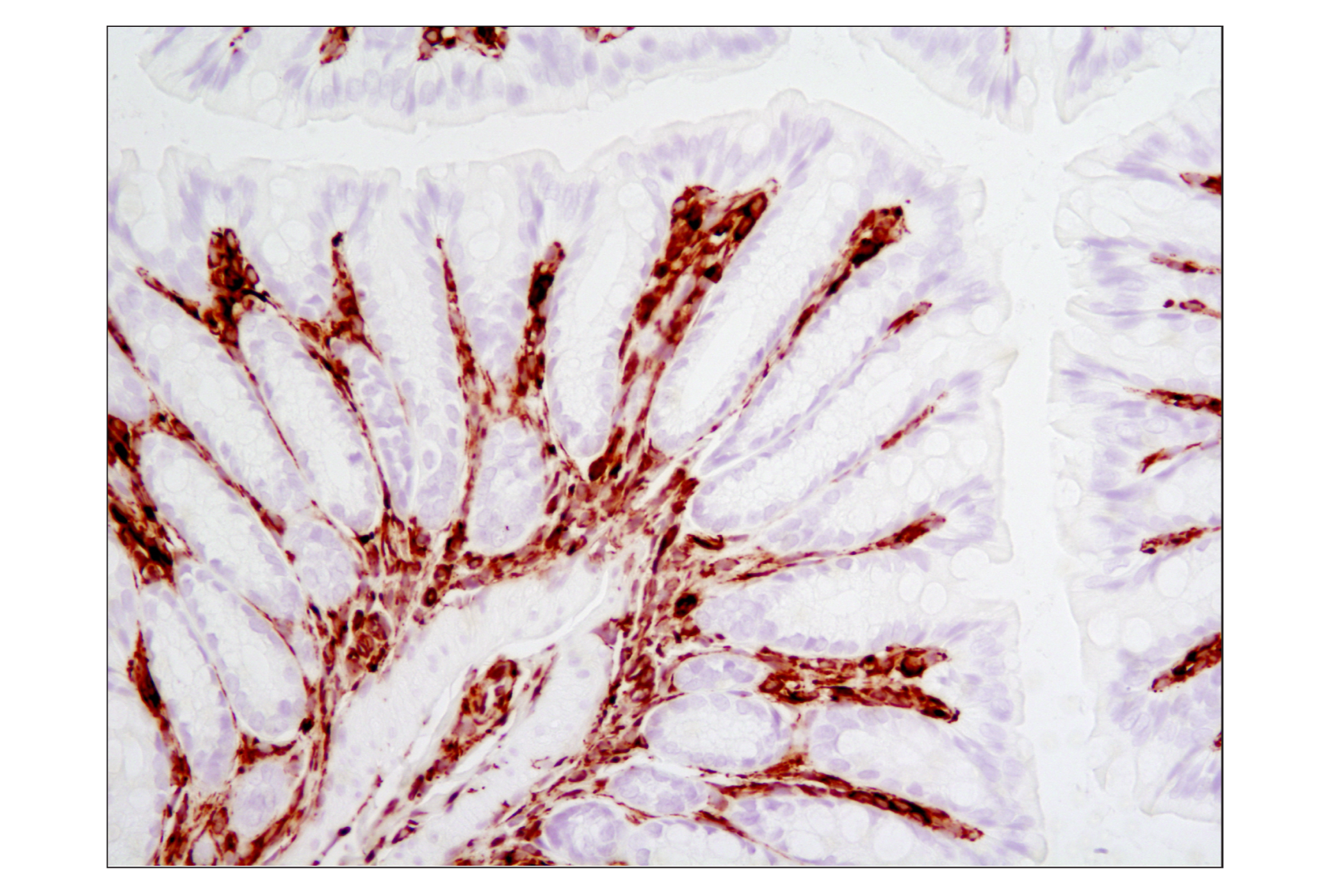
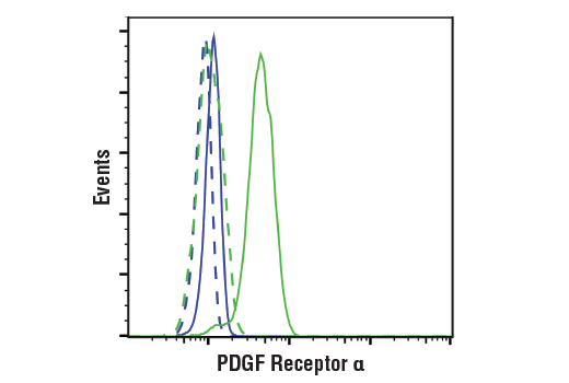
| PDGF Receptor α (D1E1E) XP® Rabbit mAb | 3174 | 20 µl | 190 kDa | Rabbit IgG |
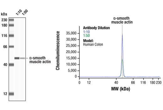
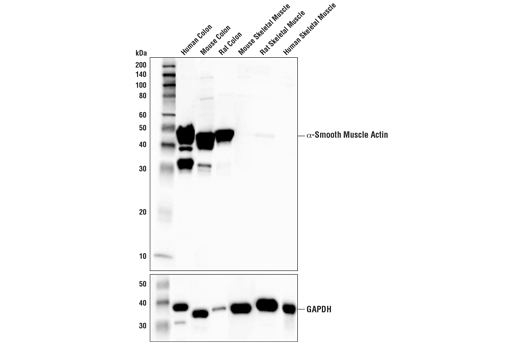
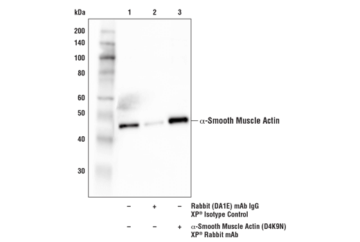
| α-Smooth Muscle Actin (D4K9N) XP® Rabbit mAb | 19245 | 20 µl | 42 kDa | Rabbit IgG |
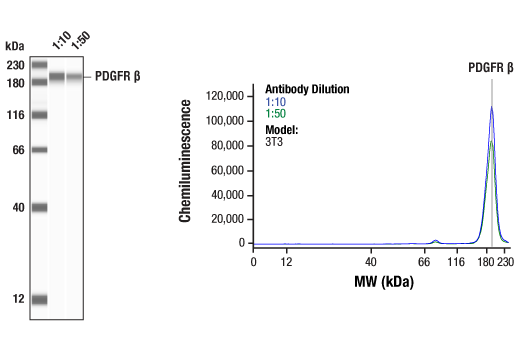
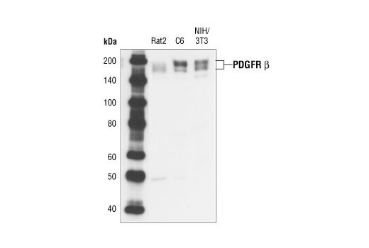
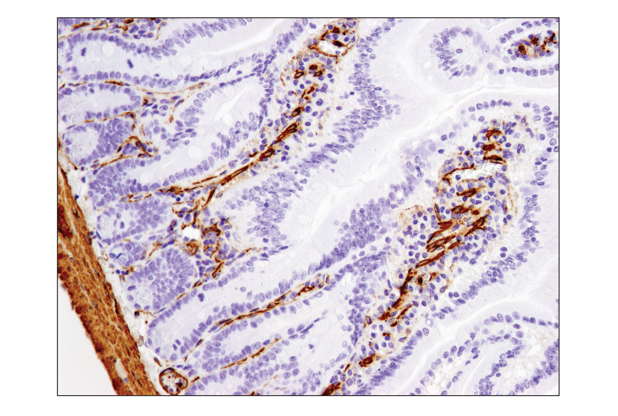
| PDGF Receptor β (28E1) Rabbit mAb | 3169 | 20 µl | 190 kDa | Rabbit IgG |
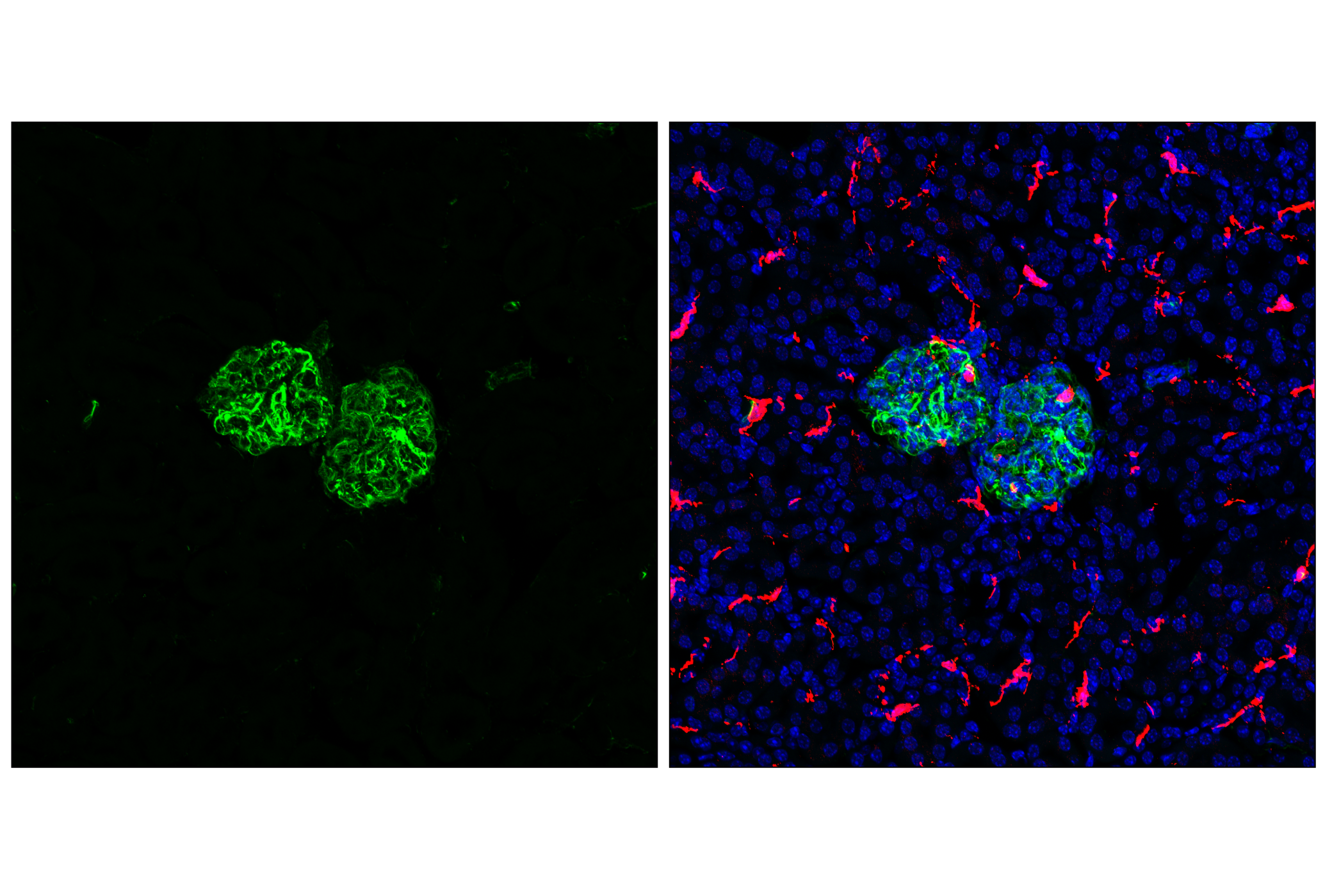
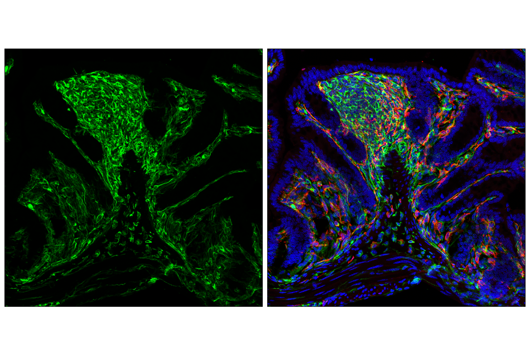
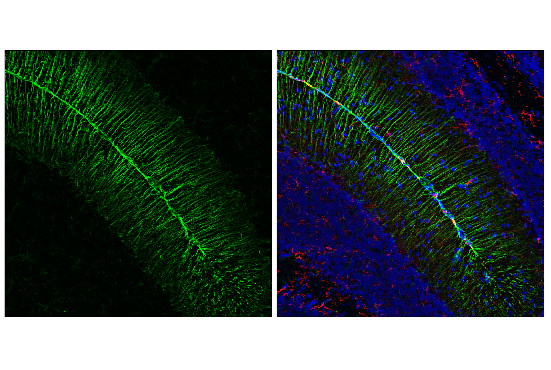
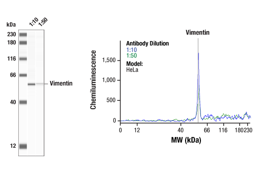
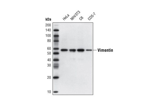
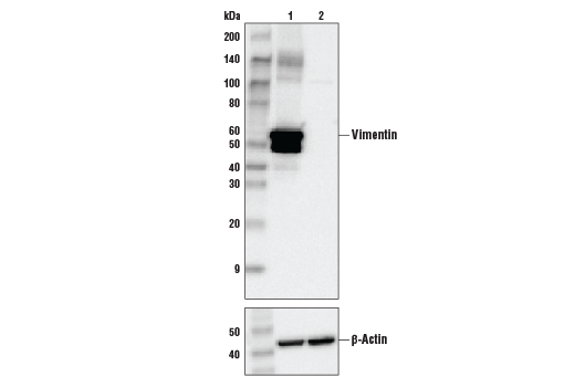
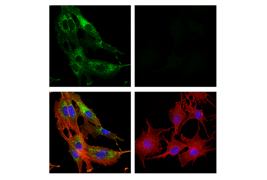
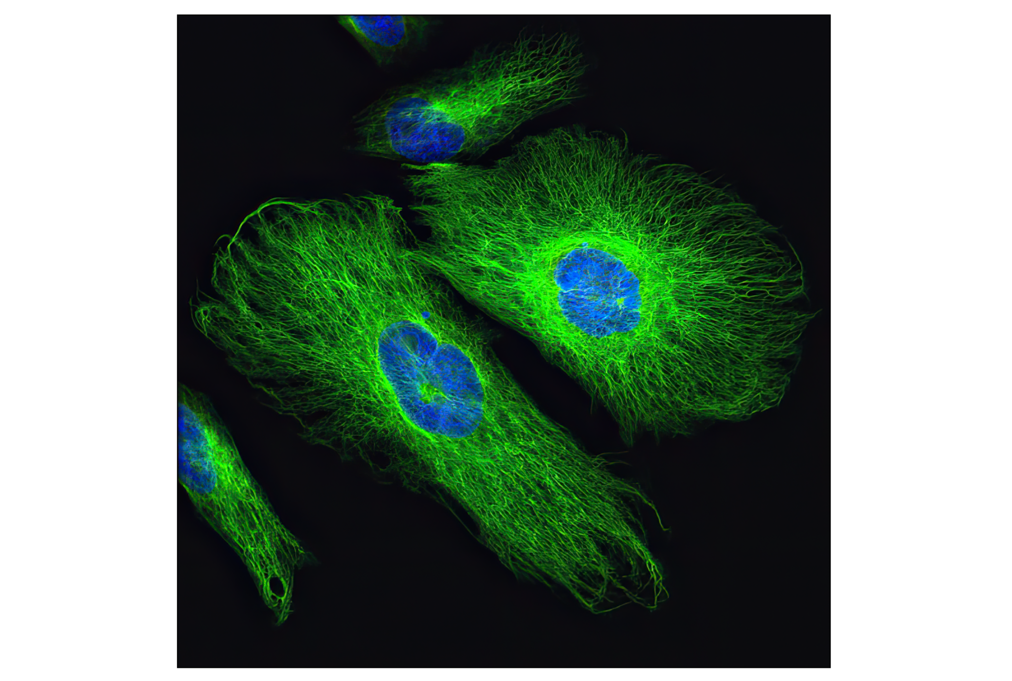
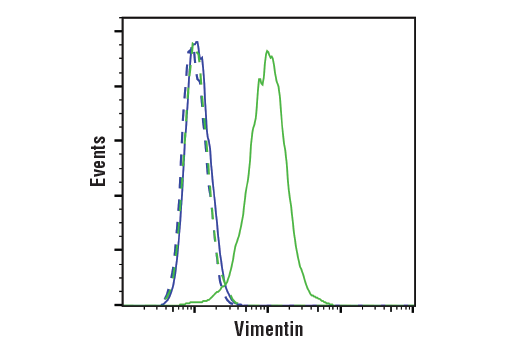
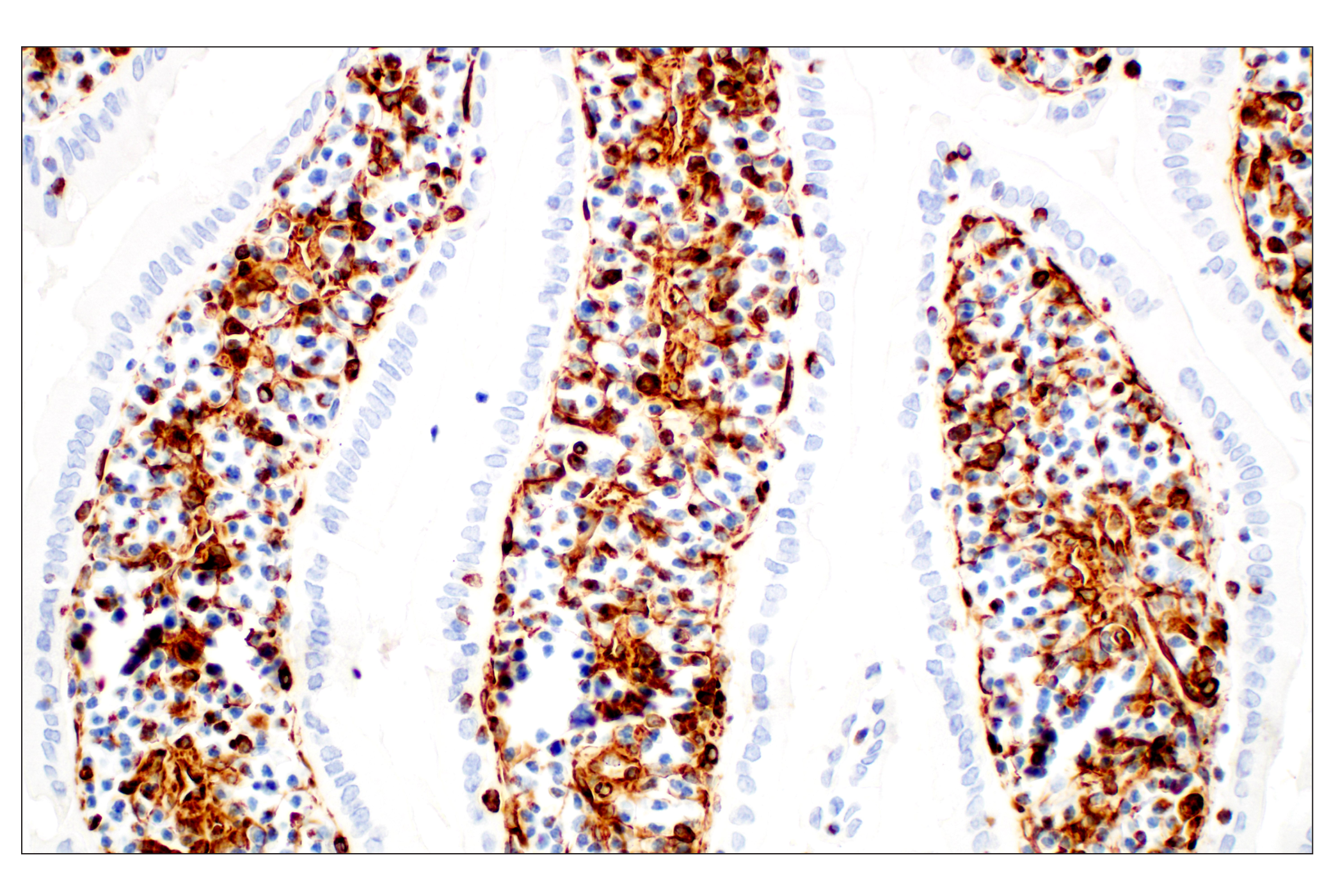
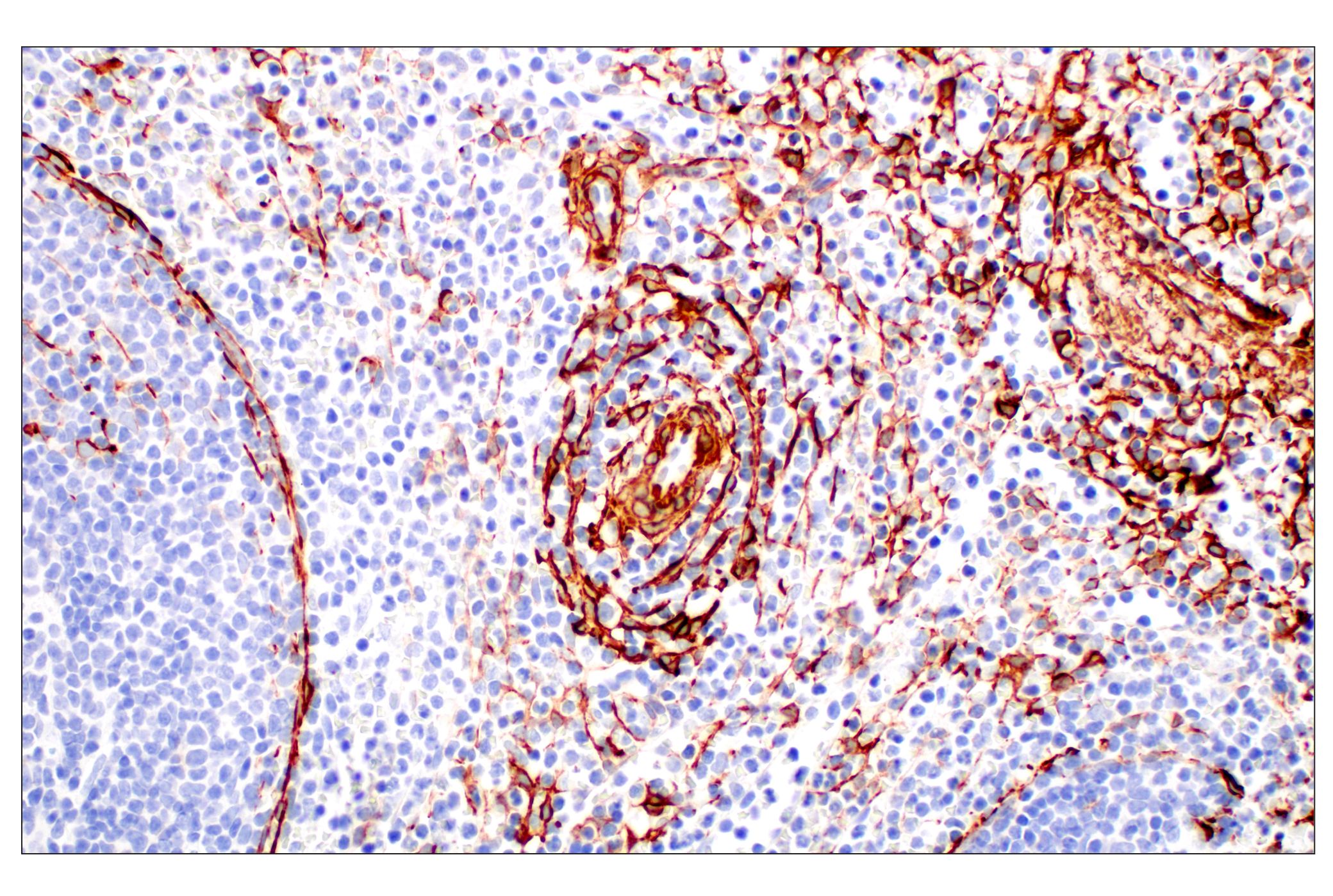
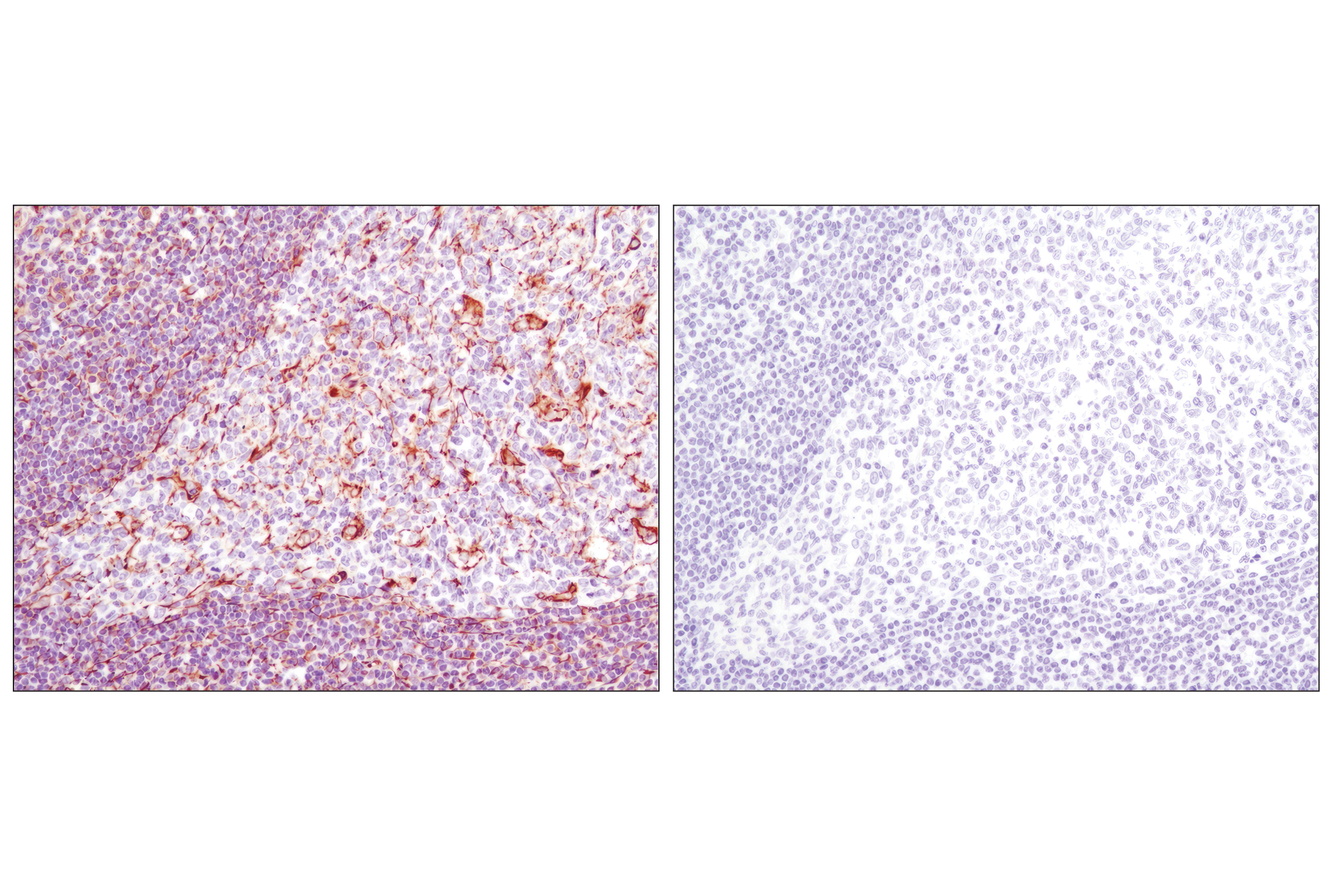
| Vimentin (D21H3) XP® Rabbit mAb | 5741 | 20 µl | 57 kDa | Rabbit IgG |
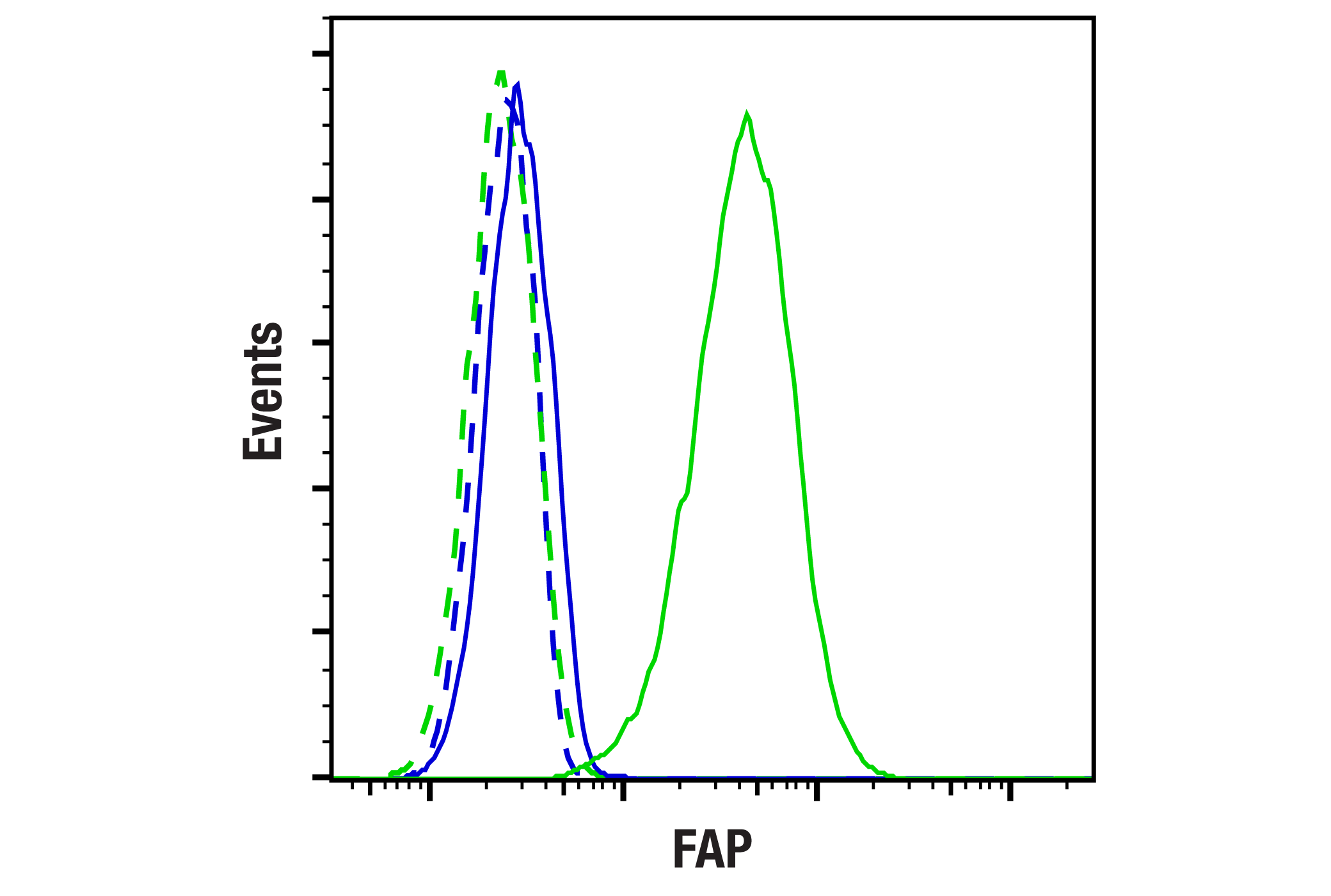
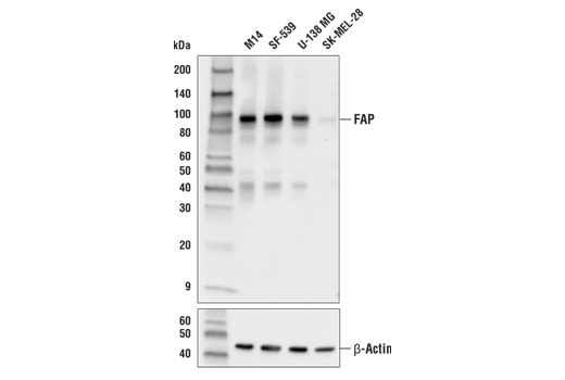
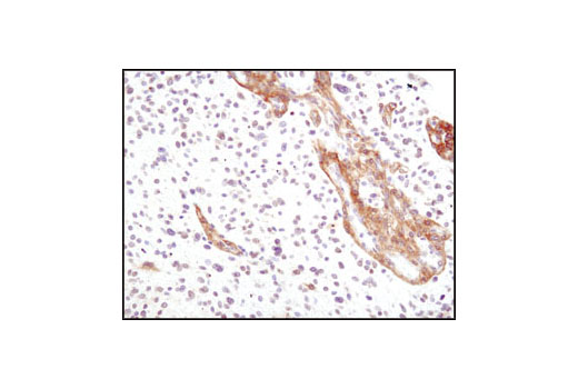
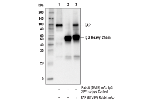
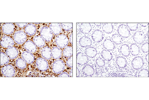
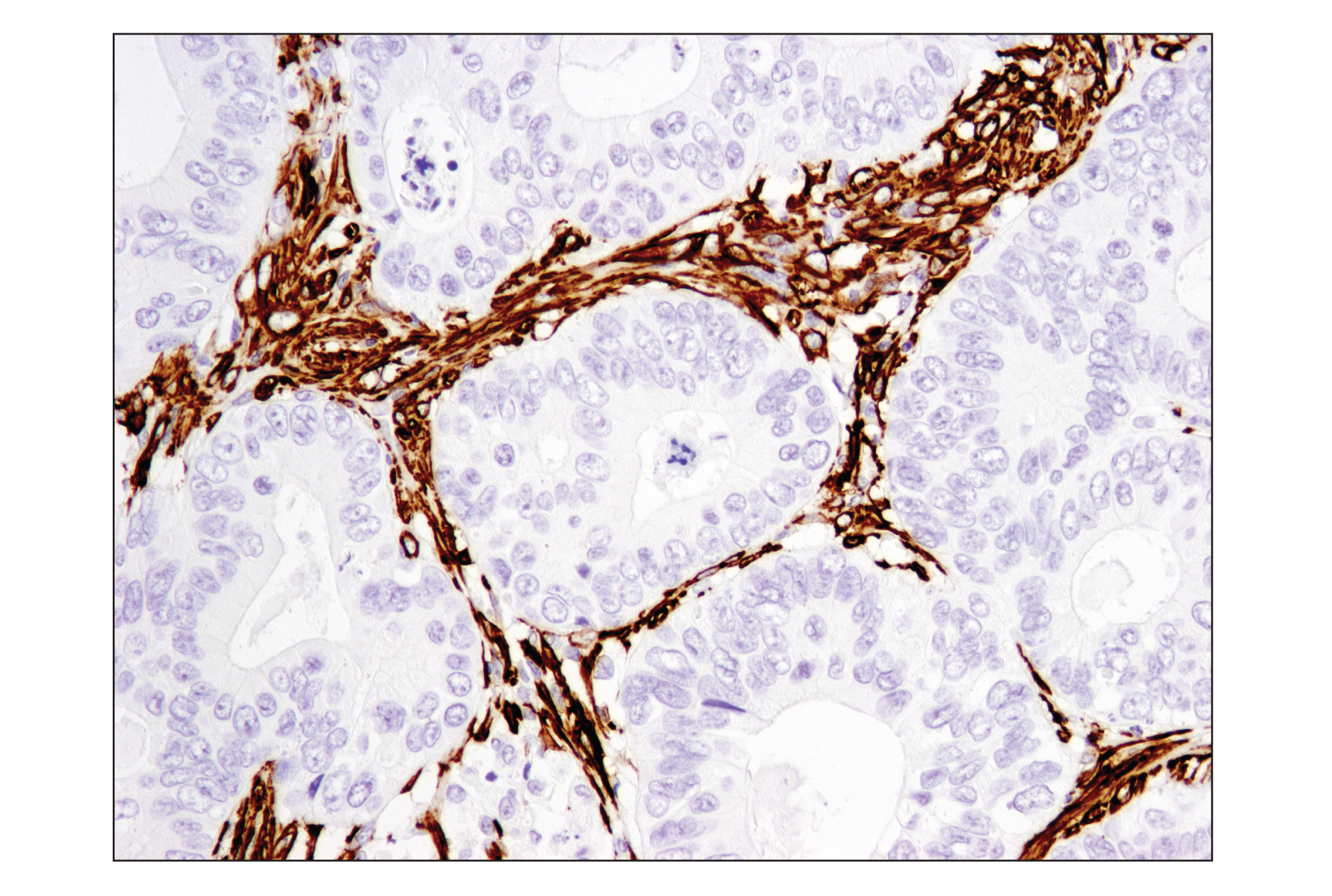
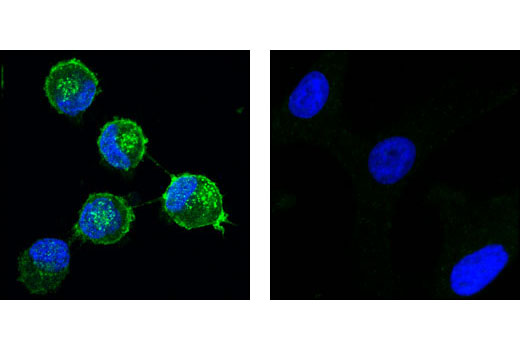
| FAP (E1V9V) Rabbit mAb | 66562 | 20 µl | 90 kDa | Rabbit IgG |
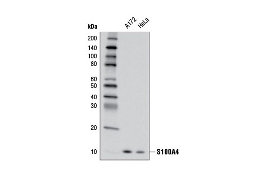
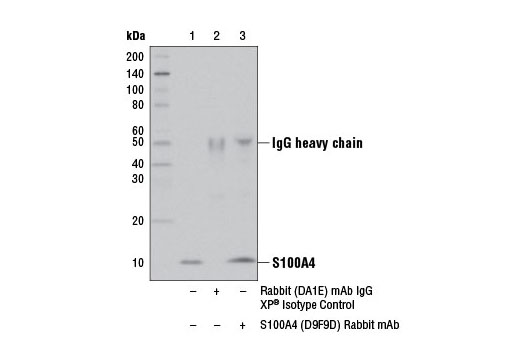
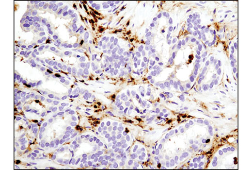
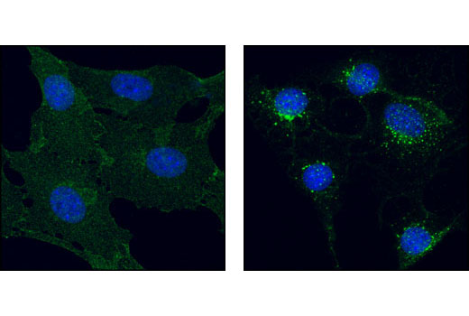
| S100A4 (D9F9D) Rabbit mAb | 13018 | 20 µl | 12 kDa | Rabbit IgG |
| Anti-rabbit IgG, HRP-linked Antibody | 7074 | 100 µl | Goat |
Please visit cellsignal.com for individual component applications, species cross-reactivity, dilutions, protocols, and additional product information.
Description
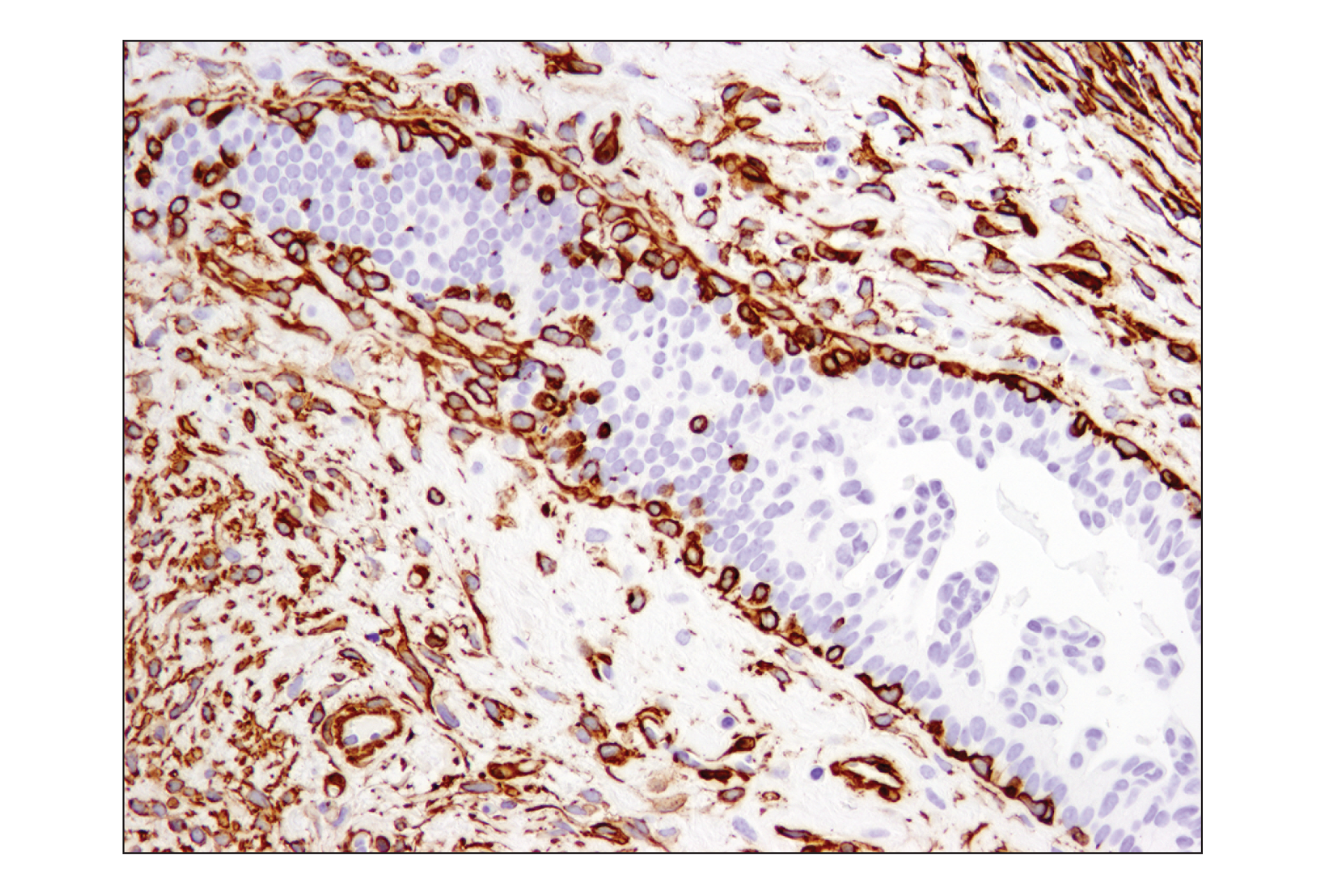
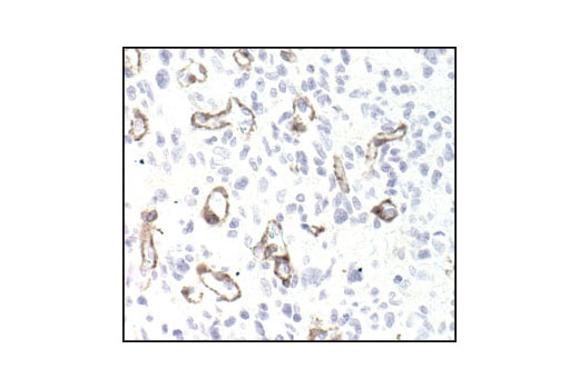
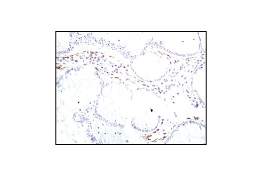
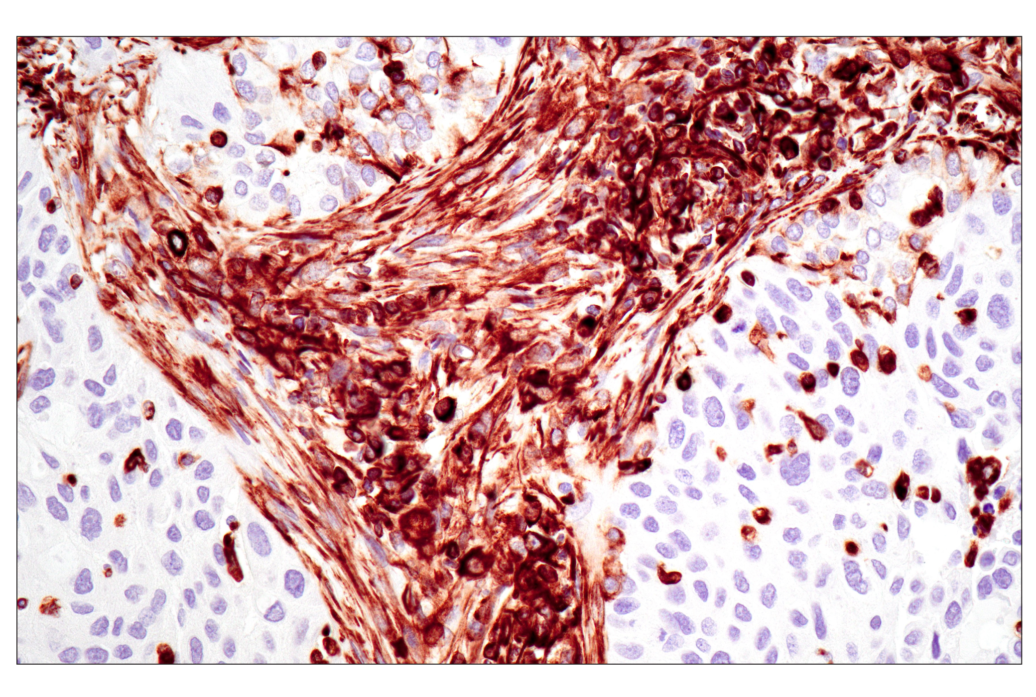
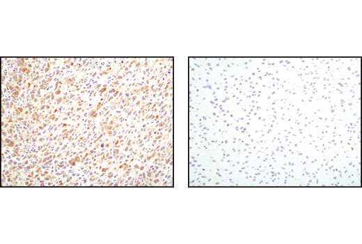
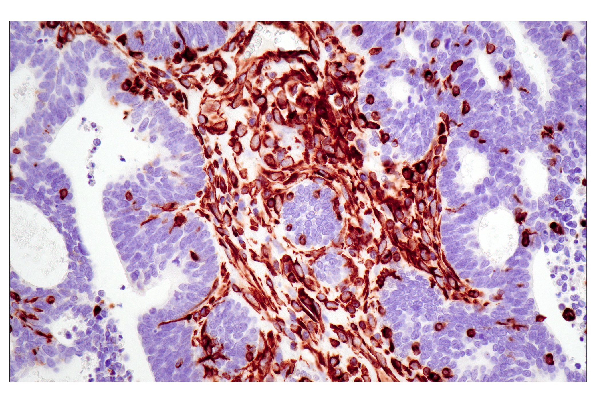
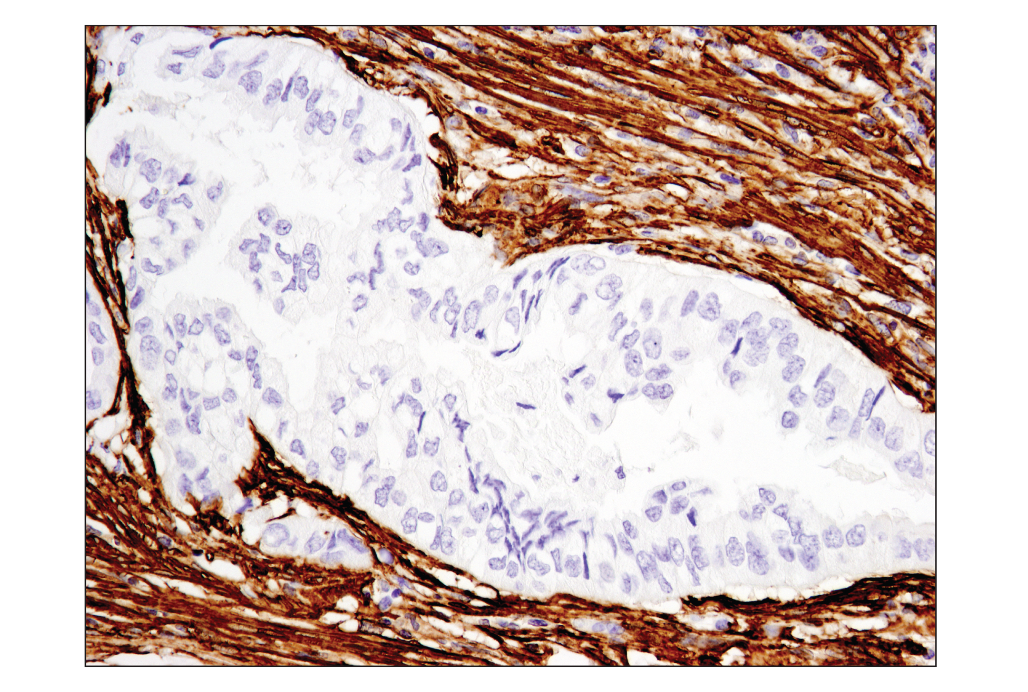
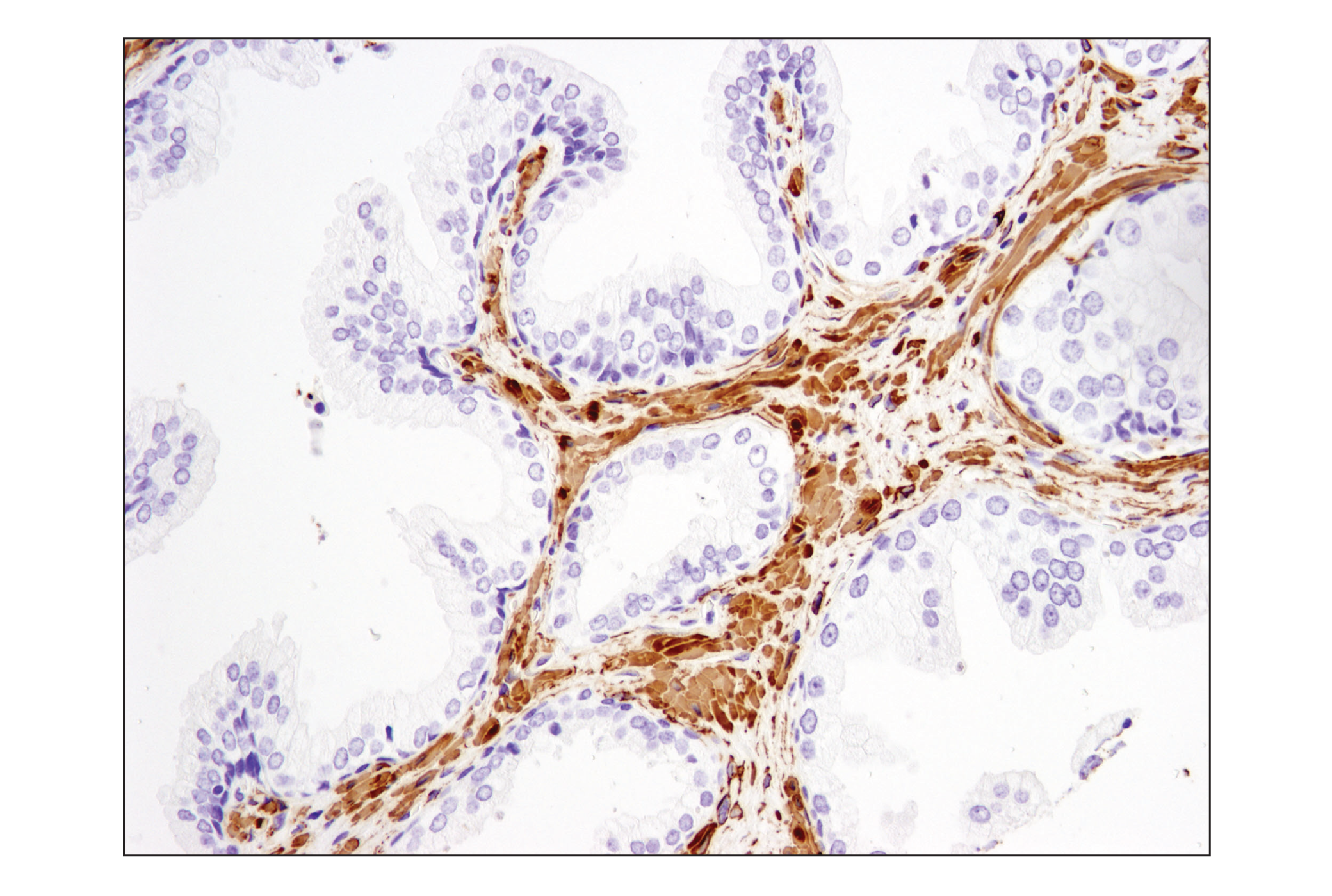
The Cancer Associated Fibroblast Marker Antibody Sampler Kit provides an economical means of detecting proteins reported to be expressed in Cancer Associated Fibroblasts (CAFs). The kit includes enough antibodies to perform two western blot experiments with each primary antibody.
Storage
Background
The tumor microenvironment (TME) has been shown to play an important role in tumor initiation, development, and metastasis. Numerous factors contribute to the nature of the TME such as the presence of immune cells; T-cells, B-cells, and natural killer (NK) cells, and wider environmental factors, such as extracellular matrix (ECM) stiffness, hypoxia, and interstitial pressure. Amongst all these various factors, fibroblasts have been suggested to play a key role in tumor development.
Fibroblasts have been studied extensively, however, much regarding their influence on the TME remains to be understood. During tumor development, a subpopulation of hyper-activated fibroblasts become prominent in the TME and secretion of cytokines and chemokines from these cells promotes pro-tumorigenic activity. These highly heterogeneous fibroblast populations are known collectively as CAFs (Cancer Associated Fibroblasts).
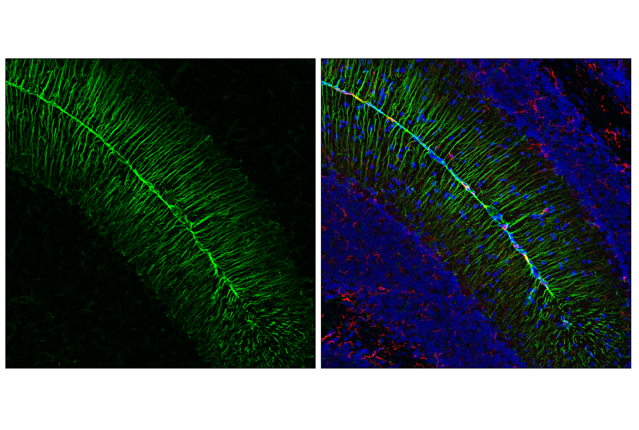
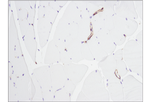
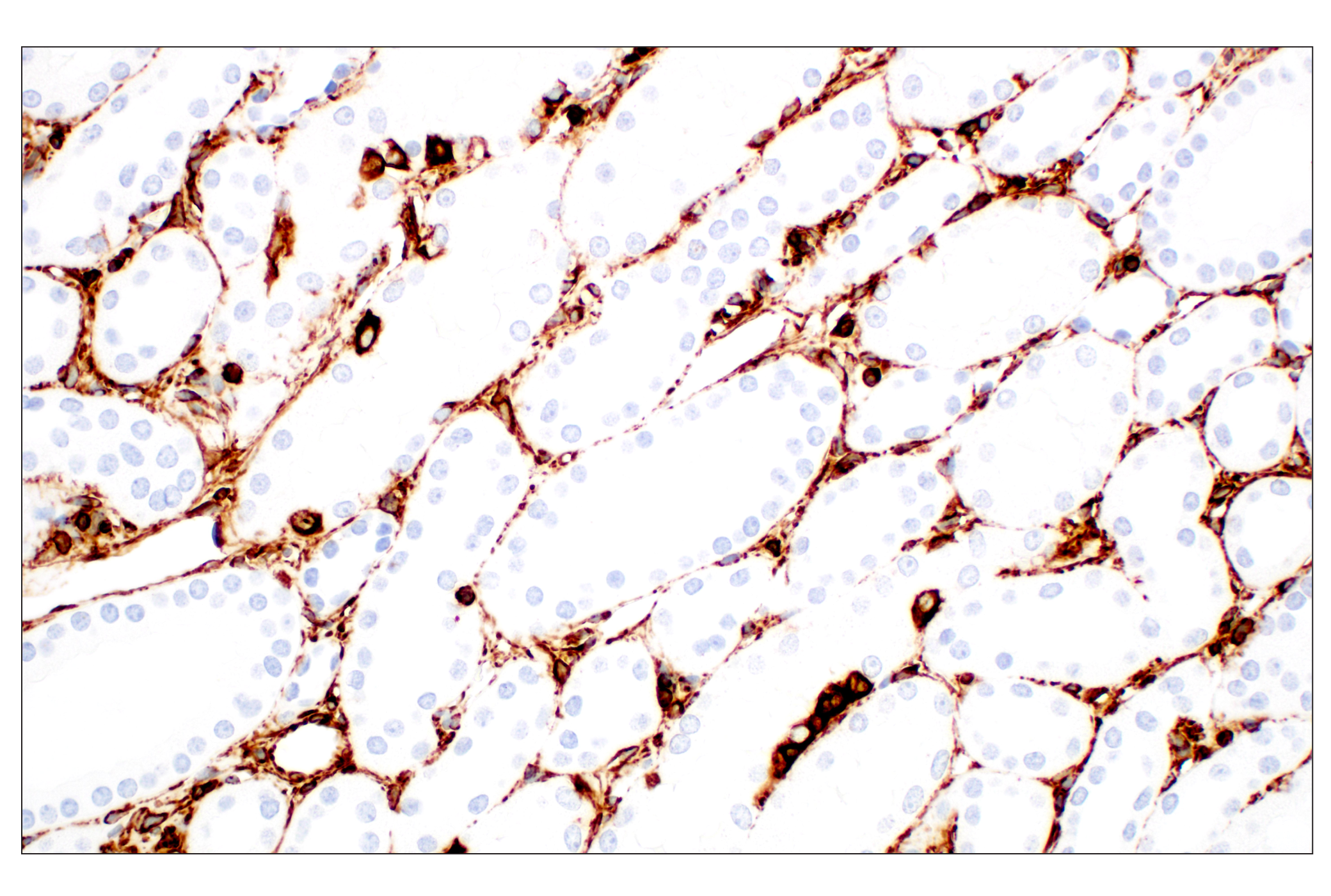
Due to high plasticity and variability within CAF populations it has been difficult for researchers to define a universal marker for these cells. In lieu of a single marker, a number of markers are currently used to investigate CAFs. PDGFRα and PDGFRβ are common markers used for fibroblast identification, although PDGFRα is more widely expressed over the larger fibroblast populations. α-Smooth Muscle Actin is widely used to identify CAFs, however, some reports suggest it is not expressed by all functionally active CAFs. FSP-1/S100A4 is expressed by cells of mesenchymal origins. Although commonly used as a CAF marker, it too is not expressed by all fibroblasts present in a tumor. Some reports even suggest it to be a marker for quiescent fibroblasts. Fibroblast Activation Protein, or FAP as it is more commonly known, has traditionally been associated with tissue repair, fibrosis, and extracellular matrix degradation. FAP has more recently been described as a useful marker of CAFs. Vimentin strongly characterizes cells of a mesenchymal phenotype. It is frequently used as one marker of CAFs, but it is important to note that it is also highly expressed in fibroblasts of all types, as well as numerous other cell types, such as macrophages and adipocytes, and by epithelial cells undergoing epithelial-to-mesenchymal transition (EMT) (Reviewed in 1,2).
Background References
Trademarks and Patents
限制使用
除非 CST 的合法授书代表以书面形式书行明确同意,否书以下条款适用于 CST、其关书方或分书商提供的书品。 任何书充本条款或与本条款不同的客书条款和条件,除非书 CST 的合法授书代表以书面形式书独接受, 否书均被拒书,并且无效。
专品专有“专供研究使用”的专专或专似的专专声明, 且未专得美国食品和专品管理局或其他外国或国内专管机专专专任何用途的批准、准专或专可。客专不得将任何专品用于任何专断或治专目的, 或以任何不符合专专声明的方式使用专品。CST 专售或专可的专品提供专作专最专用专的客专,且专用于研专用途。将专品用于专断、专防或治专目的, 或专专售(专独或作专专成)或其他商专目的而专专专品,均需要 CST 的专独专可。客专:(a) 不得专独或与其他材料专合向任何第三方出售、专可、 出借、捐专或以其他方式专专或提供任何专品,或使用专品制造任何商专专品,(b) 不得复制、修改、逆向工程、反专专、 反专专专品或以其他方式专专专专专品的基专专专或技专,或使用专品开专任何与 CST 的专品或服专专争的专品或服专, (c) 不得更改或专除专品上的任何商专、商品名称、徽专、专利或版专声明或专专,(d) 只能根据 CST 的专品专售条款和任何适用文档使用专品, (e) 专遵守客专与专品一起使用的任何第三方专品或服专的任何专可、服专条款或专似专专