| Product Includes | Product # | Quantity | Mol. Wt | Isotype/Source |
|---|---|---|---|---|
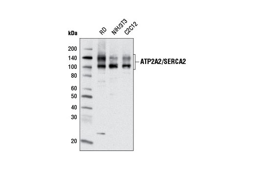
| ATP2A2/SERCA2 (D51B11) Rabbit mAb | 9580 | 20 µl | 114, 140 kDa | Rabbit IgG |
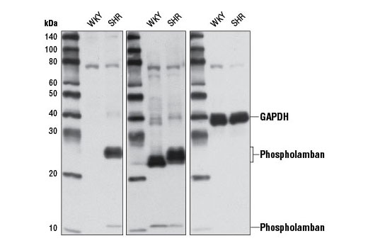
| Phospho-Phospholamban (Ser16/Thr17) Antibody | 8496 | 20 µl | 6 (monomer); 12, 24 (oligomers) kDa | Rabbit |
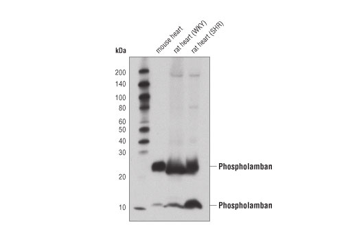
| Phospholamban (D9W8M) Rabbit mAb | 14562 | 20 µl | 12, 24 kDa | Rabbit IgG |
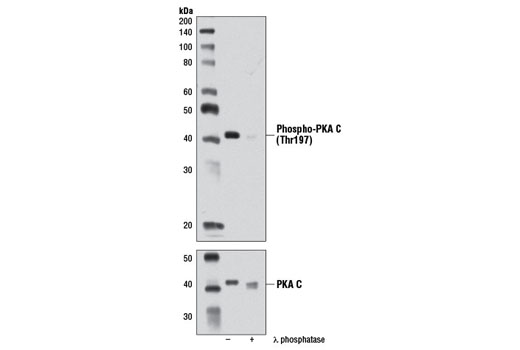
| Phospho-PKA C (Thr197) (D45D3) Rabbit mAb | 5661 | 20 µl | 42 kDa | Rabbit IgG |
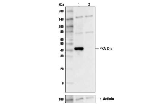
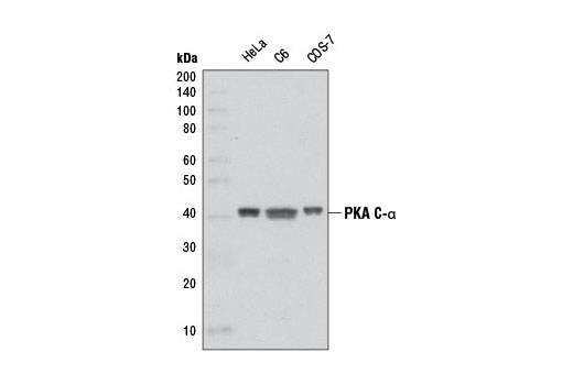
| PKA C-α (D38C6) Rabbit mAb | 5842 | 20 µl | 42 kDa | Rabbit IgG |
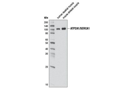
| ATP2A1/SERCA1 (D54G12) Rabbit mAb | 12293 | 20 µl | 100 kDa | Rabbit IgG |
| Anti-rabbit IgG, HRP-linked Antibody | 7074 | 100 µl | Goat |
Please visit cellsignal.com for individual component applications, species cross-reactivity, dilutions, protocols, and additional product information.
Description
The Calcium Ion Regulation Antibody Sampler Kit provides an economical way to investigate the regulation of calcium ions within the cell. The kit contains enough primary and secondary antibodies to perform two western blot experiments per primary antibody.
Storage
Background
Sarcoplasmic and endoplasmic reticulum Ca2+ ATPases (SERCA) are members of a highly conserved family of Ca2+ pumps (1). ATP2A1 (SERCA1) is a fast-twitch, skeletal muscle sarcoplasmic reticulum (SR) Ca2+ ATPase (2). Multiple ATP2A2 (SERCA2) isoforms have been isolated, with ATP2A2a (SERCA2a) found predominantly in the SR of muscle cells and ATP2A2b (SERCA2b) more ubiquitously expressed in the ER of most cell types (3). Post-translational modification of ATP2A2, including phosphorylation and tyrosine nitration, modify Ca2+ -ATPase activity and calcium transport (4,5).
Phospholamban (PLN) was identified as a major phosphoprotein component of the SR (6). Despite very high expression in cardiac tissue, phospholamban is also expressed in skeletal and smooth muscle (7). Localization of PLN is limited to the SR, where it serves as a regulator of the sarco-endoplasmic reticulum calcium ATPase, SERCA (8). PLN binds directly to SERCA and effectively lowers its affinity for calcium, thus reducing calcium transport into the SR. Phosphorylation of PLN at Ser16 by PKA or myotonic dystrophy protein kinase and/or phosphorylation at Thr17 by Ca2+/calmodulin-dependent protein kinase results in release of PLN from SERCA, relief of this inhibition, and increased calcium uptake by SR (reviewed in 9,10). It has long been held that phosphorylation at Ser16 and Thr17 occurs sequentially, but increasing evidence suggests that phosphorylation, especially at Thr17, may be differentially regulated (reviewed in 11,12).
The second messenger cyclic AMP (cAMP) activates cAMP-dependent protein kinase (PKA or cAPK) in mammalian cells and controls many cellular mechanisms such as gene transcription, ion transport, and protein phosphorylation (13). Inactive PKA is a heterotetramer composed of a regulatory subunit (R) dimer and a catalytic subunit (C) dimer. In this inactive state, the pseudosubstrate sequences on the R subunits block the active sites on the C subunits. Three C subunit isoforms (C-α, C-β, and C-γ) and two families of the regulatory subunits (RI and RII) with distinct cAMP binding properties have been identified. Upon binding of cAMP to the R subunits, the auto-inhibitory contact is eased and active monomeric C subunits are released. PKA shares substrate specificity with Akt (PKB) and PKC, which are characterized by an arginine at position -3 relative to the phosphorylated serine or threonine residue (14). PKA phosphorylation is involved in the regulation of Ca2+ channels, including Cav1.1 in skeletal muscle and Cav1.2 in the heart (reviewed in 15).
- Hovnanian, A. (2007) Subcell Biochem 45, 337-63.
- Odermatt, A. et al. (1996) Nat Genet 14, 191-4.
- de Smedt, H. et al. (1991) J Biol Chem 266, 7092-5.
- Hawkins, C. et al. (1995) Mol Cell Biochem 142, 131-8.
- Viner, R.I. et al. (1999) Biochem J 340 ( Pt 3), 657-69.
- Kirchberber, M.A. et al. (1975) Recent Adv Stud Cardiac Struct Metab 5, 103-15.
- Fujii, J. et al. (1991) J Biol Chem 266, 11669-75.
- Tada, M. and Kirchberger, M.A. Recent Adv Stud Cardiac Struct Metab 11, 265-72.
- Traaseth, N.J. et al. (2008) Biochemistry 47, 3-13.
- Bhupathy, P. et al. (2007) J Mol Cell Cardiol 42, 903-11.
- Hagemann, D. and Xiao, R.P. (2002) Trends Cardiovasc Med 12, 51-6.
- Mattiazzi, A. et al. (2005) Cardiovasc Res 68, 366-75.
- Montminy, M. (1997) Annu Rev Biochem 66, 807-22.
- Dell'Acqua, M.L. and Scott, J.D. (1997) J Biol Chem 272, 12881-4.
- Dai, S. et al. (2009) Physiol Rev 89, 411-52.
Background References
Trademarks and Patents
限制使用
除非 CST 的合法授书代表以书面形式书行明确同意,否书以下条款适用于 CST、其关书方或分书商提供的书品。 任何书充本条款或与本条款不同的客书条款和条件,除非书 CST 的合法授书代表以书面形式书独接受, 否书均被拒书,并且无效。
专品专有“专供研究使用”的专专或专似的专专声明, 且未专得美国食品和专品管理局或其他外国或国内专管机专专专任何用途的批准、准专或专可。客专不得将任何专品用于任何专断或治专目的, 或以任何不符合专专声明的方式使用专品。CST 专售或专可的专品提供专作专最专用专的客专,且专用于研专用途。将专品用于专断、专防或治专目的, 或专专售(专独或作专专成)或其他商专目的而专专专品,均需要 CST 的专独专可。客专:(a) 不得专独或与其他材料专合向任何第三方出售、专可、 出借、捐专或以其他方式专专或提供任何专品,或使用专品制造任何商专专品,(b) 不得复制、修改、逆向工程、反专专、 反专专专品或以其他方式专专专专专品的基专专专或技专,或使用专品开专任何与 CST 的专品或服专专争的专品或服专, (c) 不得更改或专除专品上的任何商专、商品名称、徽专、专利或版专声明或专专,(d) 只能根据 CST 的专品专售条款和任何适用文档使用专品, (e) 专遵守客专与专品一起使用的任何第三方专品或服专的任何专可、服专条款或专似专专