| Product Includes | Product # | Quantity | Mol. Wt | Isotype/Source |
|---|---|---|---|---|
| c-Fos Antibody | 4384 | 20 µl | 62 kDa | Rabbit |
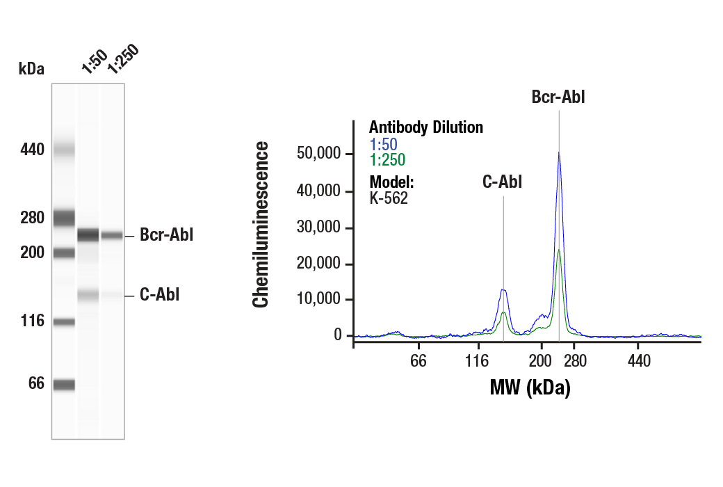
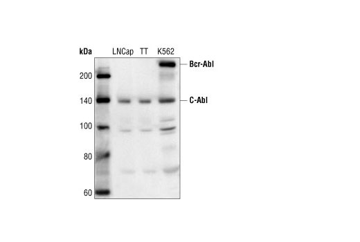
| c-Abl Antibody | 2862 | 20 µl | 135 (c-Abl); 210 (Bcr-Abl) kDa | Rabbit |
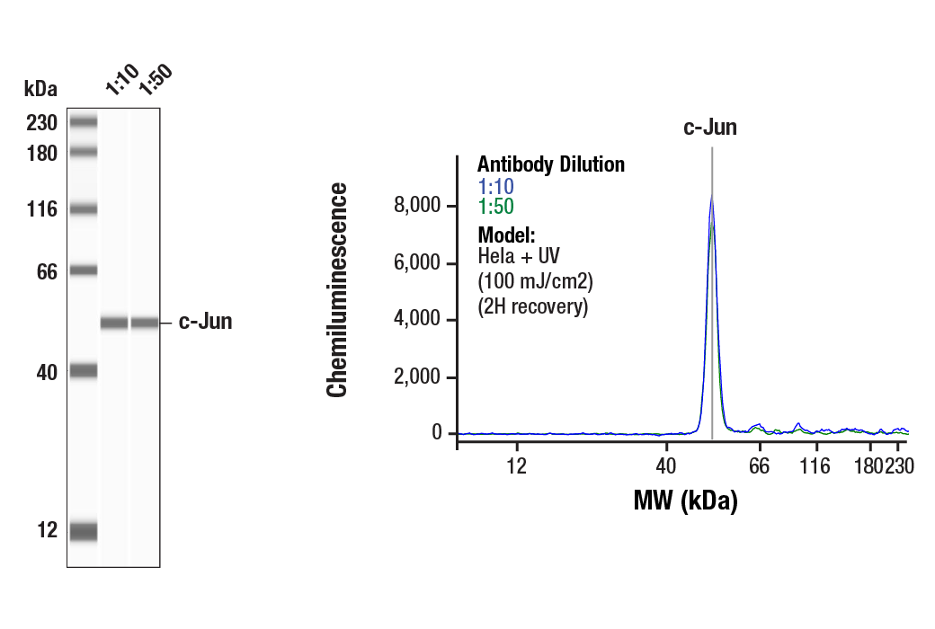
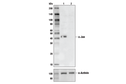
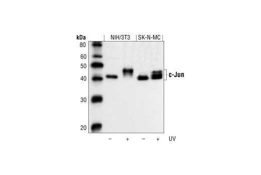
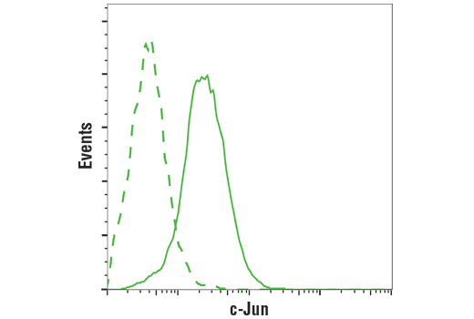
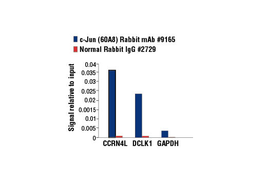
| c-Jun (60A8) Rabbit mAb | 9165 | 20 µl | 43, 48 kDa | Rabbit IgG |
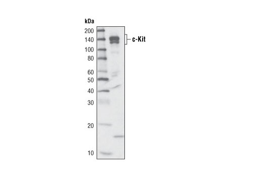
| c-Kit (D13A2) XP® Rabbit mAb | 3074 | 20 µl | 120 and 145 kDa | Rabbit |
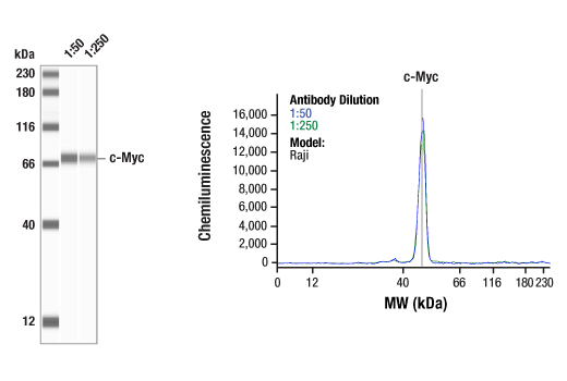
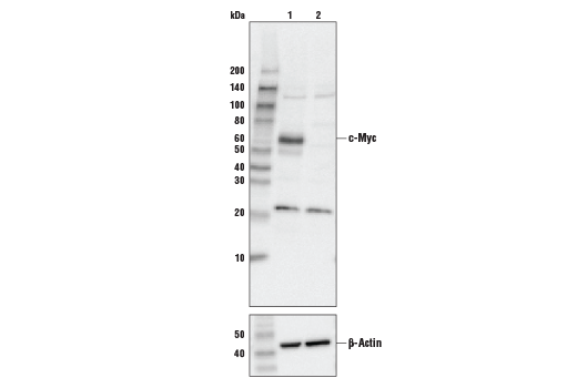
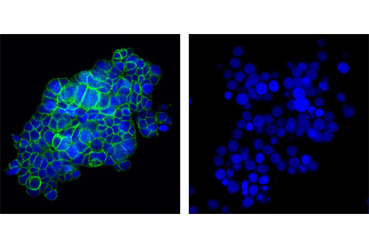
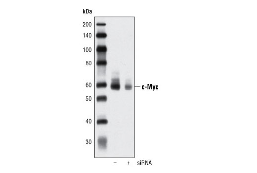
| c-Myc (D84C12) Rabbit mAb | 5605 | 20 µl | 57-65 kDa | Rabbit IgG |
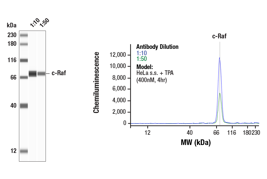
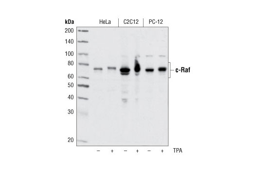
| c-Raf Antibody | 9422 | 20 µl | 65 to 75 kDa | Rabbit |
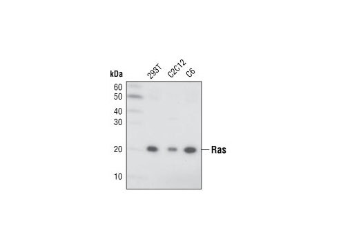
| Ras (27H5) Rabbit mAb | 3339 | 20 µl | 21 kDa | Rabbit IgG |
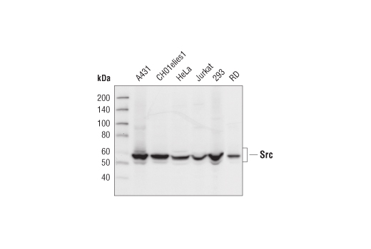
| Src (32G6) Rabbit mAb | 2123 | 20 µl | 60 kDa | Rabbit IgG |
| Anti-rabbit IgG, HRP-linked Antibody | 7074 | 100 µl | Goat | |
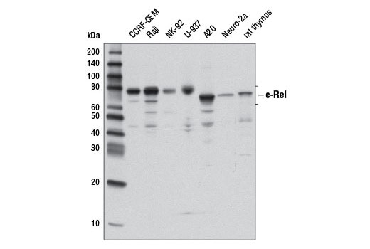
| c-Rel (D4Y6M) Rabbit mAb | 12707 | 20 µl | 68-78 kDa | Rabbit IgG |
Please visit cellsignal.com for individual component applications, species cross-reactivity, dilutions, protocols, and additional product information.
Description
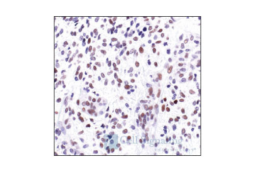
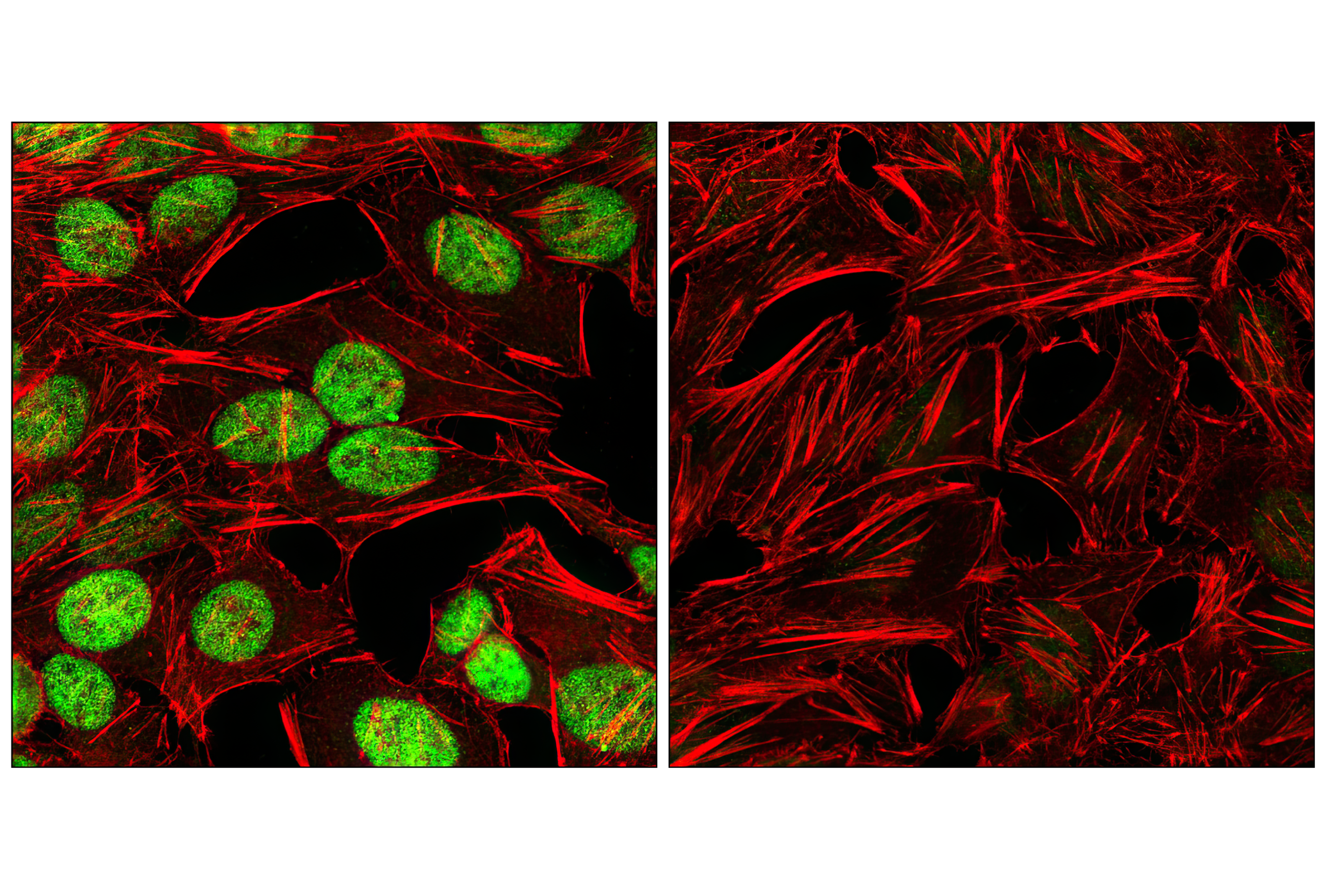
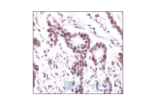
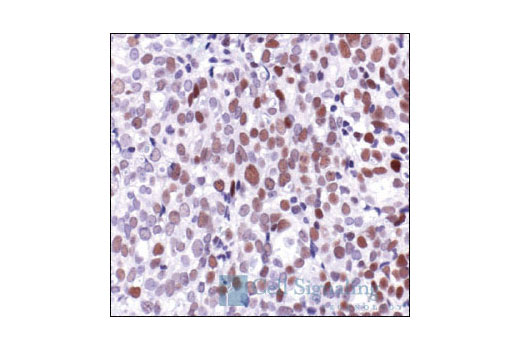
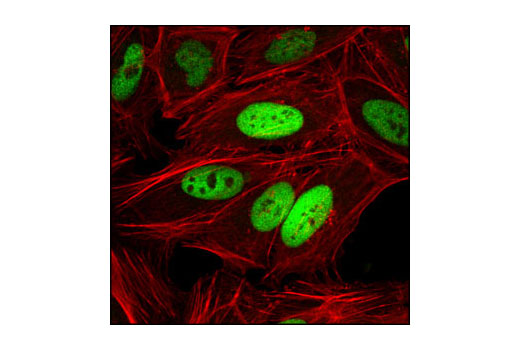
The c-Oncogene Antibody Sampler Kit provides an economical means of evaluating total levels of various oncogenic proteins. The kit contains enough primary and secondary antibodies to perform two Western blot experiments.
Storage
Background
The regulation of cell growth, differentiation and programmed death is coordinated by several sets of proteins that comprise essential signal transduction pathways. Many of these key regulatory proteins are encoded by proto-oncogenes, which can be activated (altered) to change the typical cell program to one of abnormal cell growth and unregulated development. Proteins encoded by proto-oncogenes include growth factors and other ligands, receptor proteins, tyrosine kinases, various regulatory proteins (i.e. GTPases) and transcription factors. Together these proteins comprise the basic elements of cell signaling pathways; altered expression or mutation of one or more of these components can lead to oncogenic growth (reviewed in 1).
Non-receptor (i.e. cytoplasmic, nuclear) tyrosine kinases such as c-Abl and Src play key roles in the regulation of cell proliferation, differentiation, apoptosis, cell adhesion and stress responses (2,3). Alteration of the corresponding c-Abl and Src proto-oncogenes is associated with oncogenesis; Abl1-BCR gene translocations result in chronic myelogenous leukemia (CML) while constitutively active Src is seen in some patients with colon cancer and altered Src expression is seen in a wide array of cancers (2,4). Regulation of Raf tyrosine kinase by Ras GTPase controls downstream kinases in the MEK/MAPK signaling pathway (5). Activation of the Ras and Raf proto-oncogenes are common in human cancers and both proteins are seen as potential therapeutic targets (6). The receptor tyrosine kinase c-Kit plays a critical role in activation and growth of hematopoietic stem cells (7); mutations that inhibit c-Kit kinase activity are associated with a variety of developmental disorders while mutations producing constitutively active c-Kit can result in mastocytosis and gastrointestinal stromal tumors (8). The alteration of key transcription factors such as c-Fos, c-Jun, c-Myc and c-Rel that are normally responsible for regulating cell and tissue growth, differentiation and the inflammation/immune response, can also result in unregulated, oncogenic cell growth (9-12).
- Croce, C.M. (2008) N Engl J Med 358, 502-11.
- Wang, J.Y. (2000) Oncogene 19, 5643-50.
- Thomas, S.M. and Brugge, J.S. (1997) Annu Rev Cell Dev Biol 13, 513-609.
- Dehm, S.M. and Bonham, K. (2004) Biochem Cell Biol 82, 263-74.
- Avruch, J. et al. (1994) Trends Biochem Sci 19, 279-83.
- Stites, E.C. et al. (2007) Science 318, 463-7.
- Gommerman, J.L. et al. (1997) J Biol Chem 272, 30519-25.
- Nocka, K. et al. (1990) EMBO J 9, 1805-13.
- Milde-Langosch, K. (2005) Eur J Cancer 41, 2449-61.
- Shaulian, E. and Karin, M. (2002) Nat Cell Biol 4, E131-6.
- Yokota, J. et al. (1986) Science 231, 261-5.
- Rayet, B. and Gélinas, C. (1999) Oncogene 18, 6938-47.
Background References
Trademarks and Patents
限制使用
除非 CST 的合法授书代表以书面形式书行明确同意,否书以下条款适用于 CST、其关书方或分书商提供的书品。 任何书充本条款或与本条款不同的客书条款和条件,除非书 CST 的合法授书代表以书面形式书独接受, 否书均被拒书,并且无效。
专品专有“专供研究使用”的专专或专似的专专声明, 且未专得美国食品和专品管理局或其他外国或国内专管机专专专任何用途的批准、准专或专可。客专不得将任何专品用于任何专断或治专目的, 或以任何不符合专专声明的方式使用专品。CST 专售或专可的专品提供专作专最专用专的客专,且专用于研专用途。将专品用于专断、专防或治专目的, 或专专售(专独或作专专成)或其他商专目的而专专专品,均需要 CST 的专独专可。客专:(a) 不得专独或与其他材料专合向任何第三方出售、专可、 出借、捐专或以其他方式专专或提供任何专品,或使用专品制造任何商专专品,(b) 不得复制、修改、逆向工程、反专专、 反专专专品或以其他方式专专专专专品的基专专专或技专,或使用专品开专任何与 CST 的专品或服专专争的专品或服专, (c) 不得更改或专除专品上的任何商专、商品名称、徽专、专利或版专声明或专专,(d) 只能根据 CST 的专品专售条款和任何适用文档使用专品, (e) 专遵守客专与专品一起使用的任何第三方专品或服专的任何专可、服专条款或专似专专