Revision 1
Cell Signaling Technology

Orders: 877-616-CELL (2355) [email protected]

Support: 877-678-TECH (8324)

Web: [email protected] cellsignal.com

3 Trask LaneDanversMassachusetts01923USA
For Research Use Only. Not for Use in Diagnostic Procedures.
:

:

:

:

UniProt ID:

#P05412

Entrez-Gene Id:

3725

Product Information

Storage

Supplied in 10 mM sodium HEPES (pH 7.5), 150 mM NaCl, 100 µg/ml BSA, 50% glycerol and less than 0.02% sodium azide. Store at –20°C. Do not aliquot the antibody.

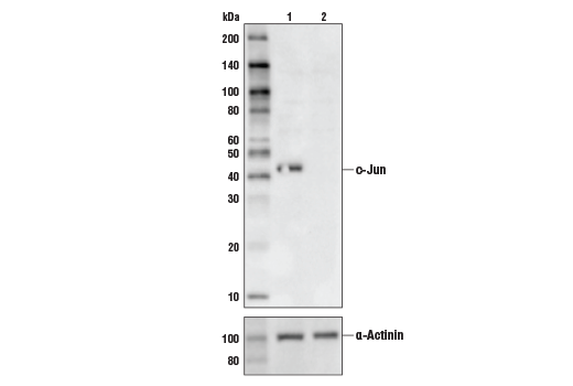
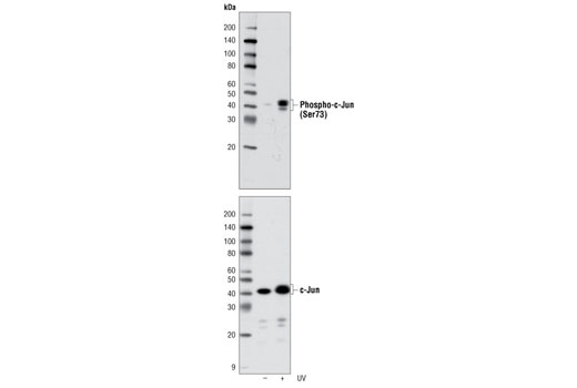
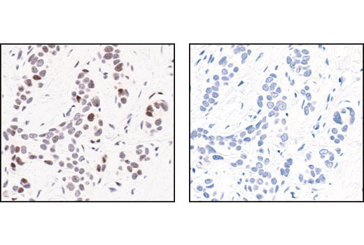
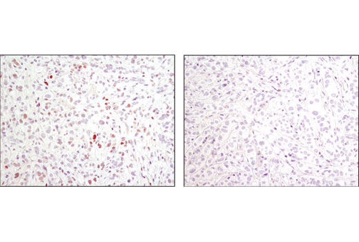
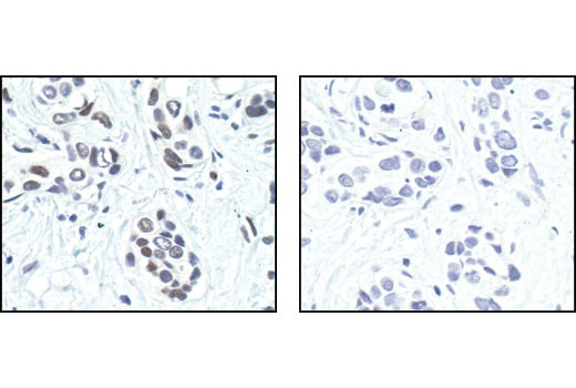
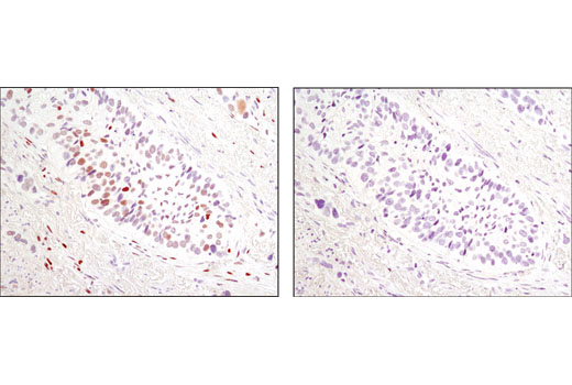
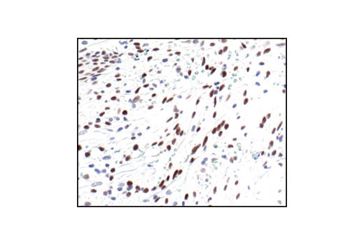
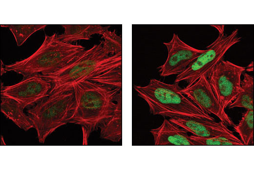
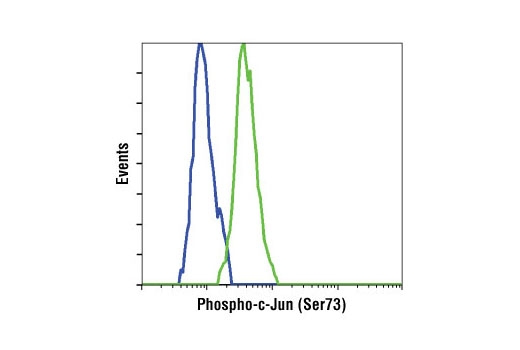
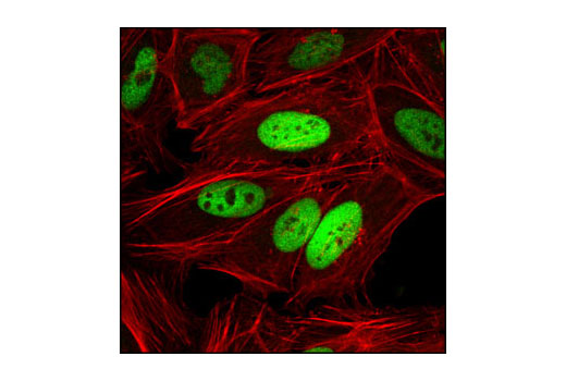
Specificity / Sensitivity

Phospho-c-Jun (Ser63) (54B3) Rabbit mAb detects endogenous levels of c-Jun only when phosphorylated at Ser63. Phospho-c-Jun (Ser73) Antibody detects endogenous levels of c-Jun only when phosphorylated at Ser73. This antibody also recognizes phosphorylation of JunD at Ser100. c-Jun (60A8) Rabbit mAb detects endogenous levels of total c-Jun protein.

Source / Purification

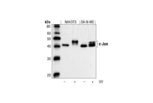
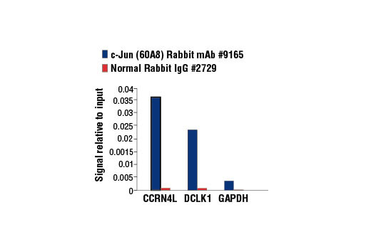
Phospho-specific polyclonal antibodies are produced by immunizing rabbits with a synthetic phosphopeptide corresponding to residues surrounding Ser63 or Ser73 of human c-Jun, and purified by protein A and peptide affinity chromatography. Monoclonal antibody is produced by immunizing animals with a synthetic peptide corresponding to the amino-terminal of human c-Jun.

Product Description

The PhosphoPlus (R) c-Jun (Ser63) and c-Jun (Ser73) Antibody Kit provides reagents and controls for rapid analysis of c-Jun phosphorylation status.
MW (kDa) 43, 50

Background

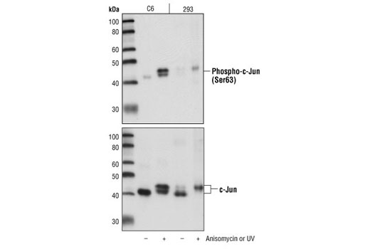
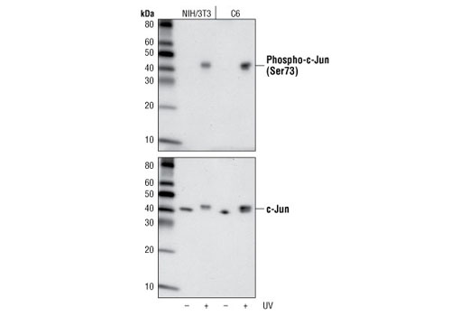
c-Jun is a member of the Jun family containing c-Jun, JunB, and JunD, and is a component of the transcription factor activator protein-1 (AP-1). AP-1 is composed of dimers of Fos, Jun, and ATF family members and binds to and activates transcription at TRE/AP-1 elements (reviewed in 1). Extracellular signals, including growth factors, chemokines, and stress, activate AP-1-dependent transcription. The transcriptional activity of c-Jun is regulated by phosphorylation at Ser63 and Ser73 through SAPK/JNK (reviewed in 2). Knockout studies in mice have shown that c-Jun is essential for embryogenesis (3), and subsequent studies have demonstrated roles for c-Jun in various tissues and developmental processes, including axon regeneration (4), liver regeneration (5), and T cell development (6). AP-1 regulated genes exert diverse biological functions, including cell proliferation, differentiation, and apoptosis, as well as transformation, invasion and metastasis, depending on cell type and context (7-9). Other target genes regulate survival, as well as hypoxia and angiogenesis (8,10). Research studies have implicated c-Jun as a promising therapeutic target for cancer, vascular remodeling, acute inflammation, and rheumatoid arthritis (11,12).

  1. Jochum, W. et al. (2001) Oncogene 20, 2401-12.
  2. Davis, R.J. (2000) Cell 103, 239-52.
  3. Hilberg, F. et al. (1993) Nature 365, 179-81.
  4. Raivich, G. et al. (2004) Neuron 43, 57-67.
  5. Behrens, A. et al. (2002) EMBO J 21, 1782-90.
  6. Riera-Sans, L. and Behrens, A. (2007) J Immunol 178, 5690-700.
  7. Leppä, S. and Bohmann, D. (1999) Oncogene 18, 6158-62.
  8. Shaulian, E. and Karin, M. (2002) Nat Cell Biol 4, E131-6.
  9. Weiss, C. and Bohmann, D. (2004) Cell Cycle 3, 111-3.
  10. Karamouzis, M.V. et al. (2007) Mol Cancer Res 5, 109-20.
  11. Kim, S. and Iwao, H. (2003) J Pharmacol Sci 91, 177-81.
  12. Dass, C.R. and Choong, P.F. (2008) Pharmazie 63, 411-4.

Species Reactivity

Species reactivity is determined by testing in at least one approved application (e.g., western blot).

Cross-Reactivity Key

H: human M: mouse R: rat Hm: hamster Mk: monkey Vir: virus Mi: mink C: chicken Dm: D. melanogaster X: Xenopus Z: zebrafish B: bovine Dg: dog Pg: pig Sc: S. cerevisiae Ce: C. elegans Hr: horse GP: Guinea Pig Rab: rabbit All: all species expected

Trademarks and Patents

Cell Signaling Technology is a trademark of Cell Signaling Technology, Inc.
PhosphoPlus is a trademark of Cell Signaling Technology, Inc.
All other trademarks are the property of their respective owners. Visit cellsignal.com/trademarks for more information.

限制使用

除非 CST 的合法授书代表以书面形式书行明确同意,否书以下条款适用于 CST、其关书方或分书商提供的书品。 任何书充本条款或与本条款不同的客书条款和条件,除非书 CST 的合法授书代表以书面形式书独接受, 否书均被拒书,并且无效。

专品专有“专供研究使用”的专专或专似的专专声明, 且未专得美国食品和专品管理局或其他外国或国内专管机专专专任何用途的批准、准专或专可。客专不得将任何专品用于任何专断或治专目的, 或以任何不符合专专声明的方式使用专品。CST 专售或专可的专品提供专作专最专用专的客专,且专用于研专用途。将专品用于专断、专防或治专目的, 或专专售(专独或作专专成)或其他商专目的而专专专品,均需要 CST 的专独专可。客专:(a) 不得专独或与其他材料专合向任何第三方出售、专可、 出借、捐专或以其他方式专专或提供任何专品,或使用专品制造任何商专专品,(b) 不得复制、修改、逆向工程、反专专、 反专专专品或以其他方式专专专专专品的基专专专或技专,或使用专品开专任何与 CST 的专品或服专专争的专品或服专, (c) 不得更改或专除专品上的任何商专、商品名称、徽专、专利或版专声明或专专,(d) 只能根据 CST 的专品专售条款和任何适用文档使用专品, (e) 专遵守客专与专品一起使用的任何第三方专品或服专的任何专可、服专条款或专似专专

Revision 1
#9260

PhosphoPlus® c-Jun (Ser63) and c-Jun (Ser73) Antibody Kit

PhosphoPlus® c-Jun (Ser63) and c-Jun (Ser73) Antibody Kit: Image 1 Expand Image

使用  c-Jun (60A8) Rabbit mAb #9165 对经过紫外线(100 mJ/cm2;恢复 2 小时)处理的 HeLa 细胞裂解物 (0.1 mg/mL) 进行 Simple Western™ 分析。虚拟泳道式图像(左图)显示一抗稀释比例为 1:10 和 1:50 时的靶标条带(如图所示)。对应的电泳图(右图)为一抗稀释比例在 1:10(蓝线)和 1:50(绿线)时沿毛细血管内分子量的化学发光结果。在还原条件下,使用 12-230 kDa 分离模块在 ProteinSimple(BioTechne 品牌)的 Jess™ Simple Western 仪器上进行该实验。

PhosphoPlus® c-Jun (Ser63) and c-Jun (Ser73) Antibody Kit: Image 2 Expand Image
使用 Phospho-c-Jun (Ser63) (54B3) Rabbit mAb(上)或 c-Jun (60A8) Rabbit mAb #9165(下),对未经或经茴香霉素处理的 C6 细胞提取物;或者,对未经或经紫外线处理的 293 细胞提取物进行蛋白质印迹分析。
PhosphoPlus® c-Jun (Ser63) and c-Jun (Ser73) Antibody Kit: Image 3 Expand Image
使用 Phospho-c-Jun (Ser73) (D47G9) XP® Rabbit mAb(上)或 c-Jun (60A8) Rabbit mAb #9165(下),对未经或经紫外线处理的 NIH/3T3 或 C6 细胞提取物进行蛋白质印迹分析。
PhosphoPlus® c-Jun (Ser63) and c-Jun (Ser73) Antibody Kit: Image 4 Expand Image
使用 CUT&RUN Assay Kit #86652 对饥饿过夜且经过Human β-Nerve Growth Factor (hβ-NGF) #5221 (50 ng/ml) 处理 2 小时的 PC-12 细胞 和 Phospho-c-Jun (Ser73) (D47G9) XP® Rabbit mAb 进行 CUT&RUN 检测。使用 DNA Library Prep Kit for Illumina® (ChIP-seq, CUT&RUN) #56795 制备 DNA 库。该图显示在 Phospho-c-Jun 的已知靶基因 Lmna 内的结合(参见包含 CUT&RUN-qPCR 数据的其他结果图)。
PhosphoPlus® c-Jun (Ser63) and c-Jun (Ser73) Antibody Kit: Image 5 Expand Image
一抗与靶标蛋白结合之后,与偶联 HRP 的二抗形成复合体。添加 LumiGLO®,在酶催化分解期间发光。
PhosphoPlus® c-Jun (Ser63) and c-Jun (Ser73) Antibody Kit: Image 6 Expand Image
一抗与靶标蛋白结合之后,与偶联 HRP 的二抗形成复合体。添加 LumiGLO®,在酶催化分解期间发光。
PhosphoPlus® c-Jun (Ser63) and c-Jun (Ser73) Antibody Kit: Image 7 Expand Image
使用 c-Jun (60A8) Rabbit mAb #9165对对照型 HeLa 细胞 (lane 1) 或 c-Jun 敲除型 HeLa 细胞 (lane 2) 的提取物进行蛋白印迹分析。c-Jun 敲除型 HeLa 细胞中没有信号,这证实了该抗体对 c-Jun 的特异性。
PhosphoPlus® c-Jun (Ser63) and c-Jun (Ser73) Antibody Kit: Image 8 Expand Image
使用 Phospho-c-Jun (Ser73) (D47G9) XP® Rabbit mAb #3270(上图)和 c-Jun (60A8) Rabbit mAb #9165 (下图),对 c-Jun 对照细胞提取物进行蛋白质印迹分析。
PhosphoPlus® c-Jun (Ser63) and c-Jun (Ser73) Antibody Kit: Image 9 Expand Image
在对照肽(左)或 Phospho-c-Jun (Ser63) II Blocking Peptide (#1020)(右)存在的情况下,使用 Phospho-c-Jun (Ser63) (54B3) Rabbit mAb 对石蜡包埋的人类乳腺癌进行免疫组织化学分析。
PhosphoPlus® c-Jun (Ser63) and c-Jun (Ser73) Antibody Kit: Image 10 Expand Image
使用 Phospho-c-Jun (Ser73) (D47G9) XP® Rabbit mAb 对石蜡包埋的人类乳腺癌(对照[左]或经 lambda 磷酸酶处理[右])进行免疫组织化学分析。
PhosphoPlus® c-Jun (Ser63) and c-Jun (Ser73) Antibody Kit: Image 11 Expand Image
使用 CUT&RUN Assay Kit #86652 对饥饿过夜且经过Human β-Nerve Growth Factor (hβ-NGF) #5221 (50 ng/ml) 处理 2 小时的 PC-12 细胞 和 Phospho-c-Jun (Ser73) (D47G9) XP® Rabbit mAb 进行 CUT&RUN 检测。使用 DNA Library Prep Kit for Illumina® (ChIP-seq, CUT&RUN) #56795 制备 DNA 库。该图显示在染色体 2(上图)内的结合,包括 Phospho-c-Jun 的已知靶基因 Lmna(下图)(参见包含 CUT&RUN-qPCR 数据的其他结果图)。
PhosphoPlus® c-Jun (Ser63) and c-Jun (Ser73) Antibody Kit: Image 12 Expand Image
使用 c-Jun (60A8) Rabbit mAb 对未经和已经 UV 处理的 NIH/3T3 和 SK-N-MC 细胞提取物进行进行蛋白质印迹分析。
PhosphoPlus® c-Jun (Ser63) and c-Jun (Ser73) Antibody Kit: Image 13 Expand Image
使用 Phospho-c-Jun (Ser63) (54B3) Rabbit mAb 对未经(左)或经(右)lambda 磷酸酶处理的石蜡包埋的人类乳腺癌进行免疫组织化学分析。
PhosphoPlus® c-Jun (Ser63) and c-Jun (Ser73) Antibody Kit: Image 14 Expand Image
在对照肽(左)或 Phospho-c-Jun (Ser73) 封闭肽(右)存在的情况下,使用 Phospho-c-Jun (Ser73) (D47G9) XP® Rabbit mAb 对石蜡包埋的人类结肠癌进行免疫组织化学分析。
PhosphoPlus® c-Jun (Ser63) and c-Jun (Ser73) Antibody Kit: Image 15 Expand Image
使用 CUT&RUN Assay Kit #86652 对饥饿过夜且经过Human β-Nerve Growth Factor (hβ-NGF) #5221 (50 ng/ml) 处理 2 小时的 PC-12 细胞 和 Phospho-c-Jun (Ser73) (D47G9) XP® Rabbit mAb 或 Rabbit (DA1E) mAb IgG XP® Isotype Control (CUT&RUN) #66362 进行 CUT&RUN 检测。使用 rat Lmna promoter primer、rat Cic intron 1 primer 和 rat Phf17 promoter primer,通过实时 PCR 定量富集 DNA。将每份样品中免疫沉淀的 DNA 的量表现为相对于所输入染色质总量(等于 1)的信号。
PhosphoPlus® c-Jun (Ser63) and c-Jun (Ser73) Antibody Kit: Image 16 Expand Image
使用 c-Jun (60A8) Rabbit mAb 对石蜡包埋的人类星形细胞瘤进行免疫组织化学分析。
PhosphoPlus® c-Jun (Ser63) and c-Jun (Ser73) Antibody Kit: Image 17 Expand Image
使用 Phospho-c-Jun (Ser63) (54B3) Rabbit mAb 对石蜡包埋的人类肺癌(显示胞核定位)进行免疫组织化学分析。
PhosphoPlus® c-Jun (Ser63) and c-Jun (Ser73) Antibody Kit: Image 18 Expand Image
使用 Phospho-c-Jun (Ser73) (D47G9) XP® Rabbit mAb 对石蜡包埋的人类肺癌进行免疫组织化学分析。
PhosphoPlus® c-Jun (Ser63) and c-Jun (Ser73) Antibody Kit: Image 19 Expand Image
使用 c-Jun (60A8) Rabbit mAb 对石蜡包埋的人类结肠癌进行免疫组织化学分析。
PhosphoPlus® c-Jun (Ser63) and c-Jun (Ser73) Antibody Kit: Image 20 Expand Image
使用 Phospho-c-Jun (Ser63) (54B3) Rabbit mAb 对石蜡包埋的 NIH/3T3 细胞聚合体(对照[左]或经茴香霉素处理过[右])进行免疫组织化学分析。
PhosphoPlus® c-Jun (Ser63) and c-Jun (Ser73) Antibody Kit: Image 21 Expand Image
使用 Phospho-c-Jun (Ser73) (D47G9) XP® Rabbit mAb #3270(绿色)对未经处理(左图)或经过 λ-磷酸酶(右图)处理的小鼠小肠进行共聚焦免疫荧光分析。肌动蛋白纤丝用 DY-554 Phalloidin 进行标记(红色)。样品用 ProLong® Gold Antifade Reagent with DAPI #8961(蓝色)封装。
PhosphoPlus® c-Jun (Ser63) and c-Jun (Ser73) Antibody Kit: Image 22 Expand Image
使用 c-Jun (60A8) Rabbit mAb 对石蜡包埋的人类卵巢癌进行免疫组织化学分析。
PhosphoPlus® c-Jun (Ser63) and c-Jun (Ser73) Antibody Kit: Image 23 Expand Image
使用 Phospho-c-Jun (Ser63) (54B3) Rabbit mAb 对石蜡包埋的人类皮肤(通常紧邻血管瘤)进行免疫组织化学分析。
PhosphoPlus® c-Jun (Ser63) and c-Jun (Ser73) Antibody Kit: Image 24 Expand Image
使用 Phospho-c-Jun (Ser73) (D47G9) XP® Rabbit mAb(绿色)对未经(左)或经 anisomycin(右)处理的 HeLa 细胞进行共聚焦免疫荧光分析。肌动蛋白纤丝用 DY-554 Phalloidin 进行标记(红色)。
PhosphoPlus® c-Jun (Ser63) and c-Jun (Ser73) Antibody Kit: Image 25 Expand Image
使用 Phospho-c-Jun (Ser73) (D47G9) XP® Rabbit mAb 对未经(蓝色)或经紫外线(绿色)处理的 HeLa 细胞进行流式细胞分析。
PhosphoPlus® c-Jun (Ser63) and c-Jun (Ser73) Antibody Kit: Image 26 Expand Image
使用 SimpleChIP® 酶解染色质免疫共沉淀试剂盒(磁珠法) #9005 对饥饿过夜并用人源 β-神经生长因子 (hβ-NGF) #5221 (50 ng/ml) 处理 2 小时的 PC-12 细胞的交联染色质与 Phospho-c-Jun (Ser73) (D47G9) XP® 兔单克隆抗体 或 c-Jun (60A8) 兔单克隆抗体 #9165 进行染色质免疫沉淀。使用 SimpleChIP® ChIP-seq DNA Library Prep Kit for Illumina® #56795 制成 DNA 库。结果图显示在磷酸化 c-Jun 和 c-Jun 的已知靶基因 Dclk1 内结合(参见包含 ChIP-qPCR 数据的其他结果图)。欲知其他 ChIP-seq 情况,请下载产品说明书。
PhosphoPlus® c-Jun (Ser63) and c-Jun (Ser73) Antibody Kit: Image 27 Expand Image
使用 c-Jun (60A8) Rabbit mAb(绿色)对 Hela 细胞进行共聚焦免疫荧光分析。肌动蛋白纤丝用 Alexa Fluor®555 phalloidin(红色)进行标记。
PhosphoPlus® c-Jun (Ser63) and c-Jun (Ser73) Antibody Kit: Image 28 Expand Image
使用 SimpleChIP® 酶解染色质免疫共沉淀试剂盒(磁珠法) #9005 对饥饿过夜并用人源 β-神经生长因子 (hβ-NGF) #5221 (50 ng/ml) 处理 2 小时的 PC-12 细胞的交联染色质与 Phospho-c-Jun (Ser73) (D47G9) XP® 兔单克隆抗体 或 c-Jun (60A8) 兔单克隆抗体 #9165 进行染色质免疫沉淀。使用 DNA Library Prep Kit for Illumina® (ChIP-seq, CUT&RUN) #56795 制备 DNA 库。结果图显示在染色体 2(上图)内的结合,包括 Phospho-c-Jun 和 c-Jun 的已知靶标基因 Dclk1(下图)(参见包含 ChIP-qPCR 数据的其他结果图)。
PhosphoPlus® c-Jun (Ser63) and c-Jun (Ser73) Antibody Kit: Image 29 Expand Image
以浓度匹配的 Rabbit (DA1E) mAb IgG XP® Isotype Control #3900(虚线)作为对照,使用 c-Jun (60A8) Rabbit mAb(实线)对 Jurkat 细胞进行流式细胞分析。Anti-rabbit IgG (H+L), F(ab')2 Fragment (Alexa Fluor® 488 Conjugate) #4412 用作二抗。
PhosphoPlus® c-Jun (Ser63) and c-Jun (Ser73) Antibody Kit: Image 30 Expand Image
使用 SimpleChIP® Enzymatic Chromatin IP Kit (Magnetic Beads) #9003 对过夜饥饿并经过 Human β-Nerve Growth Factor (hβ-NGF) #5221 (50 ng/ml) 处理 2 小时的细胞交联染色质,在加入 Phospho-c-Jun (Ser73) (D47G9) XP® Rabbit mAb or Normal Rabbit IgG #2729 后,进行染色质免疫沉淀法分析。使用大鼠 CCRN4L Promoter Primers #7983、大鼠 DCLK1 promoter primers 和 SimpleChIP® Rat GAPDH Promoter Primers #7964,通过实时 RCR SimpleChIP®对富集的 DNA 进行量化。将每份样品中免疫沉淀的 DNA 的量表现为相对于所输入染色质总量(等于 1)的信号。
PhosphoPlus® c-Jun (Ser63) and c-Jun (Ser73) Antibody Kit: Image 31 Expand Image
使用 SimpleChIP® Plus Enzymatic Chromatin IP Kit (Magnetic Beads) #9005,对饥饿过夜后以β-NGF #5221 (50ng/ml) 处理 2 小时的 PC-12 细胞中提取的交联染色质,在加入 c-Jun (60A8) Rabbit mAb 后进行染色质免疫沉淀。DNA Library Prep Kit for Illumina® (ChIP-seq, CUT&RUN) #56795。图片中显示 Dclk1 内结合,而 Dclk1 是已知的 c-Jun 靶标基因(请参阅含有 ChIP-qPCR 数据的附图)。
PhosphoPlus® c-Jun (Ser63) and c-Jun (Ser73) Antibody Kit: Image 32 Expand Image
使用 SimpleChIP® Plus Enzymatic Chromatin IP Kit (Magnetic Beads) #9005,对饥饿过夜后以β-NGF #5221 (50ng/ml) 处理 2 小时的 PC-12 细胞中提取的交联染色质,在加入 c-Jun (60A8) Rabbit mAb 后进行染色质免疫沉淀。DNA Library Prep Kit for Illumina® (ChIP-seq, CUT&RUN) #56795。结果图显示在染色体2(上图)内的结合,包括 c-Jun 的已知靶标基因 Dclk1(下图)(参见包含 ChIP-qPCR 数据的其他结果图)。
PhosphoPlus® c-Jun (Ser63) and c-Jun (Ser73) Antibody Kit: Image 33 Expand Image
使用 SimpleChIP® Enzymatic Chromatin IP Kit (Magnetic Beads) #9003,对饥饿过夜后以 β-NGF #5221 (50ng/ml) 处理 2 小时的 PC-12 细胞中提取的交联染色质,在加入 c-Jun (60A8) Rabbit mAb 或 Normal Rabbit IgG #2729 后进行染色质免疫沉淀。使用 SimpleChIP® Rat CCRN4L Promoter Primers #7983、rat DCLK1 promoter primers 和 SimpleChIP® Rat GAPDH Promoter Primers #7964,通过实时 PCR 对富集的 DNA 进行定量。将每份样品中免疫沉淀的 DNA 的量表现为相对于所输入染色质总量(等于 1)的信号。