Revision 1
#12109
Store at -20C
877-616-CELL (2355)
877-678-TECH (8324)
3 Trask Lane | Danvers | Massachusetts | 01923 | USA
For Research Use Only. Not for Use in Diagnostic Procedures.
Applications:
W, IP, FC-FP
Reactivity:
H
Sensitivity:
Endogenous
MW (kDa):
80
Source/Isotype:
Rabbit IgG
UniProt ID:
#Q6PGQ7
Entrez-Gene Id:
79866
Product Usage Information
| Application | Dilution |
|---|---|
| Western Blotting | 1:1000 |
| Immunoprecipitation | 1:50 |
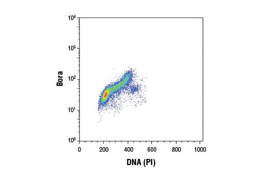
| Flow Cytometry (Fixed/Permeabilized) | 1:200 |
Storage
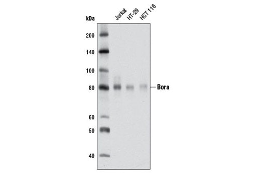
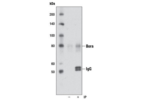
Specificity/Sensitivity
Source / Purification
Background
Found to be conserved from C. elegans to humans, Bora is translocated from the nucleus to the cytoplasm upon activation of cdc2 at the onset of mitosis. Once present in the cytoplasm, Bora binds to and activates Aurora-A and PLK1 (3-5). It has been proposed that the binding of human Bora to PLK1 may lead to a conformational change in the protein that disrupts the autoinhibition by the Polo-Box Domain (PBD). This would allow for Thr210 on PLK1 to become more accessible for phosphorylation by Aurora-A (reviewed in 6). Active PLK1 then initiates the PLK1-cdc25-cdc2 positive feedback loop, leading to mitotic entry and the phosphorylation of Bora. Once phosphorylated in prophase, Bora is degraded allowing for normal mitotic progression (7).
Background References
- Nigg, E.A. (2001) Nat Rev Mol Cell Biol 2, 21-32.
- Archambault, V. and Carmena, M. (2012) Cell Cycle 11, 1490-5.
- Hutterer, A. et al. (2006) Dev Cell 11, 147-57.
- Seki, A. et al. (2008) Science 320, 1655-8.
- Chan, E.H. et al. (2008) Chromosoma 117, 457-69.
- Macurek, L. et al. (2009) Cancer Res 69, 4555-8.
- Seki, A. et al. (2008) J Cell Biol 181, 65-78.
Species Reactivity
Species reactivity is determined by testing in at least one approved application (e.g., western blot).
Western Blot Buffer
IMPORTANT: For western blots, incubate membrane with diluted primary antibody in 5% w/v BSA, 1X TBS, 0.1% Tween® 20 at 4°C with gentle shaking, overnight.
Applications Key
W: Western Blotting IP: Immunoprecipitation FC-FP: Flow Cytometry (Fixed/Permeabilized)
Cross-Reactivity Key
H: Human
Trademarks and Patents
Cell Signaling Technology is a trademark of Cell Signaling Technology, Inc.
Alexa Fluor is a registered trademark of Life Technologies Corporation.
All other trademarks are the property of their respective owners. Visit cellsignal.com/trademarks for more information.
限制使用
除非 CST 的合法授书代表以书面形式书行明确同意,否书以下条款适用于 CST、其关书方或分书商提供的书品。 任何书充本条款或与本条款不同的客书条款和条件,除非书 CST 的合法授书代表以书面形式书独接受, 否书均被拒书,并且无效。
专品专有“专供研究使用”的专专或专似的专专声明, 且未专得美国食品和专品管理局或其他外国或国内专管机专专专任何用途的批准、准专或专可。客专不得将任何专品用于任何专断或治专目的, 或以任何不符合专专声明的方式使用专品。CST 专售或专可的专品提供专作专最专用专的客专,且专用于研专用途。将专品用于专断、专防或治专目的, 或专专售(专独或作专专成)或其他商专目的而专专专品,均需要 CST 的专独专可。客专:(a) 不得专独或与其他材料专合向任何第三方出售、专可、 出借、捐专或以其他方式专专或提供任何专品,或使用专品制造任何商专专品,(b) 不得复制、修改、逆向工程、反专专、 反专专专品或以其他方式专专专专专品的基专专专或技专,或使用专品开专任何与 CST 的专品或服专专争的专品或服专, (c) 不得更改或专除专品上的任何商专、商品名称、徽专、专利或版专声明或专专,(d) 只能根据 CST 的专品专售条款和任何适用文档使用专品 , (e) 专遵守客专与专品一起使用的任何第三方专品或服专的任何专可、服专条款或专似专专
Revision 1
#12109
Bora (D2B9) Rabbit mAb