Revision 1
#72159
Store at -20C
B Cell Signaling Antibody Sampler Kit II
1 Kit
(9 x 20 microliters)
877-616-CELL (2355)
877-678-TECH (8324)
3 Trask Lane | Danvers | Massachusetts | 01923 | USA
For Research Use Only. Not for Use in Diagnostic Procedures.
| Product Includes | Product # | Quantity | Mol. Wt | Isotype/Source |
|---|---|---|---|---|
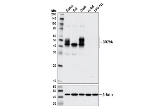
| CD79A (D1X5C) XP® Rabbit mAb | 13333 | 20 µl | 45-55 kDa | Rabbit IgG |
| Phospho-CD79A (Tyr182) Antibody | 5173 | 20 µl | 45-55 kDa | Rabbit |
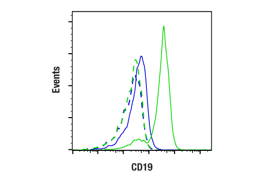
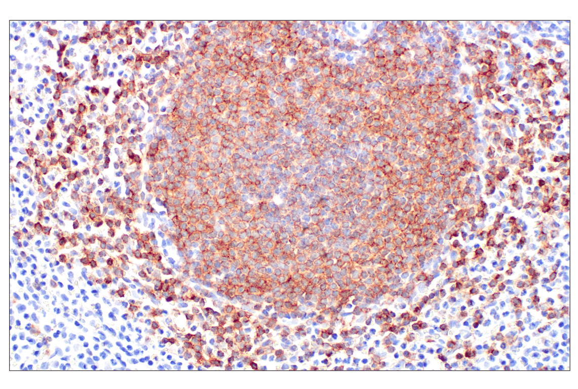
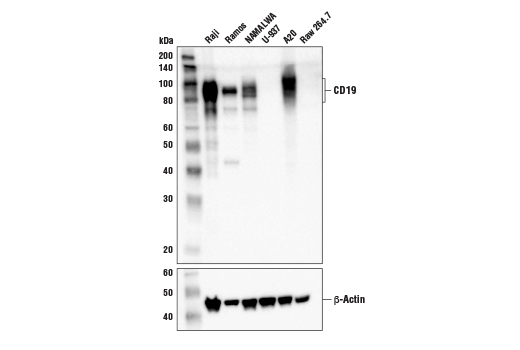
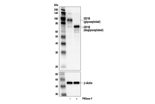
| CD19 (Intracellular Domain) (D4V4B) XP® Rabbit mAb | 90176 | 20 µl | 95 kDa | Rabbit IgG |
| Phospho-CD19 (Tyr531) Antibody | 3571 | 20 µl | 95 kDa | Rabbit |
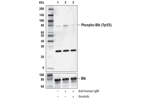
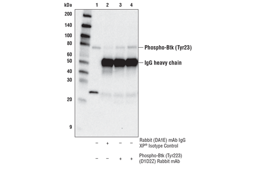
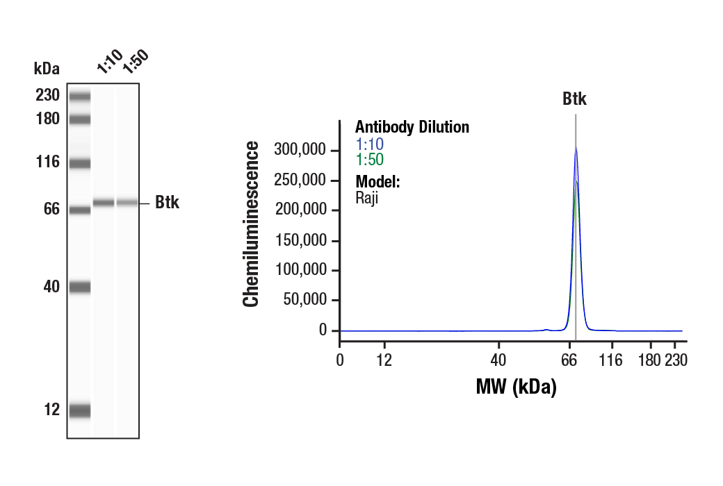
| Phospho-Btk (Tyr223) (D1D2Z) Rabbit mAb | 87457 | 20 µl | 78 kDa | Rabbit IgG |
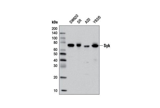
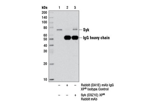
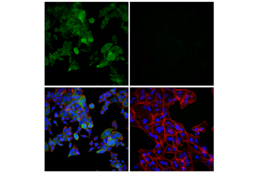
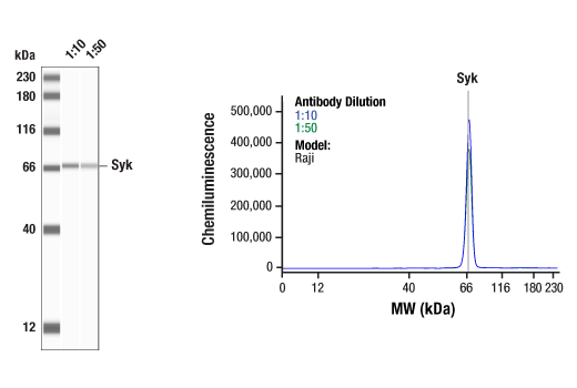
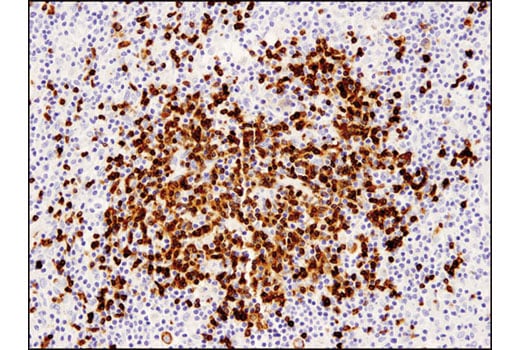
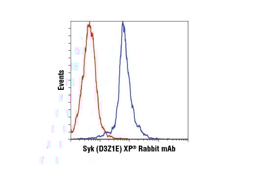
| Syk (D3Z1E) XP® Rabbit mAb | 13198 | 20 µl | 72 kDa | Rabbit IgG |
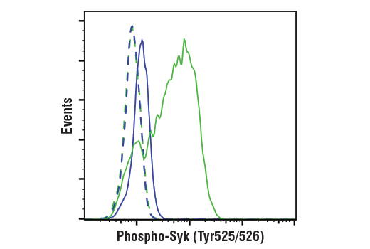
| Phospho-Syk (Tyr525/526) (C87C1) Rabbit mAb | 2710 | 20 µl | 72 kDa | Rabbit IgG |
| Phospho-BLNK (Tyr96) Antibody | 3601 | 20 µl | 68, 70 kDa | Rabbit |
| Btk (D3H5) Rabbit mAb | 8547 | 20 µl | 77 kDa | Rabbit IgG |
| Anti-rabbit IgG, HRP-linked Antibody | 7074 | 100 µl | Goat |
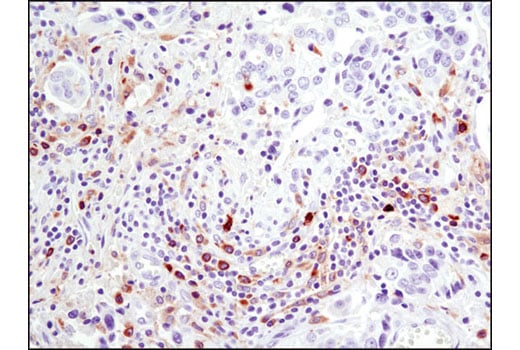
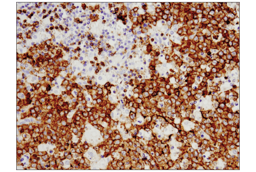
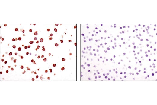
Please visit cellsignal.com for individual component applications, species cross-reactivity, dilutions, protocols, and additional product information.
Description
Storage
Background
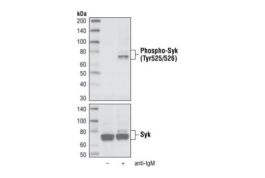
Syk is a protein tyrosine kinase that plays an important role in intracellular signal transduction in hematopoietic cells (1-3). Syk interacts with immunoreceptor tyrosine-based activation motifs (ITAMs) located in the cytoplasmic domains of immune receptors (4). It couples the activated immunoreceptors to downstream signaling events that mediate diverse cellular responses, including proliferation, differentiation, and phagocytosis (4). There is also evidence that Syk plays a role in nonimmune cells; Syk is a potential tumor suppressor in human breast carcinomas (5). Tyrosine 525 and 526 are located in the activation loop of the Syk kinase domain, and phosphorylation of Tyr525/526 of human Syk (equivalent to the Tyr519/520 of mouse Syk) is essential for Syk function (6).
Lyn, one of the Src family members, is predominantly expressed in hematopoietic cells (7). Two tyrosine residues have been reported to play a crucial role in the regulation of protein tyrosine kinases of the Src family. Autophosphorylation of Tyr396 (equivalent to Tyr416 of Src), located in the catalytic domain, correlates with enzyme activation. Csk-mediated phosphorylation of the carboxy-terminal Tyr507 (equivalent to Tyr527 of Src) inactivates the kinase. Tyrosine phosphorylation and activation of Lyn occurs upon association with cell surface receptors such as the B cell Ag receptor (BCR) and CD40 (8-10).
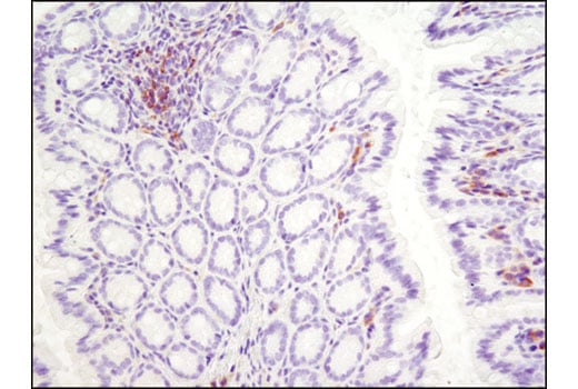
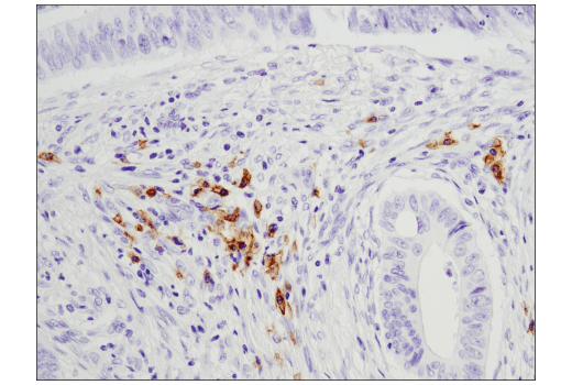
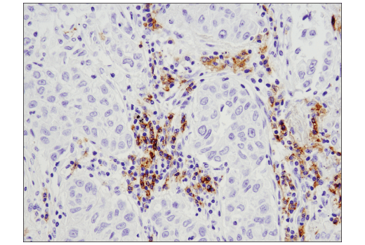
Bruton's tyrosine kinase (Btk) is a member of the Btk/Tec family of cytoplasmic tyrosine kinases. Btk plays an important role in B cell development (11,12). Activation of B cells by various ligands is accompanied by Btk membrane translocation mediated by its PH domain binding to phosphatidylinositol-3,4,5-trisphosphate (13-15). The membrane-located Btk is active and associated with transient phosphorylation of two tyrosine residues, Tyr551 and Tyr223. Tyr551 in the activation loop is transphosphorylated by the Src family tyrosine kinase, leading to autophosphorylation at Tyr223 within the SH3 domain, which is necessary for full activation (16,17).
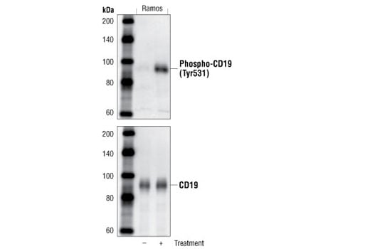
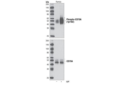
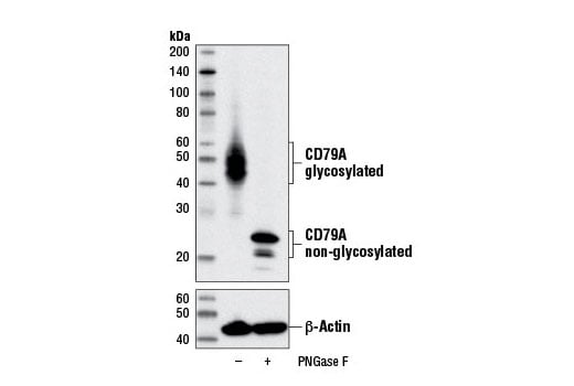
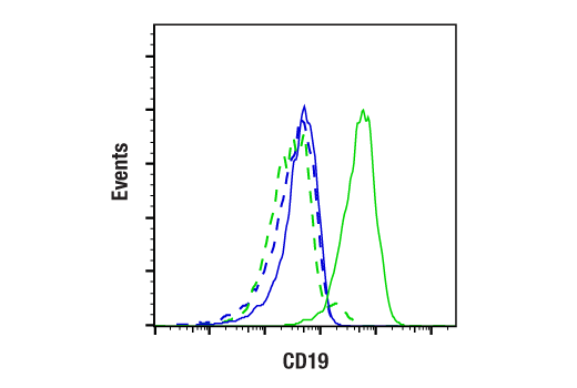
CD19 is a 95 kDa coreceptor that amplifies the signaling cascade in B cells (18). On the B cell surface, CD19 associates with CD21, CD81, and Leu-13 to exert its function. The cytoplasmic tail of CD19 has nine conserved tyrosine residues playing critical roles in CD19-mediated function by coupling signaling molecules to the receptor (18). After BCR or CD19 ligation, Tyr531 and Tyr500 of CD19 are progressively phosphorylated. This phosphorylation enables the coupling of PI3 kinase and Src family tyrosine kinase to CD19 and activates the PI3K and Src signaling pathways (19,20).
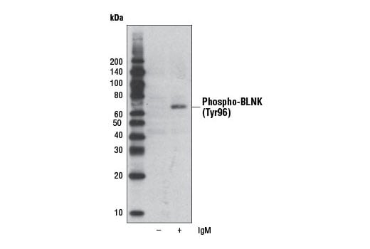
B cell linker protein (BLNK), also known as SLP-65 or BASH, is an adaptor molecule that plays key roles in B cell activation and B cell antigen receptor (BCR) engagement. BLNK acts at the interface between BCR-associated Syk and downstream signaling cascades.
Background References
- Cheng, A.M. and Chan, A.C. (1997) Curr Opin Immunol 9, 528-33.
- Chu, D.H. et al. (1998) Immunol Rev 165, 167-80.
- Yamanashi, Y. et al. (1989) Proc Natl Acad Sci U S A 86, 6538-42.
- Khan, W.N. (2001) Immunol Res 23, 147-56.
- Tedder, T.F. et al. (1997) Immunity 6, 107-18.
- Kurosaki, T. (1997) Curr Opin Immunol 9, 309-18.
- Yamanashi, Y. et al. (1991) Science 251, 192-4.
- Lewis, C.M. et al. (2001) Curr Opin Immunol 13, 317-25.
- Buhl, A.M. and Cambier, J.C. (1999) J Immunol 162, 4438-46.
- Burkhardt, A.L. et al. (1991) Proc Natl Acad Sci U S A 88, 7410-4.
- Salim, K. et al. (1996) EMBO J 15, 6241-50.
- Fujimoto, M. et al. (2000) Immunity 13, 47-57.
- Turner, M. et al. (2000) Immunol Today 21, 148-54.
- Ren, C.L. et al. (1994) J Exp Med 179, 673-80.
- Rameh, L.E. et al. (1997) J Biol Chem 272, 22059-66.
- Coopman, P.J. et al. (2000) Nature 406, 742-7.
- Várnai, P. et al. (1999) J Biol Chem 274, 10983-9.
- Rawlings, D.J. et al. (1996) Science 271, 822-5.
- Park, H. et al. (1996) Immunity 4, 515-25.
- Zhang, J. et al. (2000) J Biol Chem 275, 35442-7.
Trademarks and Patents
Cell Signaling Technology is a trademark of Cell Signaling Technology, Inc.
XP is a registered trademark of Cell Signaling Technology, Inc.
U.S. Patent No. 7,429,487, foreign equivalents, and child patents deriving therefrom.
All other trademarks are the property of their respective owners. Visit cellsignal.com/trademarks for more information.
限制使用
除非 CST 的合法授书代表以书面形式书行明确同意,否书以下条款适用于 CST、其关书方或分书商提供的书品。 任何书充本条款或与本条款不同的客书条款和条件,除非书 CST 的合法授书代表以书面形式书独接受, 否书均被拒书,并且无效。
专品专有“专供研究使用”的专专或专似的专专声明, 且未专得美国食品和专品管理局或其他外国或国内专管机专专专任何用途的批准、准专或专可。客专不得将任何专品用于任何专断或治专目的, 或以任何不符合专专声明的方式使用专品。CST 专售或专可的专品提供专作专最专用专的客专,且专用于研专用途。将专品用于专断、专防或治专目的, 或专专售(专独或作专专成)或其他商专目的而专专专品,均需要 CST 的专独专可。客专:(a) 不得专独或与其他材料专合向任何第三方出售、专可、 出借、捐专或以其他方式专专或提供任何专品,或使用专品制造任何商专专品,(b) 不得复制、修改、逆向工程、反专专、 反专专专品或以其他方式专专专专专品的基专专专或技专,或使用专品开专任何与 CST 的专品或服专专争的专品或服专, (c) 不得更改或专除专品上的任何商专、商品名称、徽专、专利或版专声明或专专,(d) 只能根据 CST 的专品专售条款和任何适用文档使用专品 , (e) 专遵守客专与专品一起使用的任何第三方专品或服专的任何专可、服专条款或专似专专
Revision 1
Revision 1
Revision 1

Revision 1
Revision 1
Revision 1
Revision 1
Revision 1
Revision 1
Revision 1
Revision 1

Revision 1
Revision 1
Revision 1
Revision 1
Revision 1