Revision 1
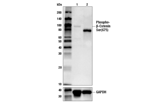
Western blot analysis of extracts from control HeLa cells (lane 1) or HeLa cells with a targeted mutation in the gene encoding β-Catenin (lane 2) using Phospho-β-Catenin (Ser675) (D2F1) XP® Rabbit mAb (upper), or GAPDH (D16H11) XP® Rabbit mAb #5174 (lower). The change in β-Catenin molecular weight in the mutated HeLa cells confirms the specificity of the antibody for β-Catenin.
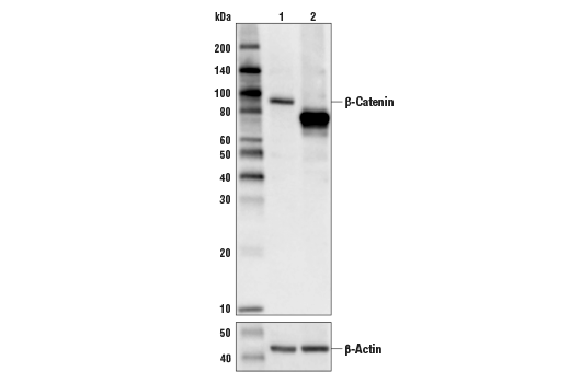
Western blot analysis of extracts from control HeLa cells (lane 1) or HeLa cells with an apparent in-frame truncation mutation in the gene encoding β-Catenin (lane 2) using β-Catenin (D10A8) XP® Rabbit mAb, #8480 (upper) or β-actin (D6A8) Rabbit mAb #8457 (lower). The change in β-Catenin molecular weight in the mutated HeLa cells is consistent with an in-frame deletion.
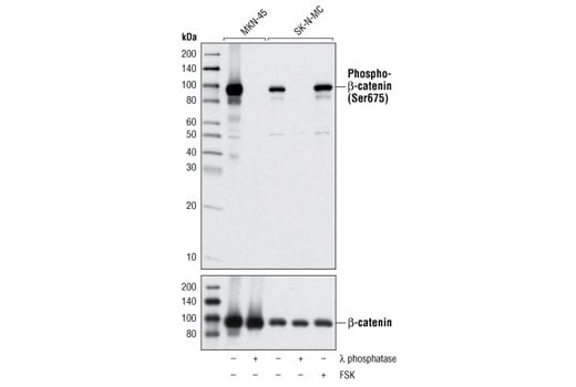
Western blot analysis of extracts from MKN-45 and SK-N-MC cells, untreated or treated with λ phosphatase for 1 hour or forskolin (FSK) for 30 minutes, using Phospho-β-Catenin (Ser675) (D2F1) XP® Rabbit mAb (upper) or β-Catenin (6B3) Rabbit mAb #9582 (lower).
Orders: 877-616-CELL (2355) • [email protected] • Support: 877-678-TECH (8324) • [email protected] •
Web:
cellsignal.com For Research Use Only. Not for Use in Diagnostic Procedures.
Revision 1
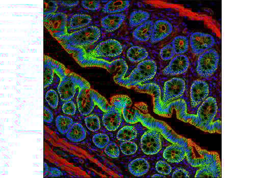
Confocal immunofluorescent analysis of rat colon using Phospho-β-Catenin (Ser675) (D2F1) XP® Rabbit mAb (green). Actin filaments have been labeled with DY-554 Phalloidin (red). Blue pseudocolor = DRAQ5® #4084 (fluorescent DNA dye).
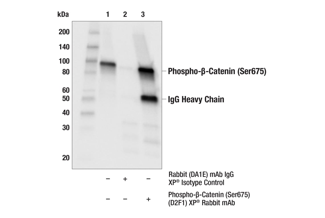
Immunoprecipitation of Phospho-β-Catenin (Ser675) protein from SW620 + Calyculin A #9902 (50nM, 30 min) cell extracts. Lane 1 is 10% input, lane 2 is Rabbit (DA1E) mAb IgG XP® Isotype Control #3900, and lane 3 is Phospho-β-Catenin (Ser675) (D2F1) XP® Rabbit mAb. Western blot analysis was performed using Phospho-β-Catenin (Ser675) (D2F1) XP® Rabbit mAb.
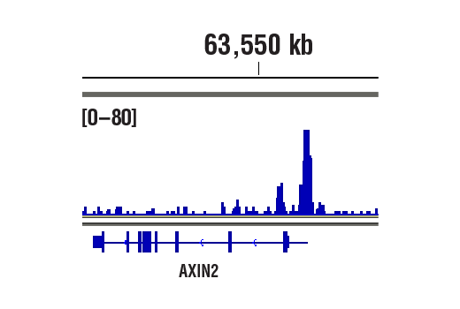
CUT&RUN was performed with HCT 116 cells and β-Catenin (D10A8) XP® Rabbit mAb, using CUT&RUN Assay Kit #86652. DNA library was prepared using DNA Library Prep Kit for Illumina® (ChIP-seq, CUT&RUN) #56795. The figure shows binding across Axin2, a known target gene of β-Catenin (see additional figure containing CUT&RUN-qPCR data).
Orders: 877-616-CELL (2355) • [email protected] • Support: 877-678-TECH (8324) • [email protected] •
Web:
cellsignal.com For Research Use Only. Not for Use in Diagnostic Procedures.
Revision 1
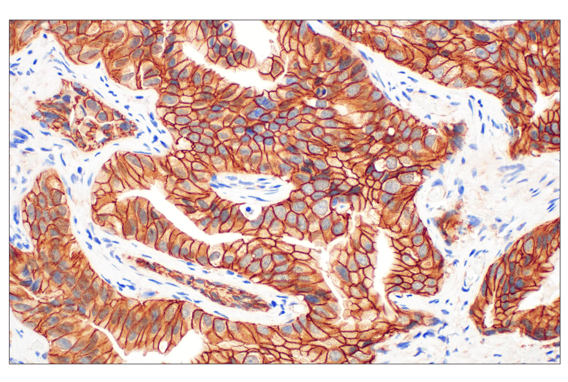
Immunohistochemical analysis of paraffin-embedded human prostate adenocarcinoma using ß-Catenin (D10A8) XP® Rabbit mAb performed on the Leica® BOND™ Rx.
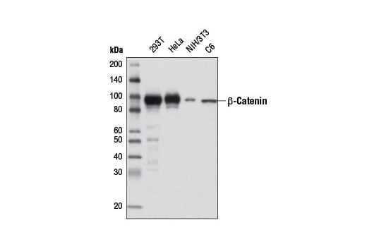
Western blot analysis of extracts from various cell lines using β-Catenin (D10A8) XP® Rabbit mAb.
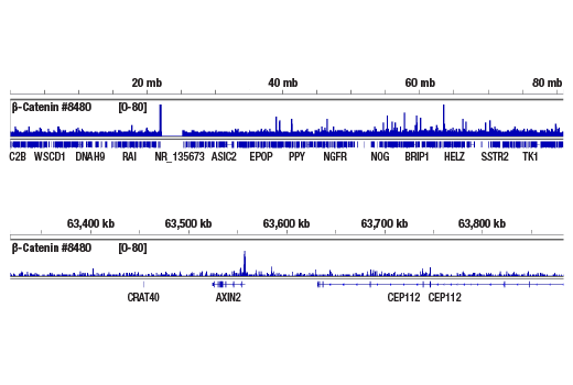
CUT&RUN was performed with HCT 116 cells and β-Catenin (D10A8) XP® Rabbit mAb, using CUT&RUN Assay Kit #86652. DNA Libraries were prepared using DNA Library Prep Kit for Illumina® (ChIP-seq, CUT&RUN) #56795. The figures show binding across chromosome 17 (upper), including Axin2 (lower), a known target gene of β-Catenin (see additional figure containing CUT&RUN-qPCR data).
Orders: 877-616-CELL (2355) • [email protected] • Support: 877-678-TECH (8324) • [email protected] •
Web:
cellsignal.com For Research Use Only. Not for Use in Diagnostic Procedures.
Revision 1
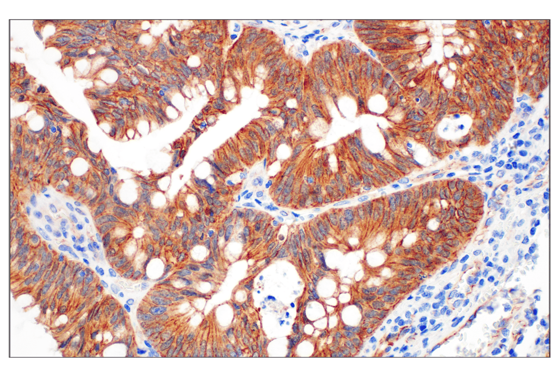
Immunohistochemical analysis of paraffin-embedded human colon adenocarcinoma using ß-Catenin (D10A8) XP® Rabbit mAb performed on the Leica® BOND™ Rx.
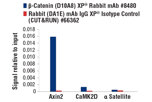
CUT&RUN was performed with HCT 116 cells and either β-Catenin (D10A8) XP® Rabbit mAb or Rabbit (DA1E) mAb IgG XP® Isotype Control (CUT&RUN) #66362, using CUT&RUN Assay Kit #86652. The enriched DNA was quantified by real-time PCR using SimpleChIP® Human Axin2 Intron 1 Primers #8973, SimpleChIP® Human CaMK2D Intron 3 Primers #5111 and SimpleChIP® Human α Satellite Repeat Primers #4486. The amount of immunoprecipitated DNA in each sample is represented as signal relative to the total amount of input chromatin, which is equivalent to one.
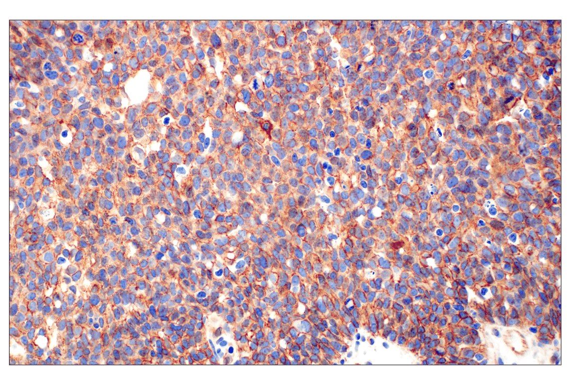
Immunohistochemical analysis of paraffin-embedded human serous adenocarcinoma of the ovary using ß-Catenin (D10A8) XP® Rabbit mAb performed on the Leica® BOND™ Rx.
Orders: 877-616-CELL (2355) • [email protected] • Support: 877-678-TECH (8324) • [email protected] •
Web:
cellsignal.com For Research Use Only. Not for Use in Diagnostic Procedures.
Revision 1
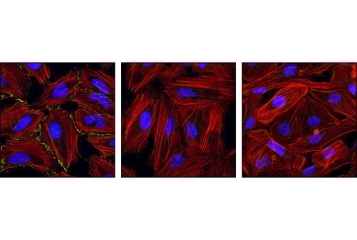
Confocal immunofluorescent analysis of HeLa cells, untreated (left), λ phosphatase-treated (middle), or untreated NCI-H28 cells (β-catenin null; right) using Phospho-β-Catenin (Ser675) (D2F1) XP® Rabbit mAb (green). Actin filaments have been labeled with DY-554 phalloidin (red). Blue pseudocolor = DRAQ5® #4084 (fluorescent DNA dye).
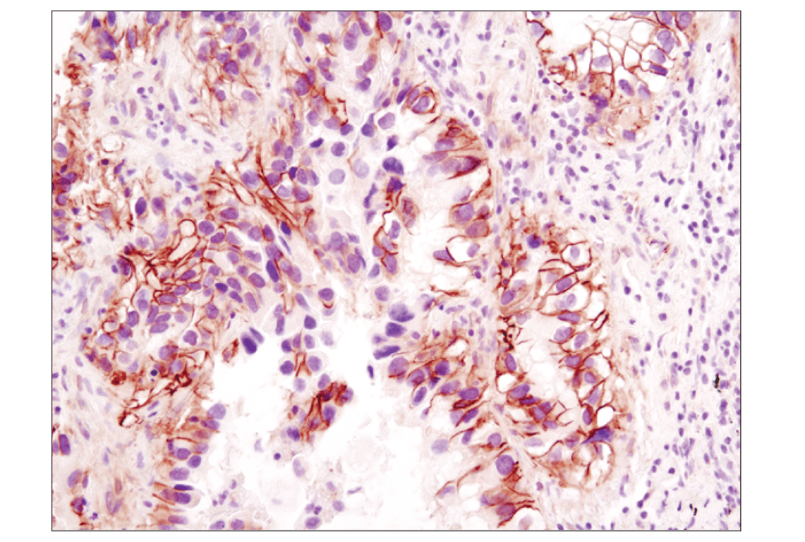
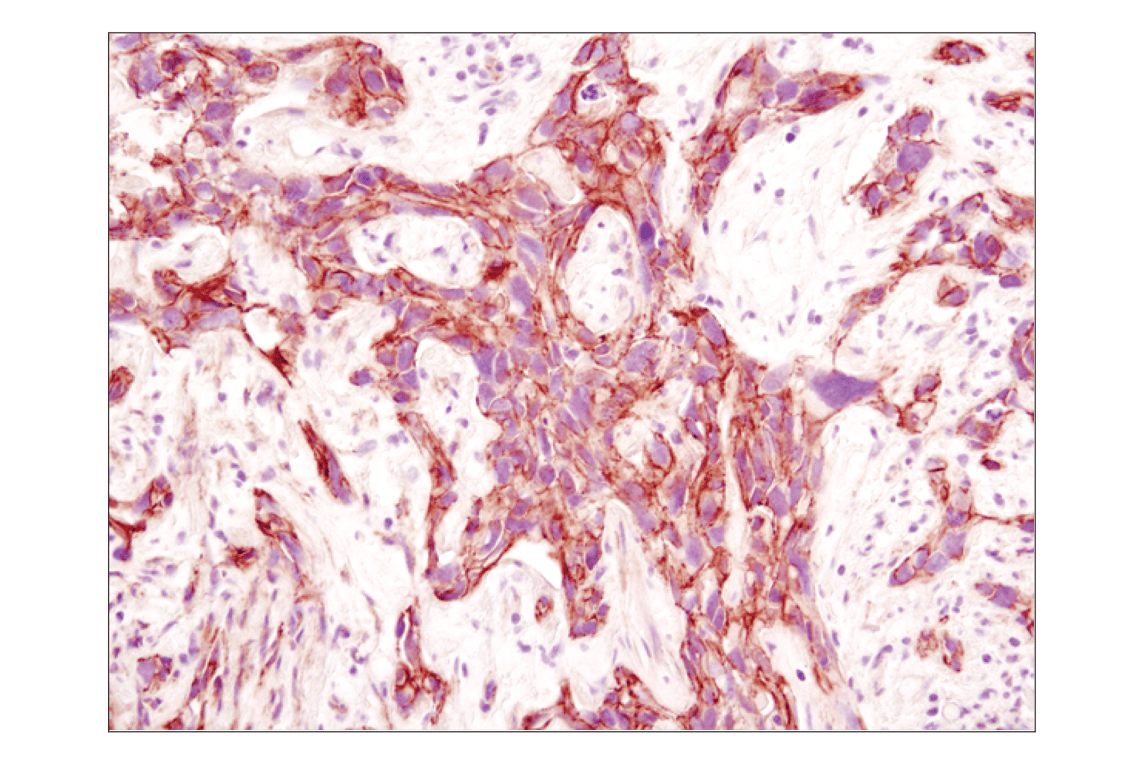
Immunohistochemical analysis of paraffin-embedded human colon adenocarcinoma using ß-Catenin (D10A8) XP® Rabbit mAb.
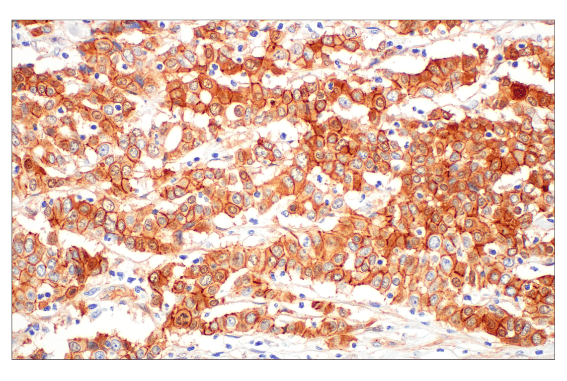
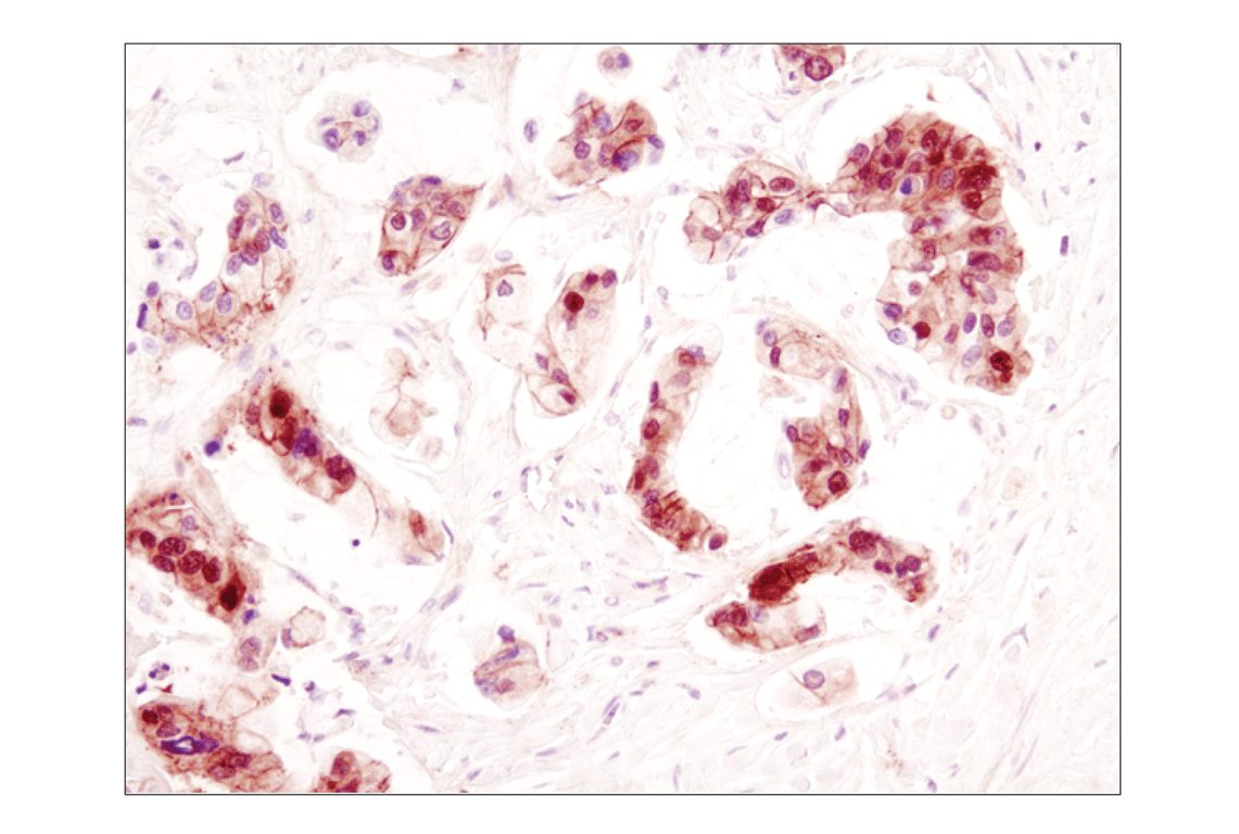
Immunohistochemical analysis of paraffin-embedded human lung carcinoma using β-Catenin (D10A8) XP® Rabbit mAb.
Orders: 877-616-CELL (2355) • [email protected] • Support: 877-678-TECH (8324) • [email protected] •
Web:
cellsignal.com For Research Use Only. Not for Use in Diagnostic Procedures.
Revision 1
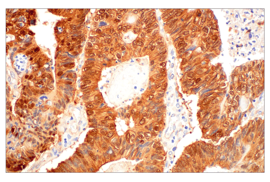
Immunohistochemical analysis of paraffin-embedded human colon adenocarcinoma using ß-Catenin (D10A8) XP® Rabbit mAb.
Immunohistochemical analysis of paraffin-embedded human colon carcinoma using β-Catenin (D10A8) XP® Rabbit mAb.
Immunohistochemical analysis of paraffin-embedded human breast carcinoma using β-Catenin (D10A8) XP® Rabbit mAb.
Orders: 877-616-CELL (2355) • [email protected] • Support: 877-678-TECH (8324) • [email protected] •
Web:
cellsignal.com For Research Use Only. Not for Use in Diagnostic Procedures.
Revision 1
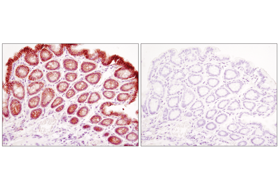
Immunohistochemical analysis of paraffin-embedded mouse colon using β-Catenin (D10A8) XP® Rabbit mAb in the presence of control peptide (left) or antigen-specific peptide (right).
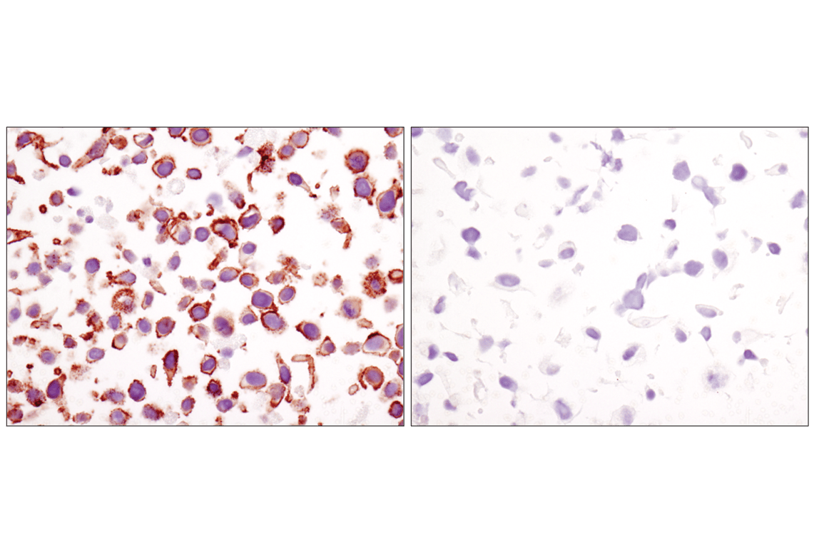
Immunohistochemical analysis of paraffin-embedded cell pellets, HeLa (left) or NCI-H28 (right), using β-Catenin (D10A8) XP® Rabbit mAb.
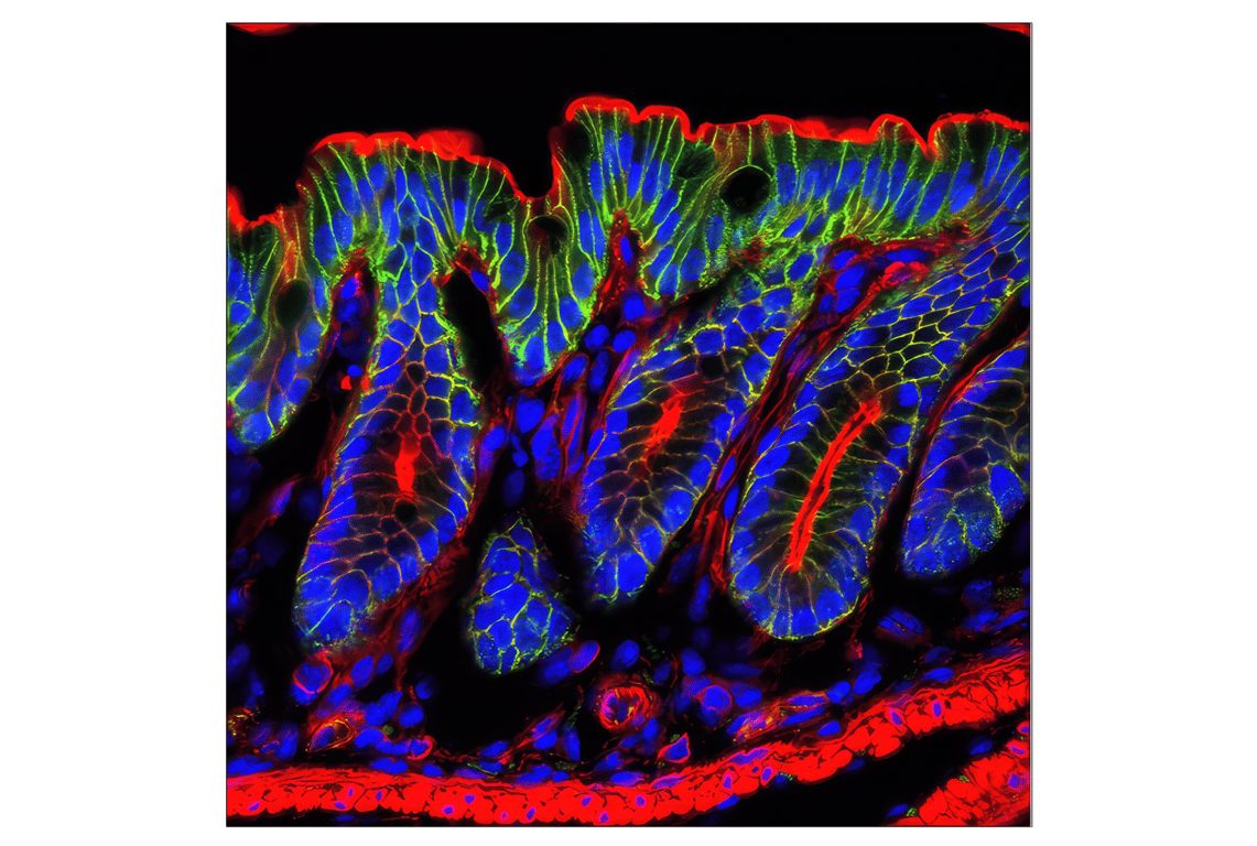
Confocal immunofluorescent analysis of mouse colon using β-Catenin (D10A8) XP® Rabbit mAb (green). Actin filaments were labeled with DY-554 phalloidin (red). Blue pseudocolor = DRAQ5® #4084 (fluorescent DNA dye).
Orders: 877-616-CELL (2355) • [email protected] • Support: 877-678-TECH (8324) • [email protected] •
Web:
cellsignal.com For Research Use Only. Not for Use in Diagnostic Procedures.
Revision 1
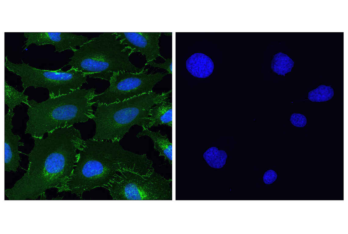
Confocal immunofluorescent analysis of HeLa (left) and NCI-H28 (right) cells using β-Catenin (D10A8) XP® Rabbit mAb (green). Blue pseudocolor = DRAQ5® #4084 (fluorescent DNA dye).
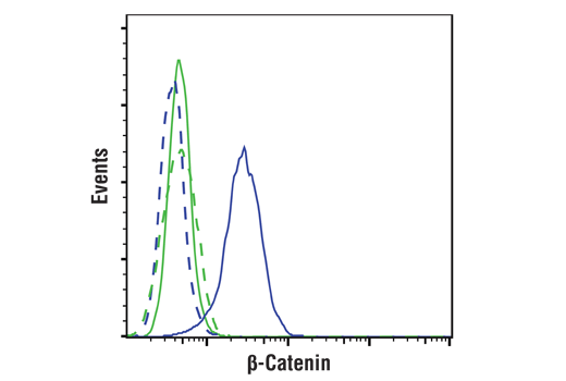
Flow cytometric analysis of NCI-H28 cells (green) and HeLa cells (blue) using β-Catenin (D10A8) XP® Rabbit mAb (solid lines) or concentration-matched Rabbit Isotype Control #3900 (dashed lines). Anti-rabbit IgG (H+L), F(ab')2 Fragment (Alexa Fluor® 488 Conjugate) #4412 was used as secondary antibody.
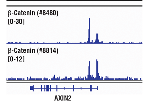
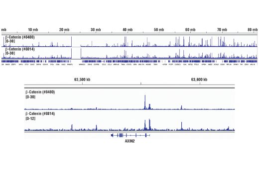
Chromatin immunoprecipitations were performed with cross-linked chromatin from HCT116 cells and either β-Catenin (D10A8) XP® Rabbit mAb or Non-phospho (Active) β-Catenin (Ser33/37/Thr41) (D13A1) Rabbit mAb #8814, using SimpleChIP® Enzymatic Chromatin IP Kit (Magnetic Beads) #9005. DNA Libraries were prepared using DNA Library Prep Kit for Illumina® (ChIP-seq, CUT&RUN) #56795. The figure shows binding across AXIN2, a known target gene of β-Catenin (see additional figure containing ChIP-qPCR data).
Orders: 877-616-CELL (2355) • [email protected] • Support: 877-678-TECH (8324) • [email protected] •
Web:
cellsignal.com For Research Use Only. Not for Use in Diagnostic Procedures.
Revision 1
Chromatin immunoprecipitations were performed with cross-linked chromatin from HCT116 cells and either β-Catenin (D10A8) XP® Rabbit mAb or Non-phospho (Active) β-Catenin (Ser33/37/Thr41) (D13A1) Rabbit mAb #8814, using SimpleChIP® Enzymatic Chromatin IP Kit (Magnetic Beads) #9005. DNA Libraries were prepared using DNA Library Prep Kit for Illumina® (ChIP-seq, CUT&RUN) #56795. The figure shows binding across chromosome 17 (upper), including AXIN2 (lower), a known target gene of β-Catenin (see additional figure containing ChIP-qPCR data).
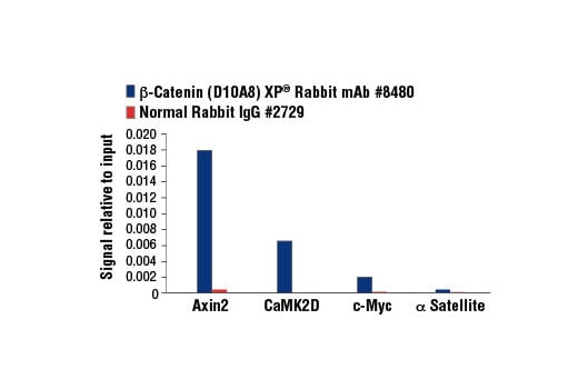
Chromatin immunoprecipitations were performed with cross-linked chromatin from HCT 116 cells and either β-Catenin (D10A8) XP® Rabbit mAb or Normal Rabbit IgG #2729 using SimpleChIP® Enzymatic Chromatin IP Kit (Magnetic Beads) #9003. The enriched DNA was quantified by real-time PCR using SimpleChIP® Human Axin2 Intron 1 Primers #8973, SimpleChIP® Human CaMK2D Intron 3 Primers #5111, human c-Myc promoter primers, and SimpleChIP® Human α Satellite Repeat Primers #4486. The amount of immunoprecipitated DNA in each sample is represented as signal relative to the total amount of input chromatin, which is equivalent to one.
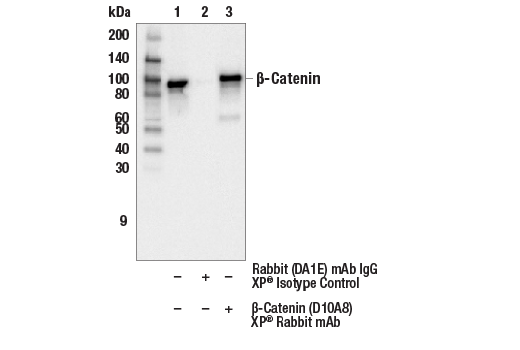
Immunoprecipitation of β-Catenin from HeLa cell extracts. Lane 1 is 10% input, lane 2 is precipitated with Rabbit (DA1E) mAb IgG XP® Isotype Control #3900, and lane 3 is β-Catenin (D10A8) XP® Rabbit mAb, #8480. Western blot was performed using β-Catenin (15B8) Mouse mAb, #37447.
Orders: 877-616-CELL (2355) • [email protected] • Support: 877-678-TECH (8324) • [email protected] •
Web:
cellsignal.com For Research Use Only. Not for Use in Diagnostic Procedures.
Revision 1
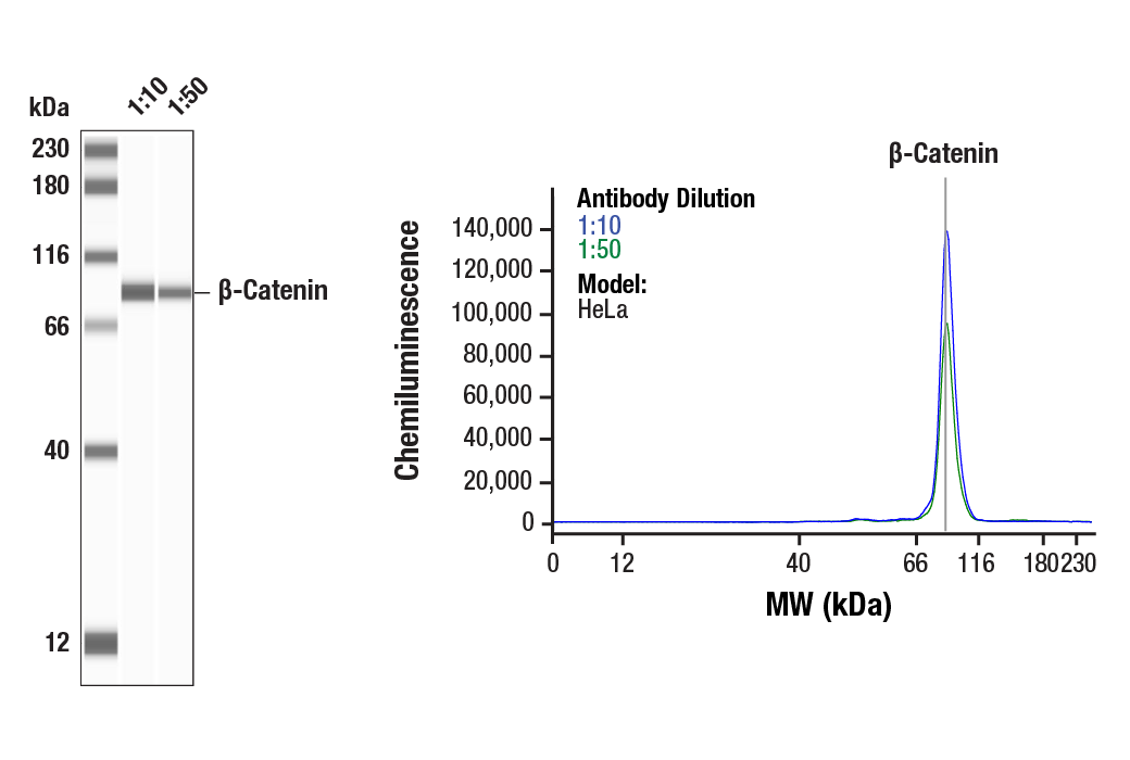
Simple Western™ analysis of lysates (0.1 mg/mL) from HeLa cells using β-Catenin (D10A8) XP® Rabbit mAb #8480. The virtual lane view (left) shows the target band (as indicated) at 1:10 and 1:50 dilutions of primary antibody. The corresponding electropherogram view (right) plots chemiluminescence by molecular weight along the capillary at 1:10 (blue line) and 1:50 (green line) dilutions of primary antibody. This experiment was performed under reducing conditions on the Jess™ Simple Western instrument from ProteinSimple, a BioTechne brand, using the 12-230 kDa separation module.
Orders: 877-616-CELL (2355) • [email protected] • Support: 877-678-TECH (8324) • [email protected] •
Web:
cellsignal.com For Research Use Only. Not for Use in Diagnostic Procedures.