| Product Includes | Product # | Quantity | Mol. Wt | Isotype/Source |
|---|---|---|---|---|
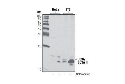
| LC3A (D50G8) XP® Rabbit mAb | 4599 | 20 µl | 14, 16 kDa | Rabbit IgG |
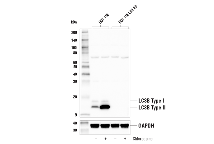
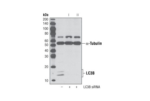
| LC3B (D11) XP® Rabbit mAb | 3868 | 20 µl | 14, 16 kDa | Rabbit IgG |
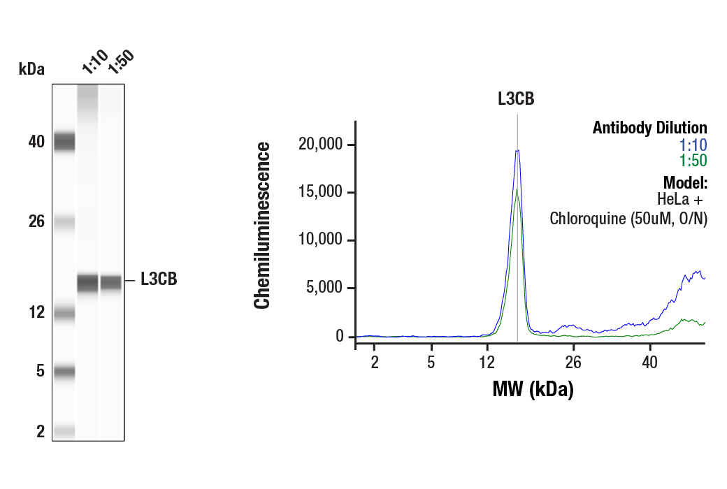
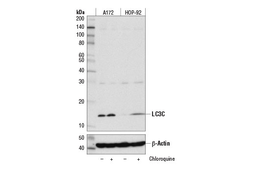
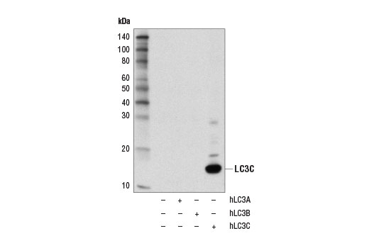
| LC3C (D3O6P) Rabbit mAb | 14736 | 20 µl | 14 kDa | Rabbit IgG |
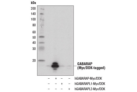
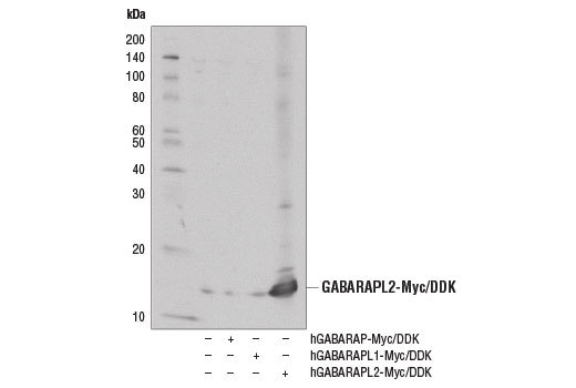
| GABARAP (E1J4E) Rabbit mAb | 13733 | 20 µl | 14, 16 kDa | Rabbit IgG |
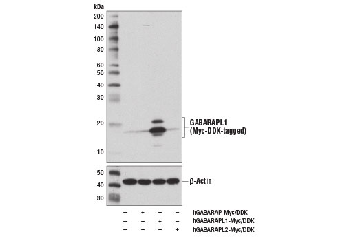
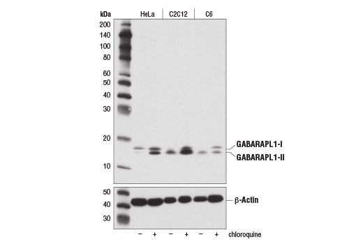
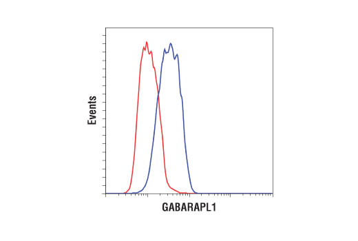
| GABARAPL1 (D5R9Y) XP® Rabbit mAb | 26632 | 20 µl | 14, 16 kDa | Rabbit IgG |
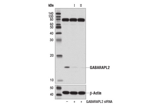
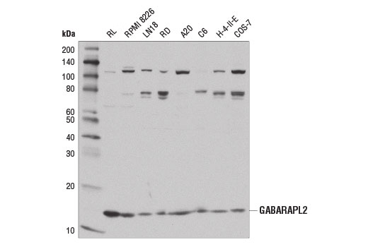
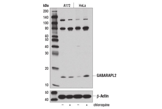
| GABARAPL2 (D1W9T) Rabbit mAb | 14256 | 20 µl | 14 kDa | Rabbit IgG |
| Anti-rabbit IgG, HRP-linked Antibody | 7074 | 100 µl | Goat |
Please visit cellsignal.com for individual component applications, species cross-reactivity, dilutions, protocols, and additional product information.
Description
The Autophagy Atg8 Family Antibody Sampler Kit provides an economical means of detecting each of the Atg8 family members. The kit contains enough primary antibody to perform at least two western blot experiments.
Storage
Background
Autophagy is a catabolic process for the autophagosomic-lysosomal degradation of bulk cytoplasmic contents (1,2). Autophagy is generally activated by conditions of nutrient deprivation, but it has also been associated with a number of physiological processes, including development, differentiation, neurodegenerative diseases, infection, and cancer (3).
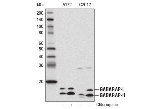
Atg8 is a ubiquitin-like protein that is critical for autophagosome formation. Atg8 is synthesized as a precursor protein that is processed by the cysteine protease Atg4, followed by lipidation with phosphatidylethanolamine (PE) in a ubiqutin-like conjugation pathway involving Atg7 and Atg3 (4). This processing of Atg8, which is described as a conversion from type-I to type-II forms, is frequently described as a marker for autophagy. The type-II form of Atg8 is incorporated into maturing autophagosomes and leads to the recruitment of additional autophagy components, including cargo receptors like SQSTM1/p62. While yeast has a single Atg8 gene, many eukaryotes have at least six orthologs, including three microtubule-associated protein 1 light chain 3 (MAP1LC3/LC3) family members (LC3A, LC3B, and LC3C) and three GABAA receptor associated protein (GABARAP) family members (GABARAP, GABARAPL1/GEC1, and GABARAPL2/GATE-16). While highly conserved, these various family members can have important differences in their post-translational processing, expression profile, and protein interactions including distinct cargo receptor. This complexity within the Atg8 family is critical for selective mechanisms of autophagy that have been reported (5, 6).
- Reggiori, F. and Klionsky, D.J. (2002) Eukaryot Cell 1, 11-21.
- Codogno, P. and Meijer, A.J. (2005) Cell Death Differ 12 Suppl 2, 1509-18.
- Levine, B. and Yuan, J. (2005) J Clin Invest 115, 2679-88.
- Ichimura, Y. et al. (2000) Nature 408, 488-92.
- Slobodkin, M.R. and Elazar, Z. (2013) Essays Biochem 55, 51-64.
- Schaaf, M.B. et al. (2016) FASEB J 30, 3961-3978.
Background References
Trademarks and Patents
限制使用
除非 CST 的合法授书代表以书面形式书行明确同意,否书以下条款适用于 CST、其关书方或分书商提供的书品。 任何书充本条款或与本条款不同的客书条款和条件,除非书 CST 的合法授书代表以书面形式书独接受, 否书均被拒书,并且无效。
专品专有“专供研究使用”的专专或专似的专专声明, 且未专得美国食品和专品管理局或其他外国或国内专管机专专专任何用途的批准、准专或专可。客专不得将任何专品用于任何专断或治专目的, 或以任何不符合专专声明的方式使用专品。CST 专售或专可的专品提供专作专最专用专的客专,且专用于研专用途。将专品用于专断、专防或治专目的, 或专专售(专独或作专专成)或其他商专目的而专专专品,均需要 CST 的专独专可。客专:(a) 不得专独或与其他材料专合向任何第三方出售、专可、 出借、捐专或以其他方式专专或提供任何专品,或使用专品制造任何商专专品,(b) 不得复制、修改、逆向工程、反专专、 反专专专品或以其他方式专专专专专品的基专专专或技专,或使用专品开专任何与 CST 的专品或服专专争的专品或服专, (c) 不得更改或专除专品上的任何商专、商品名称、徽专、专利或版专声明或专专,(d) 只能根据 CST 的专品专售条款和任何适用文档使用专品, (e) 专遵守客专与专品一起使用的任何第三方专品或服专的任何专可、服专条款或专似专专