Revision 1
Cell Signaling Technology

Orders: 877-616-CELL (2355) [email protected]

Support: 877-678-TECH (8324)

Web: [email protected] cellsignal.com

3 Trask LaneDanversMassachusetts01923USA
For Research Use Only. Not for Use in Diagnostic Procedures.
Applications:

WB, IHC-P, IF-F, IF-IC, FC-FP

REACTIVITY:

M

SENSITIVITY:

Endogenous

MW (kDa):

22

Source/Isotype:

Rabbit IgG

UniProt ID:

#Q9EPB4

Entrez-Gene Id:

66824

Product Information

Product Usage Information

This product is the carrier free version of product #67824. All data were generated using the same antibody clone in the standard formulation which contains BSA and glycerol.

This formulation is ideal for use with technologies requiring specialized or custom antibody labeling, including fluorophores, metals, lanthanides, and oligonucleotides. It is not recommended for ChIP, ChIP-seq, CUT&RUN or CUT&Tag assays. If you require a carrier free formulation for chromatin profiling, please contact us. Optimal dilutions/concentrations should be determined by the end user.

Formulation

Supplied in 1X PBS, BSA and Azide Free.

For standard formulation of this product see product #67824

Storage

Store at -20°C. This product will freeze at -20°C so it is recommended to aliquot into single-use vials to avoid multiple freeze/thaw cycles. A slight precipitate may be present and can be dissolved by gently vortexing. This will not interfere with antibody performance.

Specificity / Sensitivity

ASC/TMS1 (D2W8U) Rabbit mAb (BSA and Azide Free) recognizes endogenous levels of total ASC/TMS1 protein.

Species Reactivity:

Mouse

Source / Purification

Monoclonal antibody is produced by immunizing animals with recombinant mouse ASC/TMS1 protein.

Background

TMS1 (target of methylation-induced silencing)/ASC (apoptosis-associated speck-like protein containing a CARD), also referred to as PYCARD and CARD5, is a 22-kDa pro-apoptotic protein containing an N-terminal pyrin domain (PYD) and a C-terminal caspase recruitment domain (CARD) (1-2). The ASC/TMS1 gene was originally found to be aberrantly methylated and silenced in breast cancer cells (2), and has since been found to be silenced in a number of other cancers, including ovarian cancer (3), glioblastoma (4), melanoma (5), gastric cancer (6), lung cancer (7), and prostate cancer (8). Expression of ASC/TMS1 can be induced by pro-apoptotic/inflammatory stimuli (9). During apoptosis ASC/TMS1 is re-distributed from the cytosol to the mitochondria and associates with mitochondrial Bax to trigger cytochrome c release and subsequent apoptosis (10). ASC/TMS1 has also been found to be a critical component of inflammatory signaling where it associates with and activates caspase-1 in response to pro-inflammatory signals (11).

  1. Masumoto, J. et al. (1999) J Biol Chem 274, 33835-8.
  2. Conway, K.E. et al. (2000) Cancer Res 60, 6236-42.
  3. Terasawa, K. et al. (2004) Clin Cancer Res 10, 2000-6.
  4. Stone, A.R. et al. (2004) Am J Pathol 165, 1151-61.
  5. Guan, X. et al. (2003) Int J Cancer 107, 202-8.
  6. Moriai, R. et al. (2002) Anticancer Res 22, 4163-8.
  7. Virmani, A. et al. (2003) Int J Cancer 106, 198-204.
  8. Das, P.M. et al. (2006) Mol Cancer 5, 28.
  9. Strong, R. et al. (1991) Brain Res 542, 23-8.
  10. Ohtsuka, T. et al. (2004) Nat Cell Biol 6, 121-8.
  11. Srinivasula, S.M. et al. (2002) J Biol Chem 277, 21119-22.

Species Reactivity

Species reactivity is determined by testing in at least one approved application (e.g., western blot).

Applications Key

WB: Western Blotting IHC-P: Immunohistochemistry (Paraffin) IF-F: Immunofluorescence (Frozen) IF-IC: Immunofluorescence (Immunocytochemistry) FC-FP: Flow Cytometry (Fixed/Permeabilized)

Cross-Reactivity Key

H: human M: mouse R: rat Hm: hamster Mk: monkey Vir: virus Mi: mink C: chicken Dm: D. melanogaster X: Xenopus Z: zebrafish B: bovine Dg: dog Pg: pig Sc: S. cerevisiae Ce: C. elegans Hr: horse GP: Guinea Pig Rab: rabbit All: all species expected

Trademarks and Patents

Cell Signaling Technology is a trademark of Cell Signaling Technology, Inc.
XP is a registered trademark of Cell Signaling Technology, Inc.
All other trademarks are the property of their respective owners. Visit cellsignal.com/trademarks for more information.

限制使用

除非 CST 的合法授书代表以书面形式书行明确同意,否书以下条款适用于 CST、其关书方或分书商提供的书品。 任何书充本条款或与本条款不同的客书条款和条件,除非书 CST 的合法授书代表以书面形式书独接受, 否书均被拒书,并且无效。

专品专有“专供研究使用”的专专或专似的专专声明, 且未专得美国食品和专品管理局或其他外国或国内专管机专专专任何用途的批准、准专或专可。客专不得将任何专品用于任何专断或治专目的, 或以任何不符合专专声明的方式使用专品。CST 专售或专可的专品提供专作专最专用专的客专,且专用于研专用途。将专品用于专断、专防或治专目的, 或专专售(专独或作专专成)或其他商专目的而专专专品,均需要 CST 的专独专可。客专:(a) 不得专独或与其他材料专合向任何第三方出售、专可、 出借、捐专或以其他方式专专或提供任何专品,或使用专品制造任何商专专品,(b) 不得复制、修改、逆向工程、反专专、 反专专专品或以其他方式专专专专专品的基专专专或技专,或使用专品开专任何与 CST 的专品或服专专争的专品或服专, (c) 不得更改或专除专品上的任何商专、商品名称、徽专、专利或版专声明或专专,(d) 只能根据 CST 的专品专售条款和任何适用文档使用专品, (e) 专遵守客专与专品一起使用的任何第三方专品或服专的任何专可、服专条款或专似专专
Revision 1
#37953

ASC/TMS1 (D2W8U) Rabbit mAb (BSA and Azide Free)

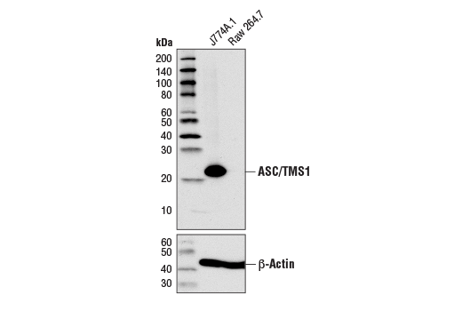
Western Blotting Image 1: ASC/TMS1 (D2W8U) Rabbit mAb (BSA and Azide Free) Expand Image
使用 ASC/TMS1 (D2W8U) Rabbit mAb(上图)或 β-Actin (D6A8) Rabbit mAb #8457(下图)对来自 J774A.1 细胞和 Raw 264.7 细胞的提取物进行蛋白质印迹分析。数据的生成使用的是该产品的常规组分产品。
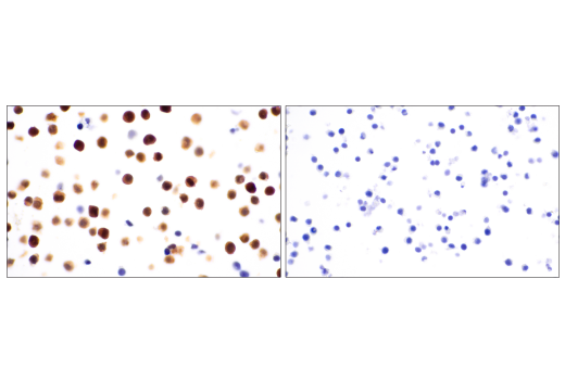
Immunohistochemistry Image 1: ASC/TMS1 (D2W8U) Rabbit mAb (BSA and Azide Free) Expand Image
使用 ASC/TMS1 (D2W8U) Rabbit mAb 对石蜡包埋的 J774A.1 细胞沉淀物(左图,阳性)或 RAW 264.7 细胞沉淀物(右图,阴性)进行免疫组织化学分析。数据的生成使用的是该产品的常规组分产品。
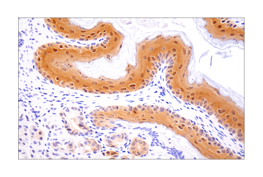
Immunohistochemistry Image 2: ASC/TMS1 (D2W8U) Rabbit mAb (BSA and Azide Free) Expand Image
使用 ASC/TMS1 (D2W8U) Rabbit mAb 对石蜡包埋的小鼠前胃组织进行免疫组织化学分析。数据的生成使用的是该产品的常规组分产品。
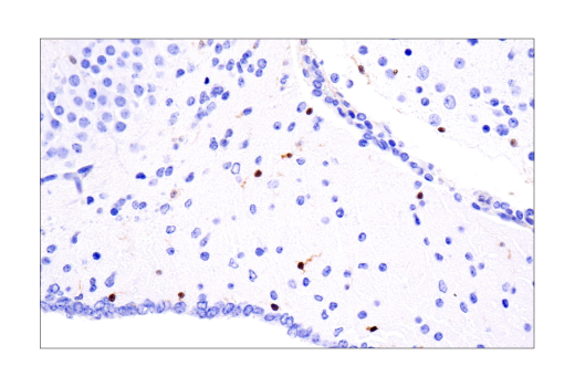
Immunohistochemistry Image 3: ASC/TMS1 (D2W8U) Rabbit mAb (BSA and Azide Free) Expand Image
使用 ASC/TMS1 (D2W8U) Rabbit mAb 对石蜡包埋的小鼠脑组织进行免疫组织化学分析。数据的生成使用的是该产品的常规组分产品。
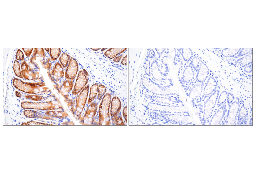
Immunohistochemistry Image 4: ASC/TMS1 (D2W8U) Rabbit mAb (BSA and Azide Free) Expand Image
与浓度匹配的 Rabbit (DA1E) mAb IgG XP® Isotype Control #3900(右图)比较,使用 ASC/TMS1 (D2W8U) Rabbit mAb(左图)对石蜡包埋的小鼠结肠组织进行免疫组织化学分析。数据的生成使用的是该产品的常规组分产品。
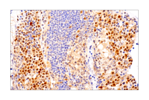
Immunohistochemistry Image 5: ASC/TMS1 (D2W8U) Rabbit mAb (BSA and Azide Free) Expand Image
使用 ASC/TMS1 (D2W8U) Rabbit mAb 对石蜡包埋的小鼠胸腺组织进行免疫组织化学分析。数据的生成使用的是该产品的常规组分产品。
Immunohistochemistry Image 6: ASC/TMS1 (D2W8U) Rabbit mAb (BSA and Azide Free) Expand Image
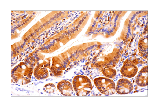
使用 ASC/TMS1 (D2W8U) Rabbit mAb 对石蜡包埋的小鼠小肠组织进行免疫组织化学分析。数据的生成使用的是该产品的常规组分产品。
Immunohistochemistry Image 7: ASC/TMS1 (D2W8U) Rabbit mAb (BSA and Azide Free) Expand Image
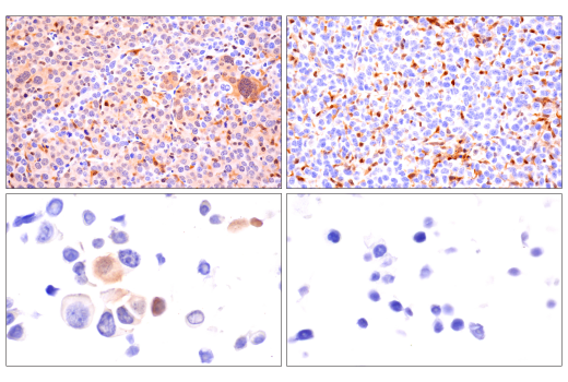
使用 ASC/TMS1 (D2W8U) Rabbit mAb 对石蜡包埋的 Renca 同基因肿瘤(左上图)、4T1 同基因乳腺肿瘤(右上图)、Renca 细胞沉淀物(左下图)和 4T1 细胞沉淀物(右下图)进行免疫组织化学分析。两种肿瘤均显示浸润的免疫细胞染色。请注意,Renca 肿瘤细胞中存在染色,而 4T1 肿瘤细胞中没有染色,这与相应细胞沉淀的染色结果一致。数据的生成使用的是该产品的常规组分产品。
Immunofluorescence Image 1: ASC/TMS1 (D2W8U) Rabbit mAb (BSA and Azide Free) Expand Image
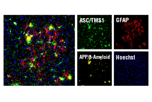
对过表达突变型人 APP695 的小鼠 Tg2576 脑细胞进行共聚焦免疫荧光分析。切片首先经 ASC/TMS1 (D2W8U) Rabbit mAb #67824(绿色)和 APP/β-Amyloid (NAB228) Mouse mAb #2450(黄色)标记。用 Mouse (G3A1) mAb IgG1 Isotype Control #5415 封闭游离二抗结合位点之后,切片用 GFAP (GA5) Mouse mAb (Alexa Fluor® 647 Conjugate) #3657(红色)孵育。胞核用 Hoechst 33342 #4082(蓝色)标记。数据的生成使用的是该产品的常规组分产品。
Immunofluorescence Image 1: ASC/TMS1 (D2W8U) Rabbit mAb (BSA and Azide Free) Expand Image
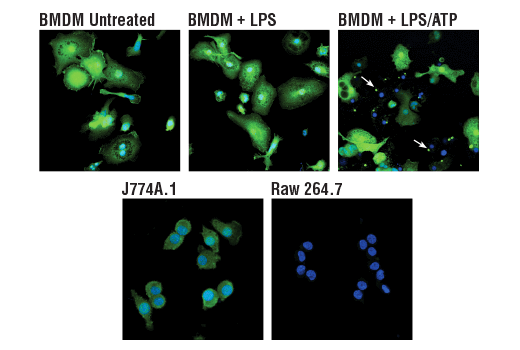
使用 ASC/TMS1 (D2W8U) Rabbit mAb(绿色)对未经处理(左上图)或经 LPS(50 ng/ml,4 小时,中图)处理或经 LPS 处理、接着经 ATP(5 mM,45 分钟,右上图)处理的小鼠原代骨髓源性巨噬细胞 (BMDM) 和 J774A.1 细胞(左下图)或 Raw 细胞 264.7(右下图)进行共聚焦免疫荧光分析。蓝色伪彩 = DRAQ5® #4084(DNA 荧光染料)。注意,经过LPS 和 ATP(白色箭头)刺激后,ASC 会转位至炎症小体。数据的生成使用的是该产品的常规组分产品。
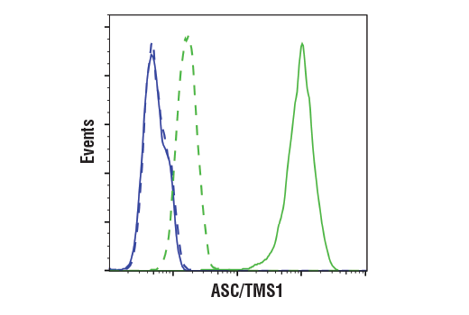
Flow Cytometry Image 1: ASC/TMS1 (D2W8U) Rabbit mAb (BSA and Azide Free) Expand Image
使用 ASC/TMS1 (D2W8U) Rabbit mAb(实线)或浓度匹配的 Rabbit (DA1E) mAb IgG XP® Isotype Control #3900(虚线)对 Raw264.7 细胞(蓝色)和 J774A.1 细胞(绿色)进行流式细胞分析。Anti-rabbit IgG (H+L), F(ab')2 Fragment (Alexa Fluor® 488 Conjugate) #4412 用作二抗。数据的生成使用的是该产品的常规组分产品。