| Product Includes | Product # | Quantity | Mol. Wt | Isotype/Source |
|---|---|---|---|---|
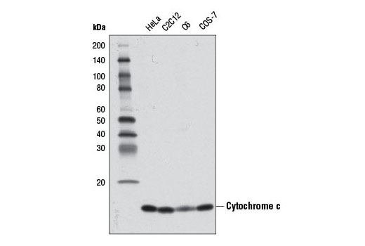
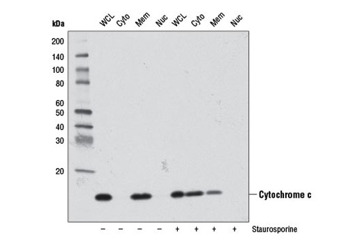
| Cytochrome c (D18C7) Rabbit mAb | 11940 | 40 µl | 14 kDa | Rabbit IgG |
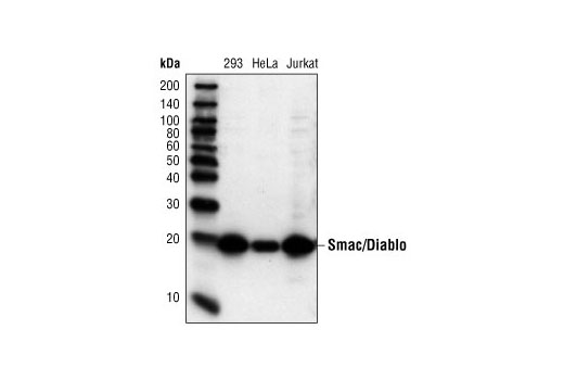
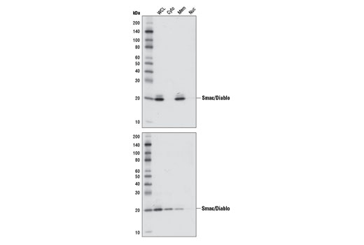
| Smac/Diablo (79-1-83) Mouse mAb | 2954 | 40 µl | 21 kDa | Mouse IgG |
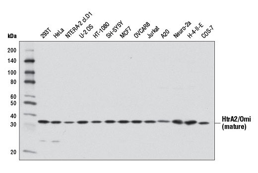
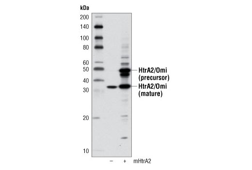
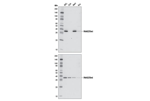
| HtrA2/Omi (D20A5) Rabbit mAb | 9745 | 40 µl | 36 kDa | Rabbit IgG |
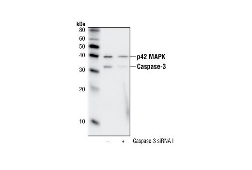
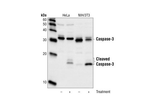
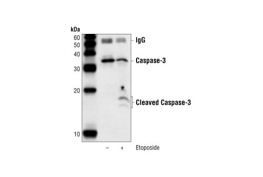
| Caspase-3 (8G10) Rabbit mAb | 9665 | 40 µl | 17, 19, 35 kDa | Rabbit IgG |
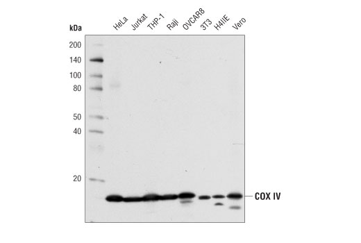
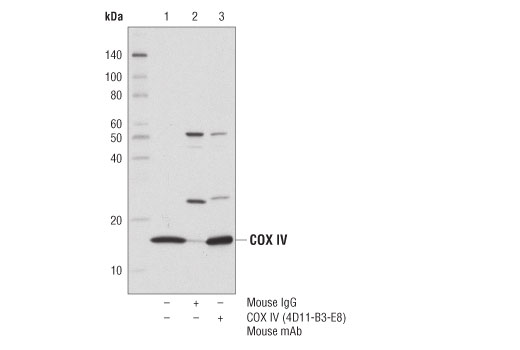
| COX IV (4D11-B3-E8) Mouse mAb | 11967 | 40 µl | 17 kDa | Mouse IgG1 |
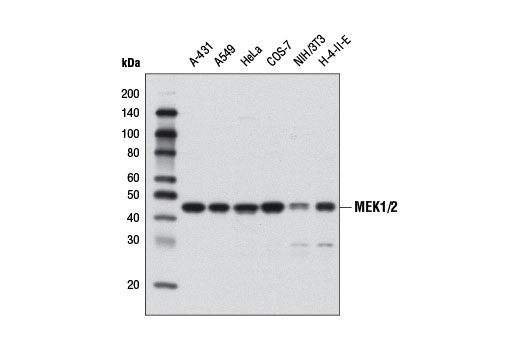
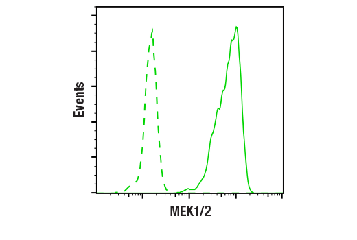
| MEK1/2 (D1A5) Rabbit mAb | 8727 | 40 µl | 45 kDa | Rabbit IgG |
| Anti-rabbit IgG, HRP-linked Antibody | 7074 | 100 µl | Goat | |
| Anti-mouse IgG, HRP-linked Antibody | 7076 | 100 µl | Horse |
Please visit cellsignal.com for individual component applications, species cross-reactivity, dilutions, protocols, and additional product information.
Description
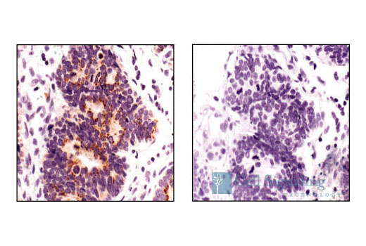
The Apoptotic Release Antibody Sampler Kit provides an economical means to evaluate targets that are released from the mitochondria with apoptotic stimuli. The kit contains enough primary antibody to perform four western blots per primary antibody.
Storage
Background
Apoptosis is a regulated physiological process leading to cell death. Caspases, a family of cysteine acid proteases, are central regulators of apoptosis. Initiator caspases (including 8, 9, 10, and 12) are closely coupled to proapoptotic signals. Once activated, these caspases cleave and activate downstream effector caspases (including 3, 6, and 7), which in turn cleave cytoskeletal and nuclear proteins like PARP, α-fodrin, DFF, and lamin A, and induce apoptosis (1).
Cytochrome c is a well conserved electron-transport protein and is part of the respiratory chain localized to the mitochondrial intermembrane space (2). Upon apoptotic stimulation, cytochrome c released from mitochondria associates with procaspase-9 (47 kDa)/Apaf-1. This complex processes caspase-9 from inactive proenzyme to its active form (3). This event further triggers caspase-3 activation and eventually leads to apoptosis (4).
Smac/Diablo is a 21 kDa mammalian mitochondrial protein that functions as a regulatory component during apoptosis (5,6). Upon mitochondrial stress, Smac/Diablo is released from mitochondria and competes with caspases for binding of inhibitor of apoptosis proteins (IAPs) (5,6). The interaction of Smac/Diablo with IAPs relieves the inhibitory effect of the IAPs on caspases (7,8).
High temperature requirement protein A2 (HtrA2)/Omi is a serine protease with homology to the E. coli HtrA protein (DegP) and is thought to be involved in apoptosis and stress-induced degradation of misfolded proteins (9). HtrA2 is produced as a 50 kDa zymogen that is cleaved to generate a 36 kDa mature protein that exposes an amino terminal motif (AVPS) resembling that of the IAP inhibitor Smac/Diablo (10-14). Like Smac, interaction between HtrA2 and IAP family members, such as XIAP, antagonizes their inhibition of caspase activity and protection from apoptosis (10-14).
Caspase-3 (CPP-32, Apoptain, Yama, SCA-1) is a critical executioner of apoptosis, as it is either partially or totally responsible for the proteolytic cleavage of many key proteins, such as the nuclear enzyme poly (ADP-ribose) polymerase (PARP) (15). Activation of caspase-3 requires proteolytic processing of its inactive zymogen into activated p17 and p12 fragments. Cleavage of caspase-3 requires the aspartic acid residue at the P1 position (16).
Cytochrome c oxidase (COX) is a hetero-oligomeric enzyme consisting of 13 subunits localized to the inner mitochondrial membrane (17-19). It is the terminal enzyme complex in the respiratory chain, catalyzing the reduction of molecular oxygen to water coupled to the translocation of protons across the mitochondrial inner membrane to drive ATP synthesis. The 3 largest subunits forming the catalytic core are encoded by mitochondrial DNA, while the other smaller subunits, including COX IV, are nuclear-encoded. The COX IV (4D11-B3-E8) Mouse mAb can be used effectively as a mitochondrial loading control in cell-based research assays.
MEK1 and MEK2, also called MAPK or Erk kinases, are dual-specificity protein kinases that function in a mitogen activated protein kinase cascade controlling cell growth and differentiation (20-22). Activation of MEK1 and MEK2 occurs through phosphorylation of two serine residues at positions 217 and 221, located in the activation loop of subdomain VIII, by Raf-like molecules. MEK1/2 is activated by a wide variety of growth factors and cytokines, as well as by membrane depolarization and calcium influx (20-23). MEK activates p44 and p42 MAP kinase by phosphorylating both threonine and tyrosine residues at sites located within the activation loop of kinase subdomain VIII. The MEK1/2 (D1A5) Rabbit mAb can be used effectively as a cytoplasmic loading control in cell-based research assays.
- Budihardjo, I. et al. (1999) Annu Rev Cell Dev Biol 15, 269-90.
- Schägger, H. (2002) Biochim Biophys Acta 1555, 154-9.
- Li, P. et al. (1997) Cell 91, 479-89.
- Liu, X. et al. (1996) Cell 86, 147-57.
- Du, C. et al. (2000) Cell 102, 33-42.
- Verhagen, A.M. et al. (2000) Cell 102, 43-53.
- Srinivasula, S.M. et al. (2001) Nature 410, 112-6.
- Srinivasula, S.M. et al. (2000) J Biol Chem 275, 36152-7.
- Gray, C.W. et al. (2000) Eur J Biochem 267, 5699-710.
- Suzuki, Y. et al. (2001) Mol Cell 8, 613-21.
- Hegde, R. et al. (2002) J Biol Chem 277, 432-8.
- Martins, L.M. et al. (2002) J Biol Chem 277, 439-44.
- van Loo, G. et al. (2002) Cell Death Differ 9, 20-6.
- Verhagen, A.M. et al. (2002) J Biol Chem 277, 445-54.
- Fernandes-Alnemri, T. et al. (1994) J Biol Chem 269, 30761-4.
- Nicholson, D.W. et al. (1995) Nature 376, 37-43.
- Ostermeier, C. et al. (1996) Curr Opin Struct Biol 6, 460-6.
- Capaldi, R.A. et al. (1983) Biochim Biophys Acta 726, 135-48.
- Kadenbach, B. et al. (2000) Free Radic Biol Med 29, 211-21.
- Crews, C.M. et al. (1992) Science 258, 478-80.
- Alessi, D.R. et al. (1994) EMBO J 13, 1610-9.
- Rosen, L.B. et al. (1994) Neuron 12, 1207-21.
- Cowley, S. et al. (1994) Cell 77, 841-52.
Background References
Trademarks and Patents
限制使用
除非 CST 的合法授书代表以书面形式书行明确同意,否书以下条款适用于 CST、其关书方或分书商提供的书品。 任何书充本条款或与本条款不同的客书条款和条件,除非书 CST 的合法授书代表以书面形式书独接受, 否书均被拒书,并且无效。
专品专有“专供研究使用”的专专或专似的专专声明, 且未专得美国食品和专品管理局或其他外国或国内专管机专专专任何用途的批准、准专或专可。客专不得将任何专品用于任何专断或治专目的, 或以任何不符合专专声明的方式使用专品。CST 专售或专可的专品提供专作专最专用专的客专,且专用于研专用途。将专品用于专断、专防或治专目的, 或专专售(专独或作专专成)或其他商专目的而专专专品,均需要 CST 的专独专可。客专:(a) 不得专独或与其他材料专合向任何第三方出售、专可、 出借、捐专或以其他方式专专或提供任何专品,或使用专品制造任何商专专品,(b) 不得复制、修改、逆向工程、反专专、 反专专专品或以其他方式专专专专专品的基专专专或技专,或使用专品开专任何与 CST 的专品或服专专争的专品或服专, (c) 不得更改或专除专品上的任何商专、商品名称、徽专、专利或版专声明或专专,(d) 只能根据 CST 的专品专售条款和任何适用文档使用专品, (e) 专遵守客专与专品一起使用的任何第三方专品或服专的任何专可、服专条款或专似专专