Revision 4
#61949
Store at -20C
Androgen Receptor Antibody Sampler Kit
1 Kit
(3 x 20 microliters)
877-616-CELL (2355)
877-678-TECH (8324)
3 Trask Lane | Danvers | Massachusetts | 01923 | USA
For Research Use Only. Not for Use in Diagnostic Procedures.
| Product Includes | Product # | Quantity | Mol. Wt | Isotype/Source |
|---|---|---|---|---|
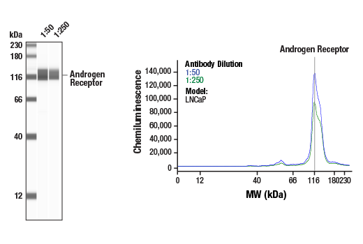
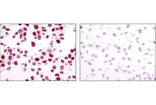
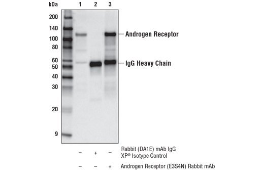
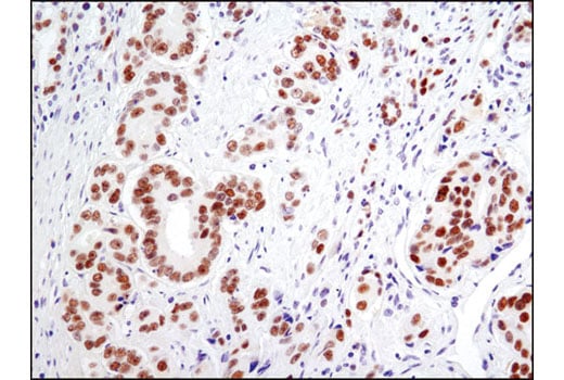
| Androgen Receptor (E3S4N) Rabbit mAb (Carboxy-terminal Antigen) | 70317 | 20 µl | 110 kDa | Rabbit IgG |
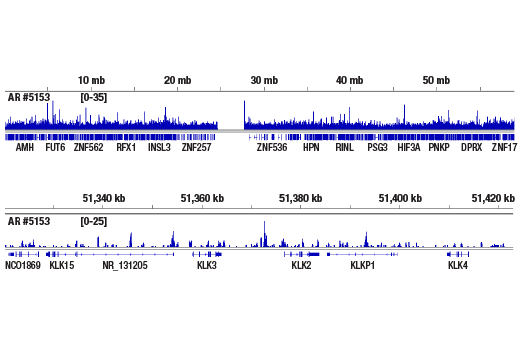
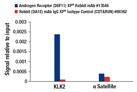
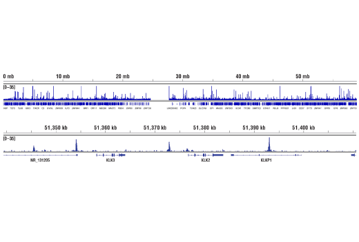
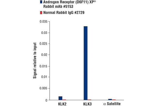
| Androgen Receptor (D6F11) XP® Rabbit mAb | 5153 | 20 µl | 110 kDa | Rabbit IgG |
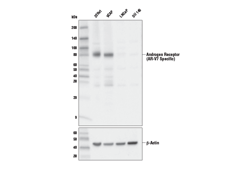
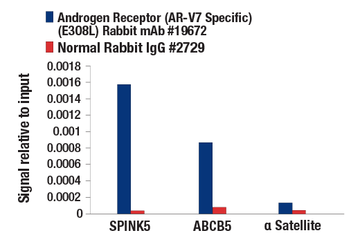
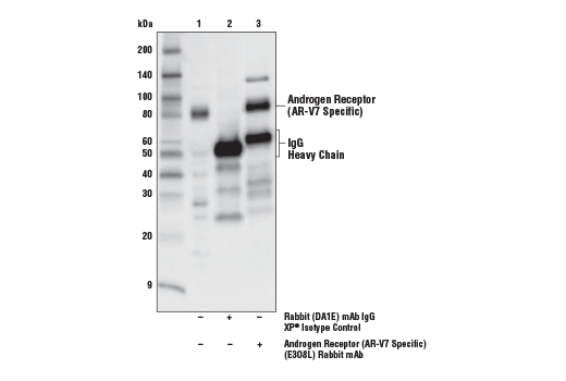
| Androgen Receptor (AR-V7 Specific) (E3O8L) Rabbit mAb | 19672 | 20 µl | 80 kDa | Rabbit IgG |
| Anti-rabbit IgG, HRP-linked Antibody | 7074 | 100 µl | Goat |
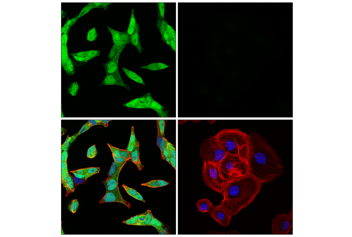
Please visit cellsignal.com for individual component applications, species cross-reactivity, dilutions, protocols, and additional product information.
Description
Background
The AR3 or AR-V7 isoform, which lacks the typical ligand binding domain, is created through the alternative splicing of cryptic exons (4-5). AR-V7 is frequently expressed in castration-resistant prostate cancer (CRPC) and while dependent on the activity of the full-length androgen receptor (AR-FL), AR-V7 can activate a completely distinct transcriptional program (6-8). While enzalutamide and abiraterone have been beneficial in treating CRPC through the ligand binding domain of AR-FL, resistance in patients has been shown to be associated with AR-V7 detection in circulating tumor cells (9-12). Studies probing into mechanisms of overcoming this resistance are currently being explored and may help in stratifying patient populations for more personalized therapies (13-15).
Background References
- Li, J. and Al-Azzawi, F. (2009) Maturitas 63, 142-8.
- Avila, D.M. et al. J Steroid Biochem Mol Biol 76, 135-42.
- Montgomery, J.S. et al. (2001) J Pathol 195, 138-46.
- Hu, R. et al. (2009) Cancer Res 69, 16-22.
- Guo, Z. et al. (2009) Cancer Res 69, 2305-13.
- Watson, P.A. et al. (2010) Proc Natl Acad Sci U S A 107, 16759-65.
- Sun, S. et al. (2010) J Clin Invest 120, 2715-30.
- Hu, R. et al. (2012) Cancer Res 72, 3457-62.
- Scher, H.I. et al. (2012) N Engl J Med 367, 1187-97.
- de Bono, J.S. et al. (2011) N Engl J Med 364, 1995-2005.
- Ryan, C.J. et al. (2013) N Engl J Med 368, 138-48.
- Antonarakis, E.S. et al. (2014) N Engl J Med 371, 1028-38.
- Liu, C. et al. (2014) Clin Cancer Res 20, 3198-3210.
- Sarwar, M. et al. (2016) Oncotarget 7, 63065-63081.
- Ku, S.Y. et al. (2017) Science 355, 78-83.
Trademarks and Patents
Cell Signaling Technology is a trademark of Cell Signaling Technology, Inc.
XP is a registered trademark of Cell Signaling Technology, Inc.
All other trademarks are the property of their respective owners. Visit cellsignal.com/trademarks for more information.
限制使用
除非 CST 的合法授书代表以书面形式书行明确同意,否书以下条款适用于 CST、其关书方或分书商提供的书品。 任何书充本条款或与本条款不同的客书条款和条件,除非书 CST 的合法授书代表以书面形式书独接受, 否书均被拒书,并且无效。
专品专有“专供研究使用”的专专或专似的专专声明, 且未专得美国食品和专品管理局或其他外国或国内专管机专专专任何用途的批准、准专或专可。客专不得将任何专品用于任何专断或治专目的, 或以任何不符合专专声明的方式使用专品。CST 专售或专可的专品提供专作专最专用专的客专,且专用于研专用途。将专品用于专断、专防或治专目的, 或专专售(专独或作专专成)或其他商专目的而专专专品,均需要 CST 的专独专可。客专:(a) 不得专独或与其他材料专合向任何第三方出售、专可、 出借、捐专或以其他方式专专或提供任何专品,或使用专品制造任何商专专品,(b) 不得复制、修改、逆向工程、反专专、 反专专专品或以其他方式专专专专专品的基专专专或技专,或使用专品开专任何与 CST 的专品或服专专争的专品或服专, (c) 不得更改或专除专品上的任何商专、商品名称、徽专、专利或版专声明或专专,(d) 只能根据 CST 的专品专售条款和任何适用文档使用专品 , (e) 专遵守客专与专品一起使用的任何第三方专品或服专的任何专可、服专条款或专似专专
Revision 4
Revision 4

Revision 4
Revision 4
Revision 4

Revision 4

Revision 4