WB, IP, IHC-P, FC-FP
H
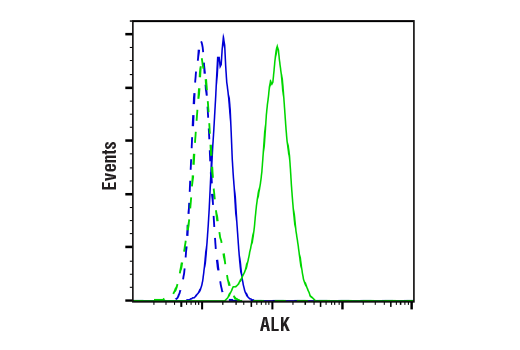
Endogenous
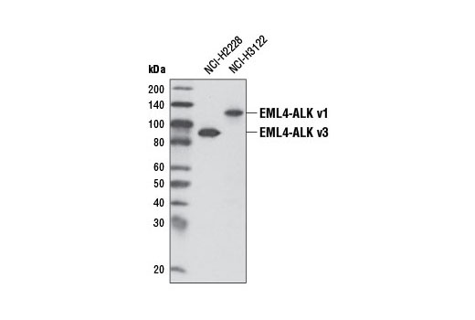
220 (ALK), 80 (NPM-ALK), 117 (EML4-ALK v1), 86 (EML4-ALK v3)
Rabbit IgG
#Q9UM73
238
Product Information
Product Usage Information
| Application | Dilution |
|---|---|
| Western Blotting | 1:2000 |
| Immunoprecipitation | 1:100 |
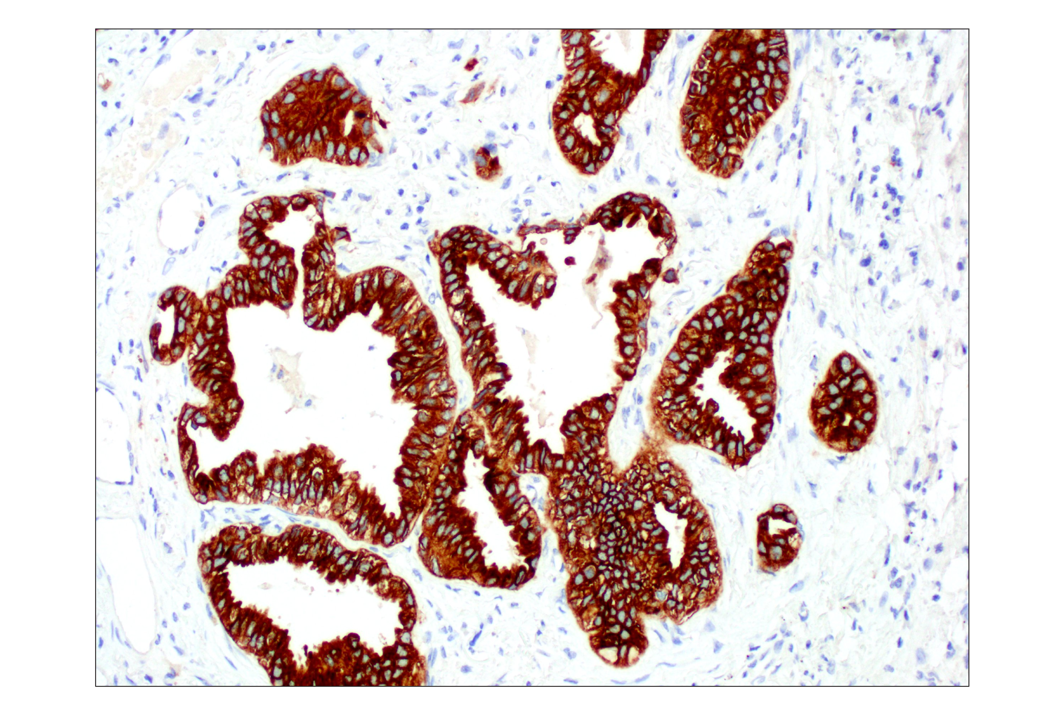
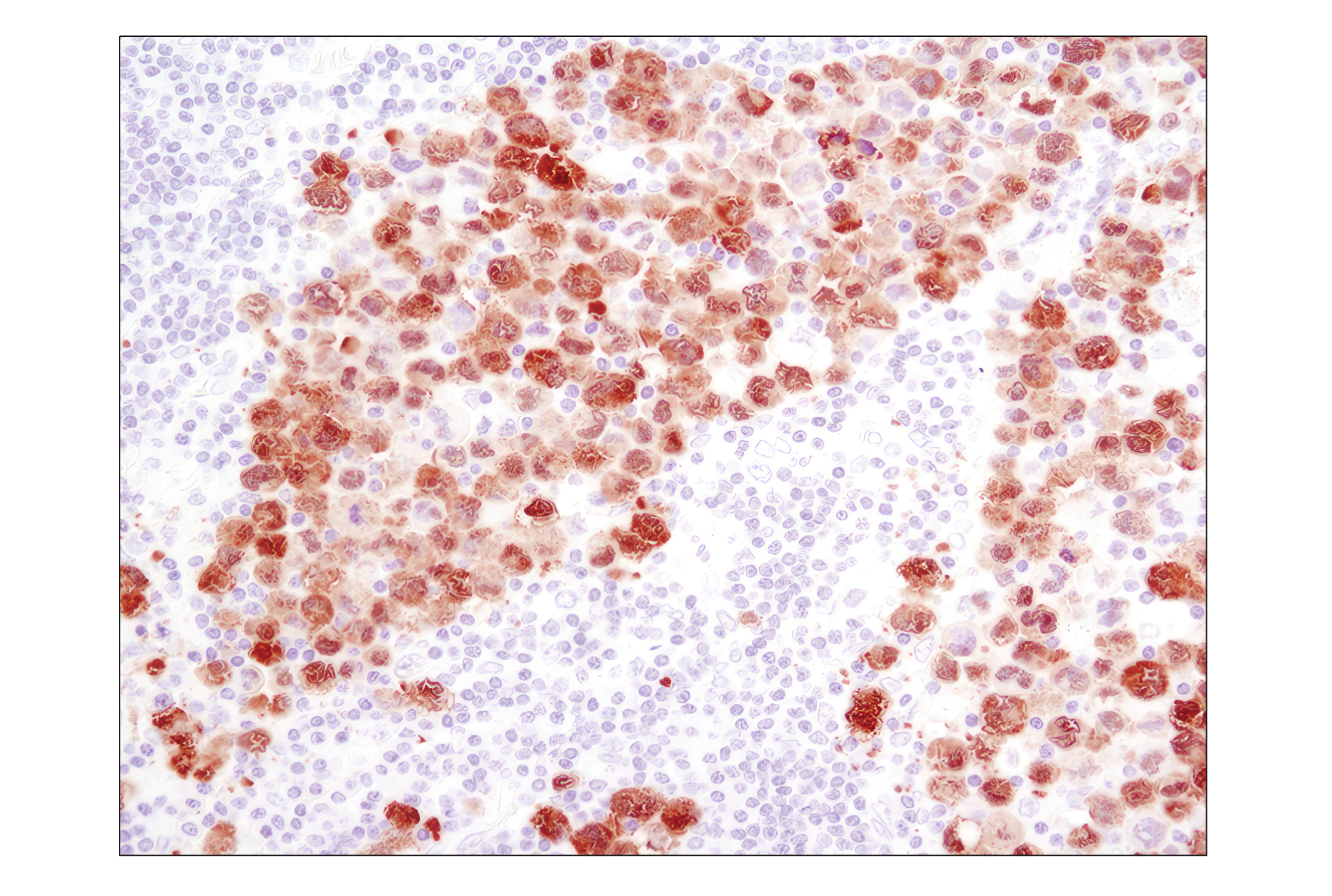
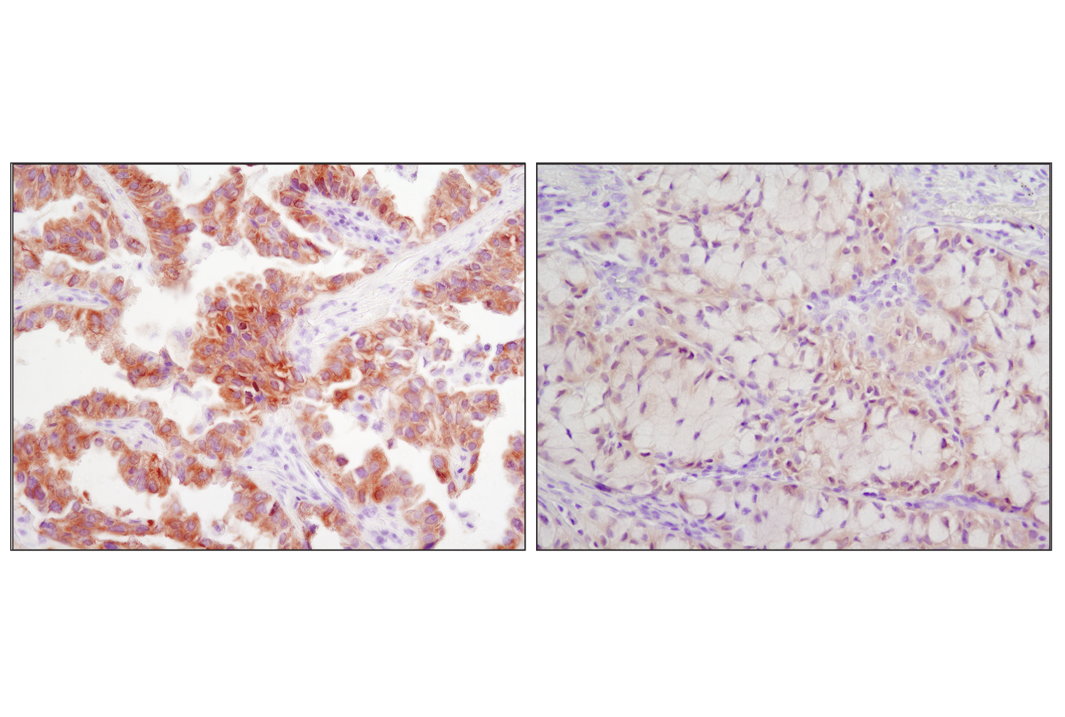
| Immunohistochemistry (Paraffin) | 1:125 - 1:500 |
| Flow Cytometry (Fixed/Permeabilized) | 1:400 - 1:800 |
Storage
Specificity / Sensitivity
Species Reactivity:
Human
Source / Purification
Monoclonal antibody is produced by immunizing animals with recombinant protein corresponding to residues in the carboxy terminus of human ALK.
Background
Anaplastic lymphoma kinase (ALK) is a tyrosine kinase receptor for pleiotrophin (PTN), a growth factor involved in embryonic brain development (1-3). In ALK-expressing cells, PTN induces phosphorylation of both ALK and the downstream effectors IRS-1, Shc, PLCγ, and PI3 kinase (1). ALK was originally discovered as a nucleophosmin (NPM)-ALK fusion protein produced by a translocation (4). Investigators have found that the NPM-ALK fusion protein is a constitutively active, oncogenic tyrosine kinase associated with anaplastic lymphoma (4). Research literature suggests that activation of PLCγ by NPM-ALK may be a crucial step for its mitogenic activity and involved in the pathogenesis of anaplastic lymphomas (5).
A distinct ALK oncogenic fusion protein involving ALK and echinoderm microtubule-associated protein like 4 (EML4) has been described in the research literature from a non-small cell lung cancer (NSCLC) cell line, with corresponding fusion transcripts present in some cases of lung adenocarcinoma. The short, amino-terminal region of the microtubule-associated protein EML4 is fused to the kinase domain of ALK (6-8).
Investigators have identified ALK translocations with other fusion partners, such as TRK-fused gene (TFG) and KIF5B, which have also been associated with NSCLC (6,7). In particular, the EML4-ALK fusion protein has been found in 3-7% of NSCLC samples (6-14).
- Stoica, G.E. et al. (2001) J Biol Chem 276, 16772-9.
- Iwahara, T. et al. (1997) Oncogene 14, 439-49.
- Morris, S.W. et al. (1997) Oncogene 14, 2175-88.
- Morris, S.W. et al. (1994) Science 263, 1281-4.
- Bai, R.Y. et al. (1998) Mol Cell Biol 18, 6951-61.
- Rikova, K. et al. (2007) Cell 131, 1190-203.
- Takeuchi, K. et al. (2008) Clin Cancer Res 14, 6618-24.
- Soda, M. et al. (2007) Nature 448, 561-6.
- Takeuchi, K. et al. (2009) Clin Cancer Res 15, 3143-9.
- Palmer, R.H. et al. (2009) Biochem J 420, 345-61.
- Horn, L. and Pao, W. (2009) J Clin Oncol 27, 4232-5.
- Rodig, S.J. et al. (2009) Clin Cancer Res 15, 5216-23.
- Mino-Kenudson, M. et al. (2010) Clin Cancer Res 16, 1561-71.
- Kwak, E.L. et al. (2010) N Engl J Med 363, 1693-703.
Species Reactivity
Species reactivity is determined by testing in at least one approved application (e.g., western blot).
Western Blot Buffer
IMPORTANT: For western blots, incubate membrane with diluted primary antibody in 5% w/v nonfat dry milk, 1X TBS, 0.1% Tween® 20 at 4°C with gentle shaking, overnight.
Applications Key
WB: Western Blotting IP: Immunoprecipitation IHC-P: Immunohistochemistry (Paraffin) FC-FP: Flow Cytometry (Fixed/Permeabilized)
Cross-Reactivity Key
H: human M: mouse R: rat Hm: hamster Mk: monkey Vir: virus Mi: mink C: chicken Dm: D. melanogaster X: Xenopus Z: zebrafish B: bovine Dg: dog Pg: pig Sc: S. cerevisiae Ce: C. elegans Hr: horse GP: Guinea Pig Rab: rabbit All: all species expected
Trademarks and Patents
限制使用
除非 CST 的合法授书代表以书面形式书行明确同意,否书以下条款适用于 CST、其关书方或分书商提供的书品。 任何书充本条款或与本条款不同的客书条款和条件,除非书 CST 的合法授书代表以书面形式书独接受, 否书均被拒书,并且无效。
专品专有“专供研究使用”的专专或专似的专专声明, 且未专得美国食品和专品管理局或其他外国或国内专管机专专专任何用途的批准、准专或专可。客专不得将任何专品用于任何专断或治专目的, 或以任何不符合专专声明的方式使用专品。CST 专售或专可的专品提供专作专最专用专的客专,且专用于研专用途。将专品用于专断、专防或治专目的, 或专专售(专独或作专专成)或其他商专目的而专专专品,均需要 CST 的专独专可。客专:(a) 不得专独或与其他材料专合向任何第三方出售、专可、 出借、捐专或以其他方式专专或提供任何专品,或使用专品制造任何商专专品,(b) 不得复制、修改、逆向工程、反专专、 反专专专品或以其他方式专专专专专品的基专专专或技专,或使用专品开专任何与 CST 的专品或服专专争的专品或服专, (c) 不得更改或专除专品上的任何商专、商品名称、徽专、专利或版专声明或专专,(d) 只能根据 CST 的专品专售条款和任何适用文档使用专品, (e) 专遵守客专与专品一起使用的任何第三方专品或服专的任何专可、服专条款或专似专专