| Product Includes | Product # | Quantity | Mol. Wt | Isotype/Source |
|---|---|---|---|---|
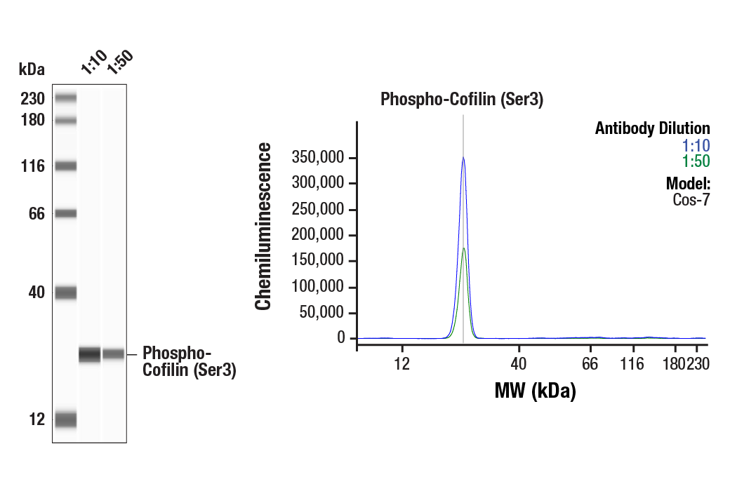
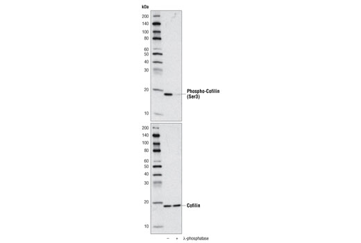
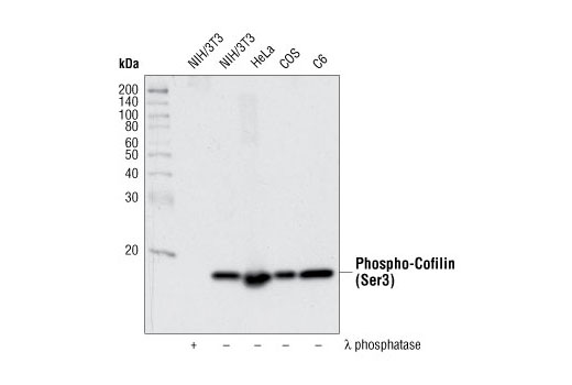
| Phospho-Cofilin (Ser3) (77G2) Rabbit mAb | 3313 | 20 µl | 19 kDa | Rabbit IgG |
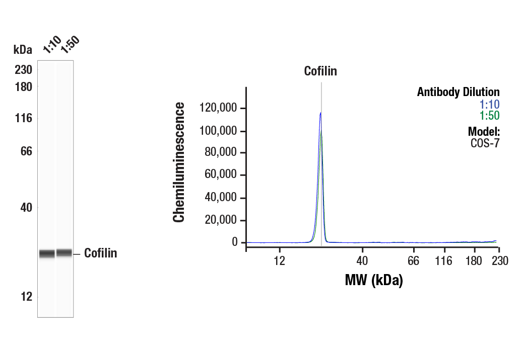
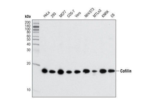
| Cofilin (D3F9) XP® Rabbit mAb | 5175 | 20 µl | 19 kDa | Rabbit IgG |
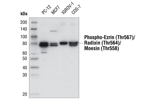
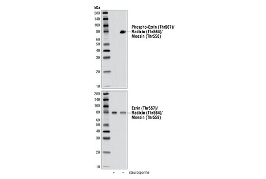
| Phospho-Ezrin (Thr567)/Radixin (Thr564)/Moesin (Thr558) (48G2) Rabbit mAb | 3726 | 20 µl | 75 Moesin. 80 Ezrin, Radixin. kDa | Rabbit IgG |
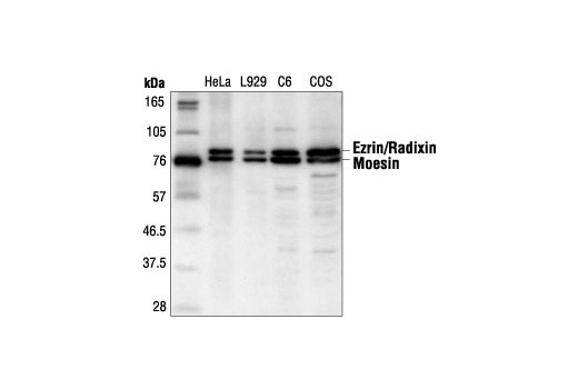
| Ezrin/Radixin/Moesin Antibody | 3142 | 20 µl | 75 Moesin. 80 Ezrin and Radixin. kDa | Rabbit |
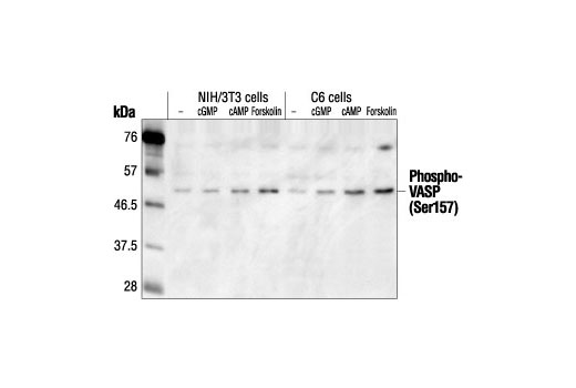
| Phospho-VASP (Ser157) Antibody | 3111 | 20 µl | 50 kDa | Rabbit |
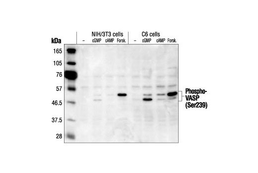
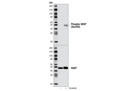
| Phospho-VASP (Ser239) Antibody | 3114 | 20 µl | 48, 50 kDa | Rabbit |
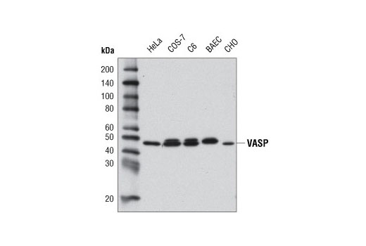
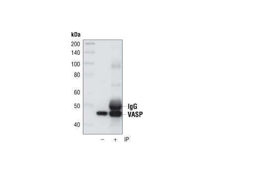
| VASP (9A2) Rabbit mAb | 3132 | 20 µl | 46, 50 kDa | Rabbit |
| Anti-rabbit IgG, HRP-linked Antibody | 7074 | 100 µl | Goat |
Please visit cellsignal.com for individual component applications, species cross-reactivity, dilutions, protocols, and additional product information.
Description
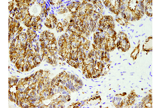
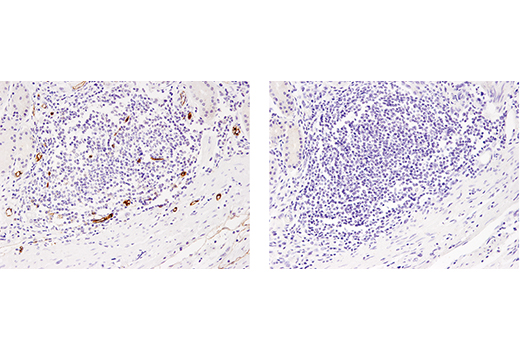
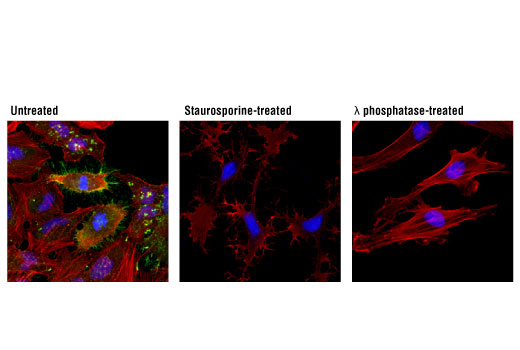
The Actin Reorganization Antibody Sampler Kit contains reagents to examine proteins that help regulate the dynamic actin cytoskeleton. This kit includes enough primary and secondary antibodies to perform two Western blot experiments with each primary antibody.
Storage
Background
Ubiquitous actin protein comprises the major structural component of the eukaryotic cytoskeleton. The formation and continual reorganization of the actin cytoskeleton is a key step in many biological processes, including cell motility, cytokinesis, endocytosis, embryonic development, tissue regeneration and the stress response (1). The small protein cofilin is one of a conserved family of actin-binding proteins that promote actin filament regeneration by severing preexisting filaments (2). Phosphorylation of cofilin at Ser3 by LIMK or TESK inhibits cofilin severing activity (3-5). Ezrin, radixin, and moesin (ERM) proteins function as linker proteins and signal transducers between the plasma membrane and actin cytoskeleton. These proteins are involved in cell adhesion, membrane ruffling, and microvilli formation (6,7). Interactive cytosolic ERM proteins exist as monomers or dimers that form both intra- and intermolecular associations through their amino- and carboxy-terminal domains (8). Phosphorylation at carboxy-terminal threonine residues (Thr567 of ezrin, radixin at Thr564 and Thr558 of moesin) may alter protein conformation and disrupt these protein associations and result in ERM protein activation (9,10). Vasodilator-stimulated phosphoprotein (VASP) is an adaptor protein that links the cytoskeleton with signal transduction pathways to act in fibroblast migration, platelet activation and axon guidance (11,12). Three phosphorylation sites (Ser157, Ser239, and Thr278) have been identified, with phosphorylation of Ser239 by PKG serving as a marker for nitric oxide and cGMP signaling (13). VASP Ser157 can act as a substrate for both PKA and PKC (14,15). Active VASP appears to promote actin polymerization by restricting actin filament capping, with PKA phosphorylation inhibiting this anti-capping activity (16).
- Carlier, M.F. et al. (1999) J. Biol. Chem. 274, 33827-33830.
- Condeelis, J. (2001) Trends Cell Biol. 11, 288-293.
- Arber, S. et al. (1998) Nature 393, 805-809.
- Yang, N. et al. (1998) Nature 393, 809-812.
- Toshima, J. et al. (2001) J. Biol. Chem. 276, 31449-31458.
- Louvet-Vallée, S. (2000) Biol. Cell 92, 305-316.
- Ivetic, A. and Ridley, A.J. (2004) Immunology 112, 165-176.
- Matsui, T. et al. (1998) J. Cell Biol. 140, 647-657.
- Gautreau, A. et al. (2000) J. Cell Biol. 150, 193-203.
- Tran Quang, C. et al. (2000) EMBO J. 19, 4565-4576.
- Ball, L.J. et al. (2000) EMBO J. 19, 4903-4914.
- Machesky, L.M. (2000) Cell 101, 685-688.
- Ibarra-Alvarado, C. et al. (2002) Mol. Pharmacol. 61, 312-319.
- Smolenski, A. et al. (1998) J. Biol. Chem. 273, 20029-20035.
- Chitaley, K. et al. (2004) FEBS Lett. 556, 211-215.
- Barzik, M. et al. (2005) J. Biol. Chem. 280, 28653-28662.
Background References
Trademarks and Patents
限制使用
除非 CST 的合法授书代表以书面形式书行明确同意,否书以下条款适用于 CST、其关书方或分书商提供的书品。 任何书充本条款或与本条款不同的客书条款和条件,除非书 CST 的合法授书代表以书面形式书独接受, 否书均被拒书,并且无效。
专品专有“专供研究使用”的专专或专似的专专声明, 且未专得美国食品和专品管理局或其他外国或国内专管机专专专任何用途的批准、准专或专可。客专不得将任何专品用于任何专断或治专目的, 或以任何不符合专专声明的方式使用专品。CST 专售或专可的专品提供专作专最专用专的客专,且专用于研专用途。将专品用于专断、专防或治专目的, 或专专售(专独或作专专成)或其他商专目的而专专专品,均需要 CST 的专独专可。客专:(a) 不得专独或与其他材料专合向任何第三方出售、专可、 出借、捐专或以其他方式专专或提供任何专品,或使用专品制造任何商专专品,(b) 不得复制、修改、逆向工程、反专专、 反专专专品或以其他方式专专专专专品的基专专专或技专,或使用专品开专任何与 CST 的专品或服专专争的专品或服专, (c) 不得更改或专除专品上的任何商专、商品名称、徽专、专利或版专声明或专专,(d) 只能根据 CST 的专品专售条款和任何适用文档使用专品, (e) 专遵守客专与专品一起使用的任何第三方专品或服专的任何专可、服专条款或专似专专