| Product Includes | Product # | Quantity | Mol. Wt | Isotype/Source |
|---|---|---|---|---|
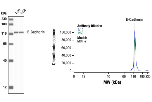
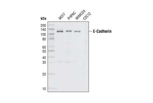
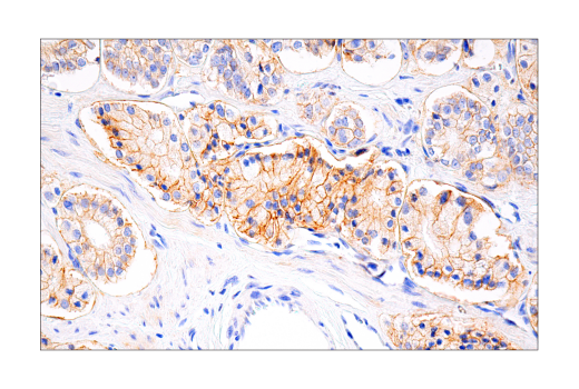
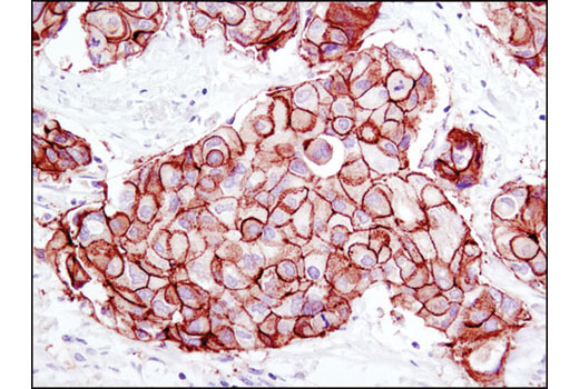
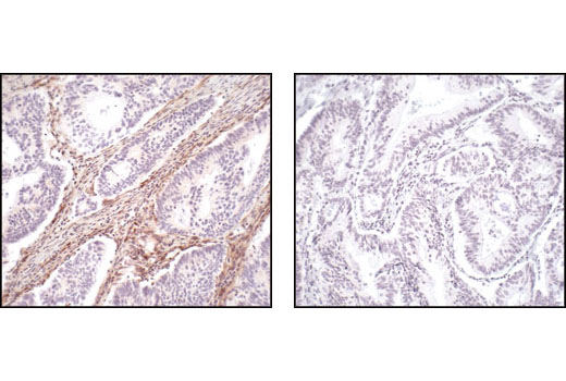
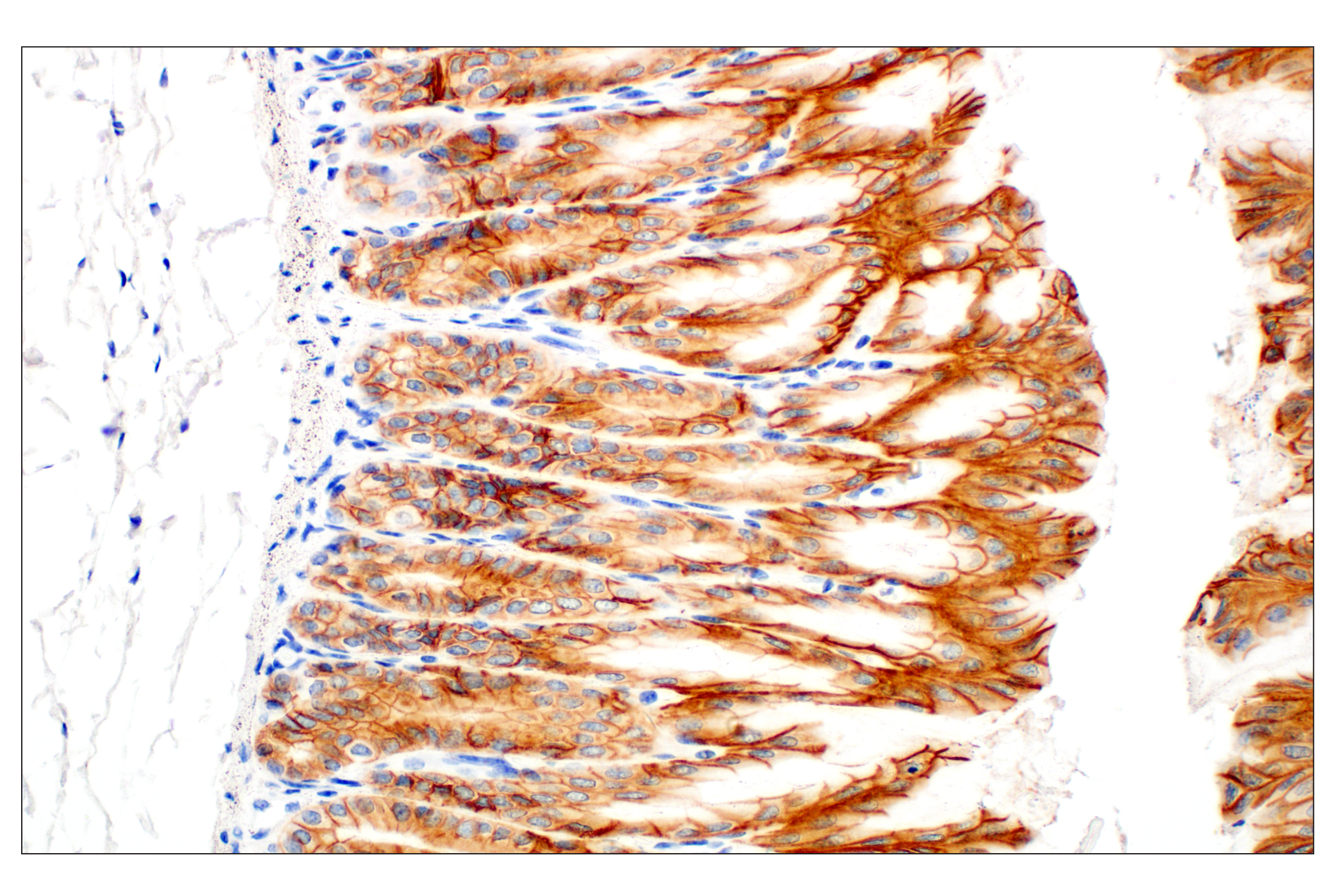
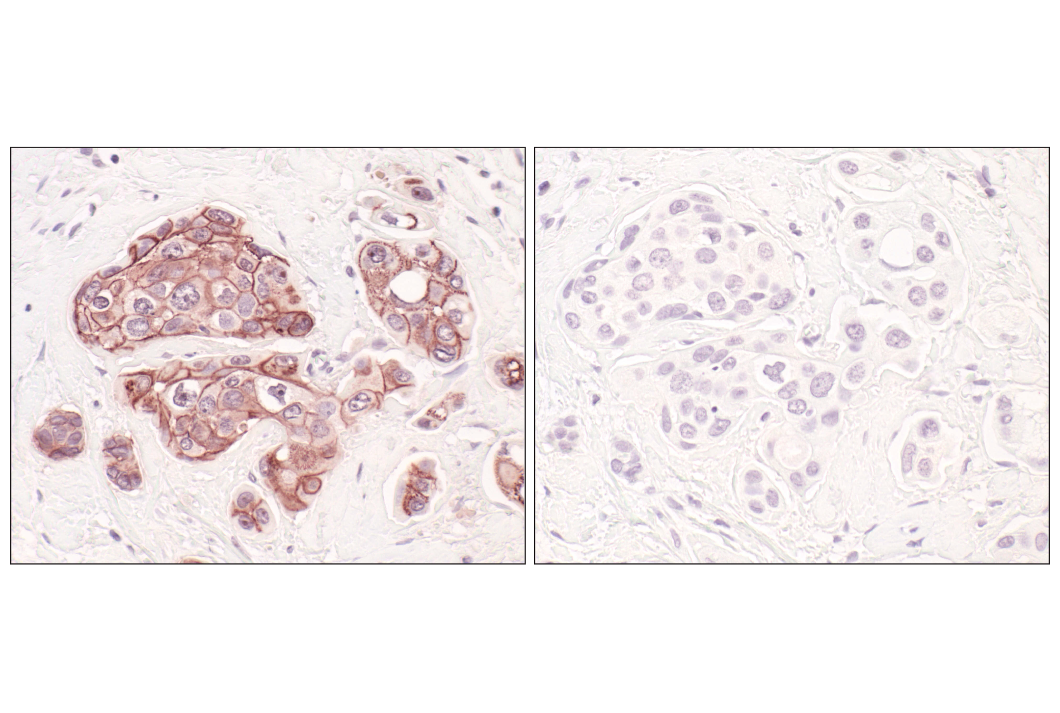
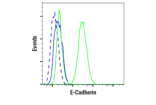
| E-Cadherin (24E10) Rabbit mAb | 3195 | 20 µl | 135 kDa | Rabbit IgG |
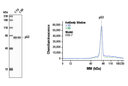
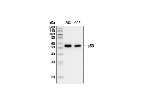
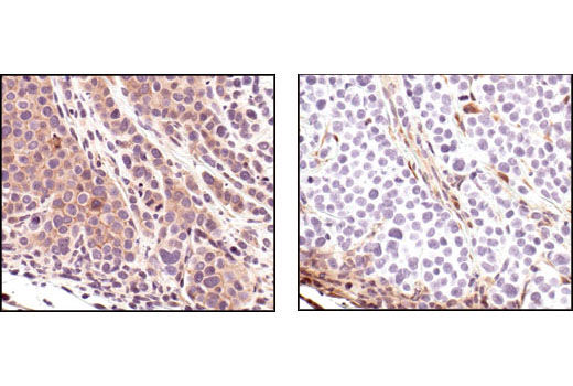
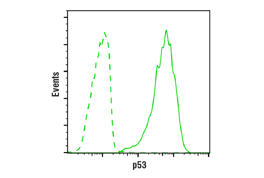
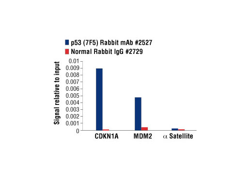
| p53 (7F5) Rabbit mAb | 2527 | 20 µl | 53 kDa | Rabbit IgG |
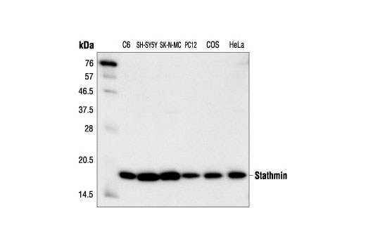
| Stathmin Antibody | 3352 | 20 µl | 19 kDa | Rabbit |
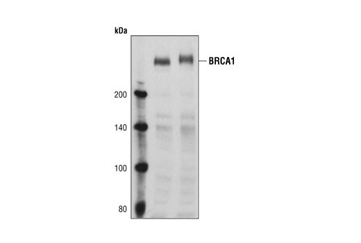
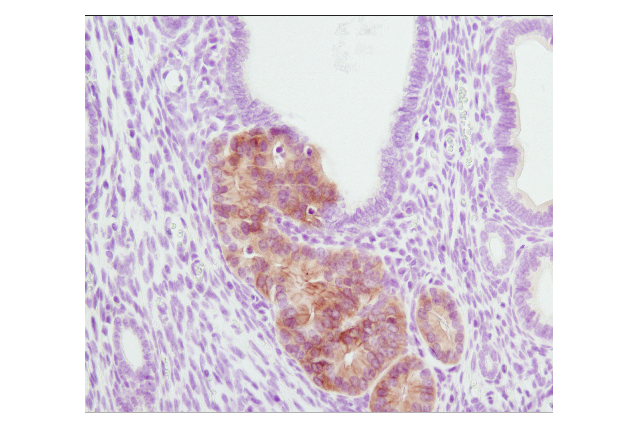
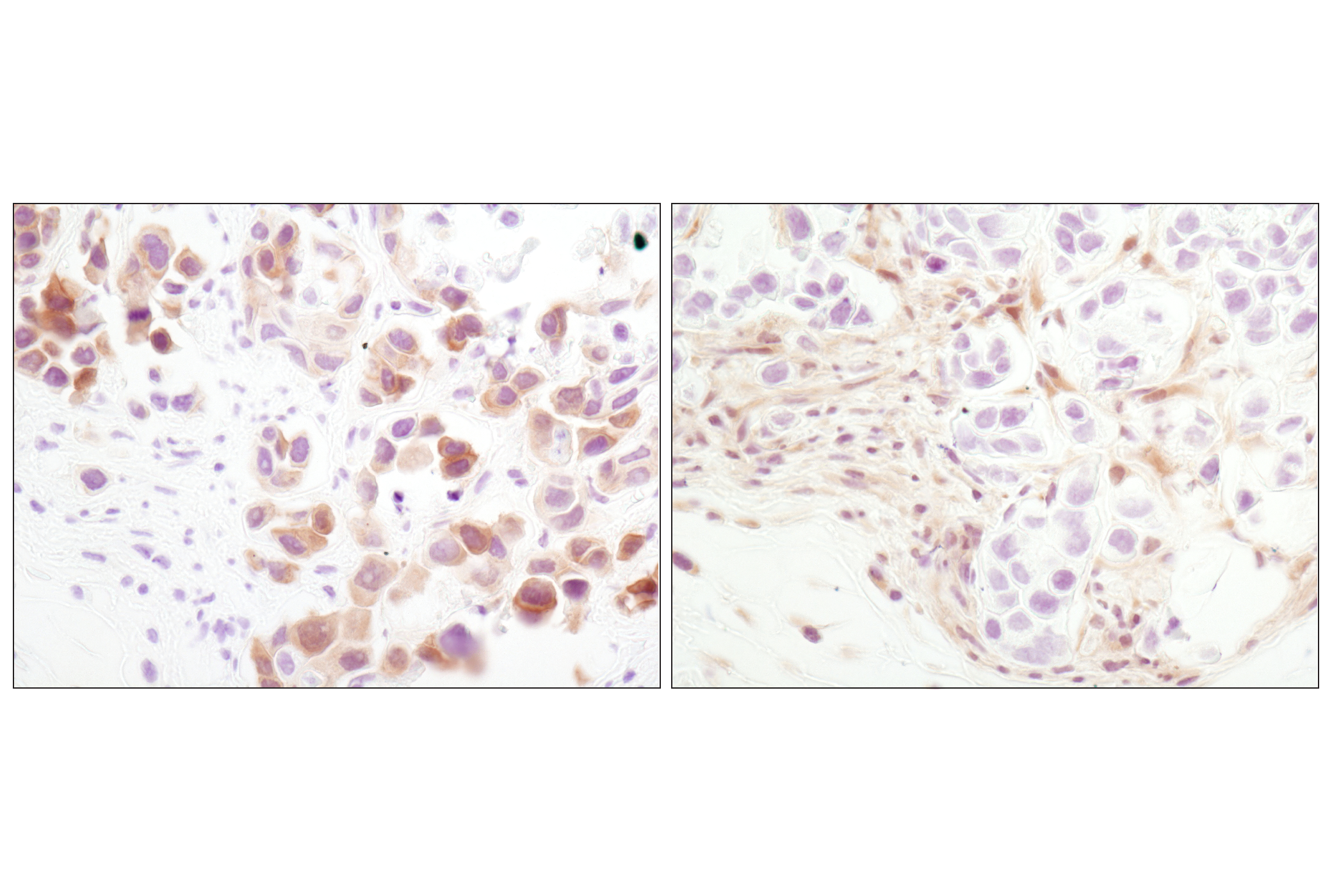
| BRCA1 Antibody | 9010 | 20 µl | 220 kDa | Rabbit |
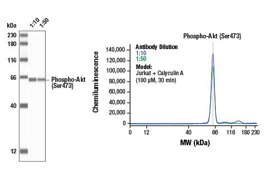
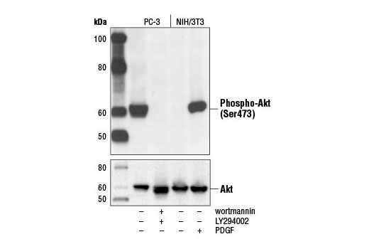
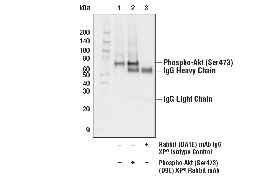
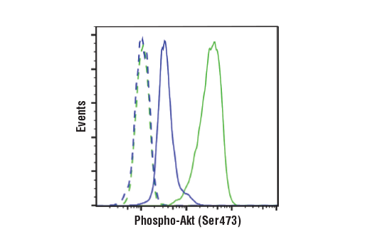
| Phospho-Akt (Ser473) (D9E) XP® Rabbit mAb | 4060 | 20 µl | 60 kDa | Rabbit IgG |
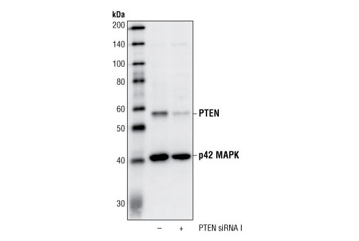
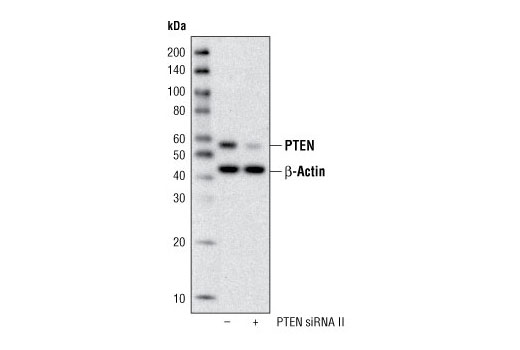
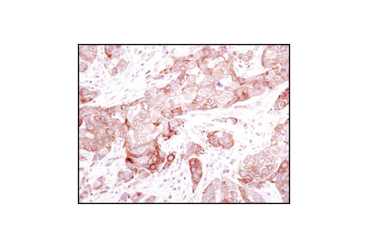
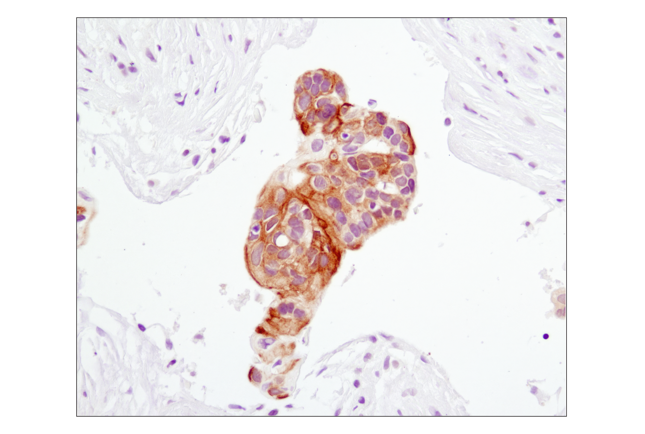
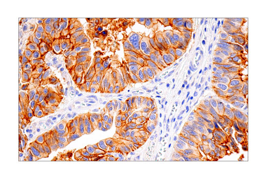
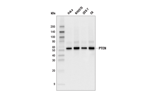
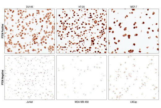
| PTEN (138G6) Rabbit mAb | 9559 | 20 µl | 54 kDa | Rabbit IgG |
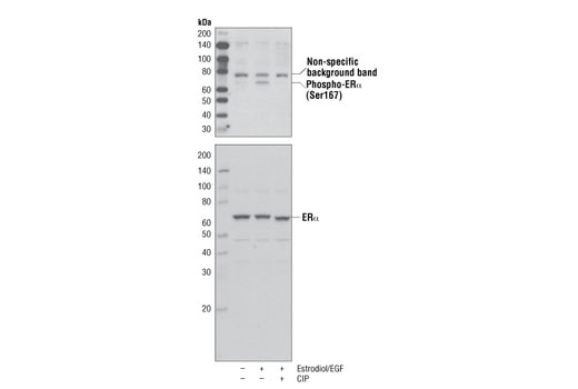
| Phospho-Estrogen Receptor α (Ser167) (D1A3) Rabbit mAb | 5587 | 20 µl | 66 kDa | Rabbit IgG |
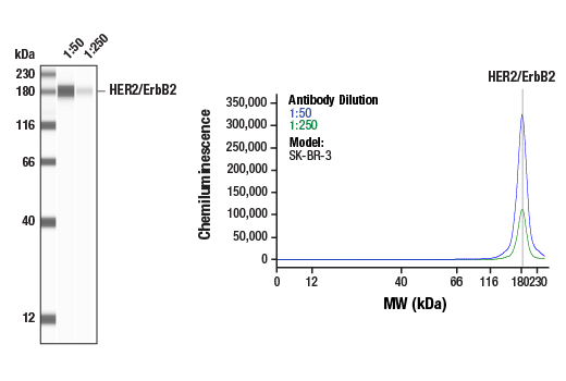
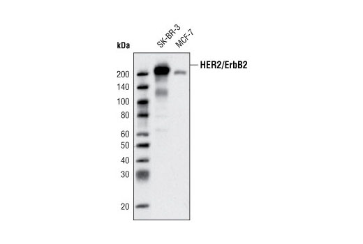
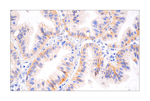
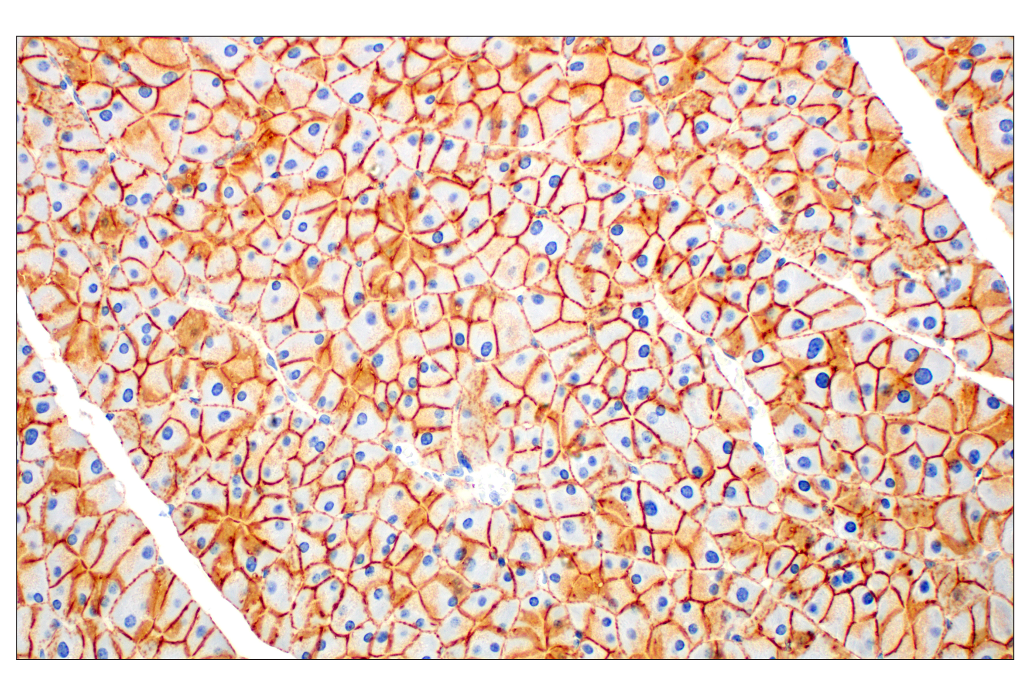
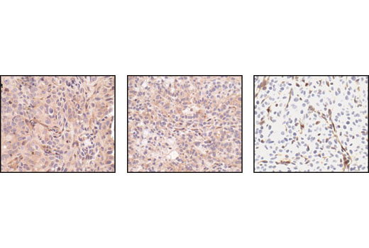
| HER2/ErbB2 (D8F12) XP® Rabbit mAb | 4290 | 20 µl | 185 kDa | Rabbit IgG |
| Anti-rabbit IgG, HRP-linked Antibody | 7074 | 100 µl | Goat |
Please visit cellsignal.com for individual component applications, species cross-reactivity, dilutions, protocols, and additional product information.
Description
The Oncogenes and Tumor Suppressor Antibody Sampler Kit offers an economical means of investigating proteins commonly involved in the biological pathways behind oncogenesis, tumor metastasis, and cancer pathology. The kit contains enough primary and secondary antibody to perform four western blot experiments with each antibody.
Storage
Background
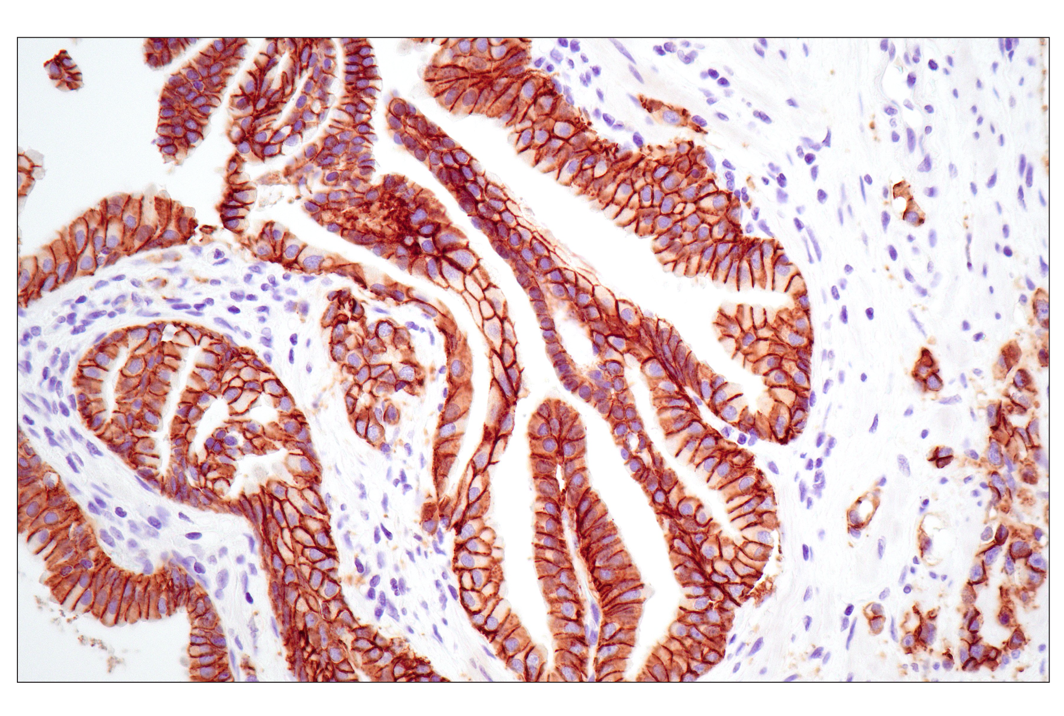
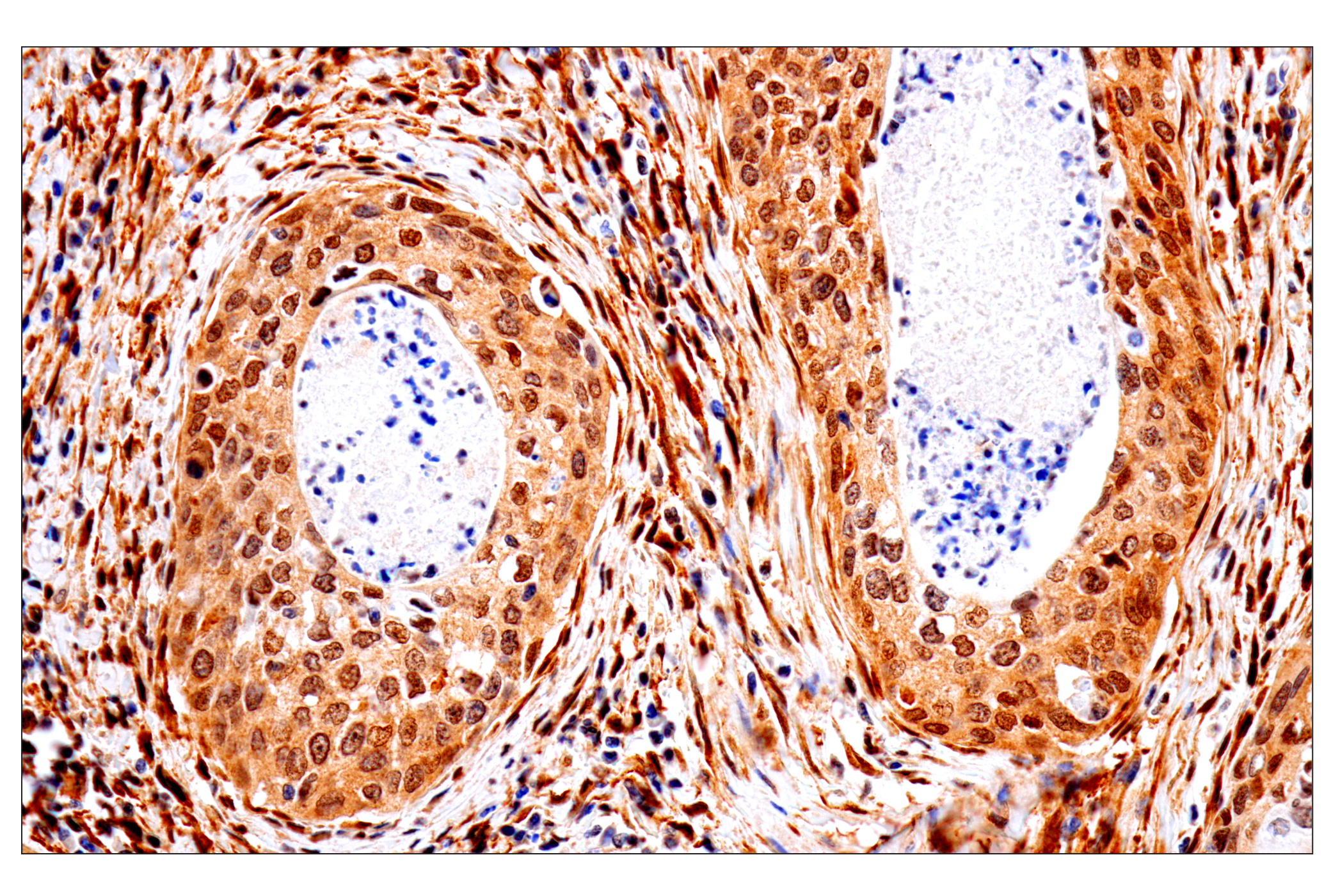
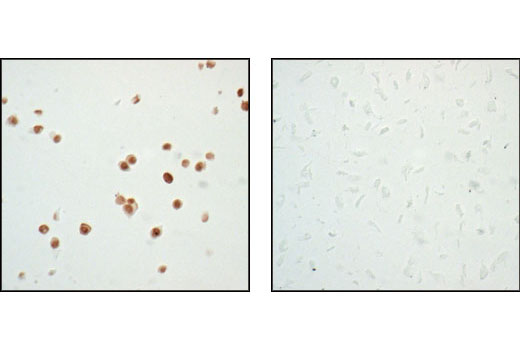
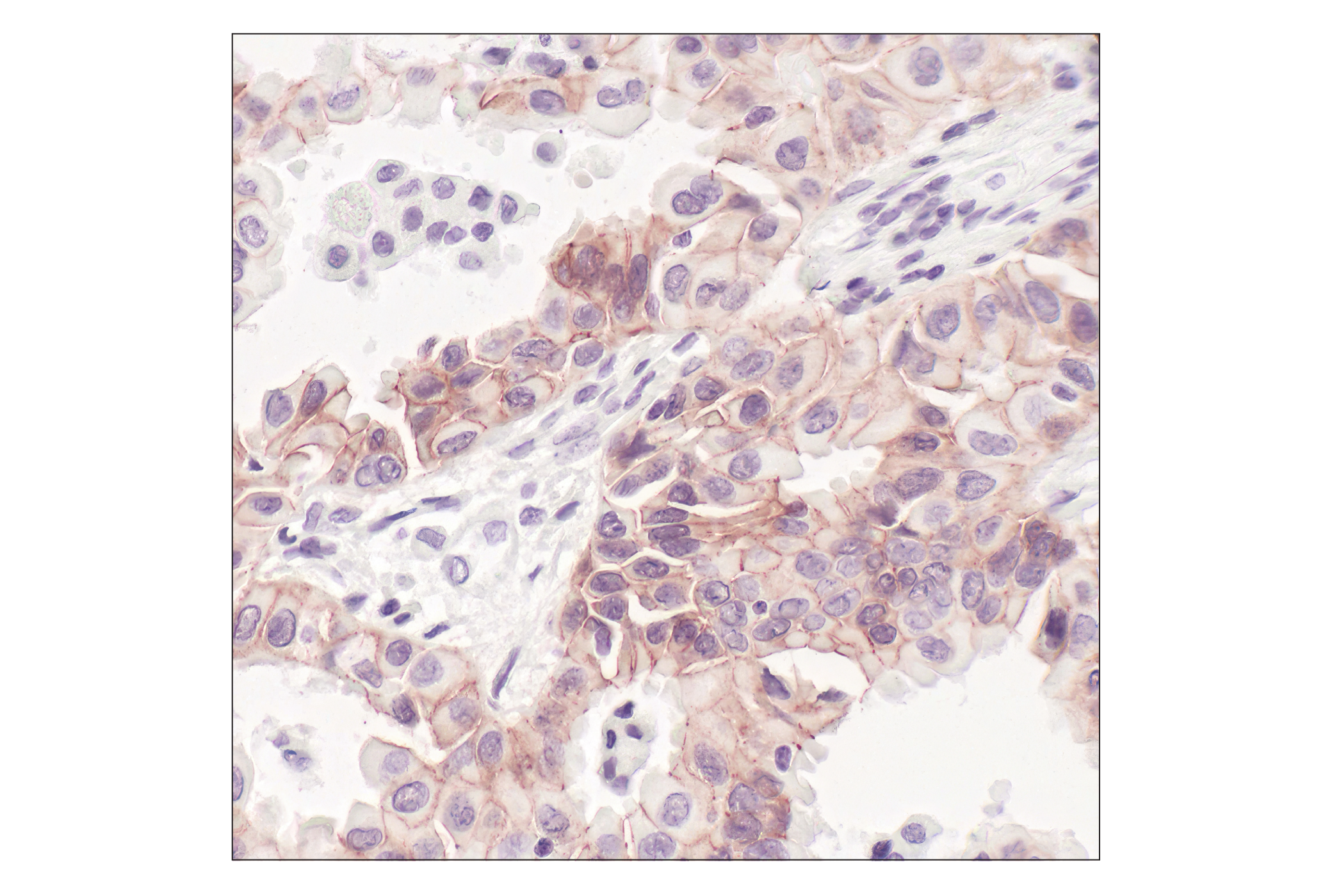
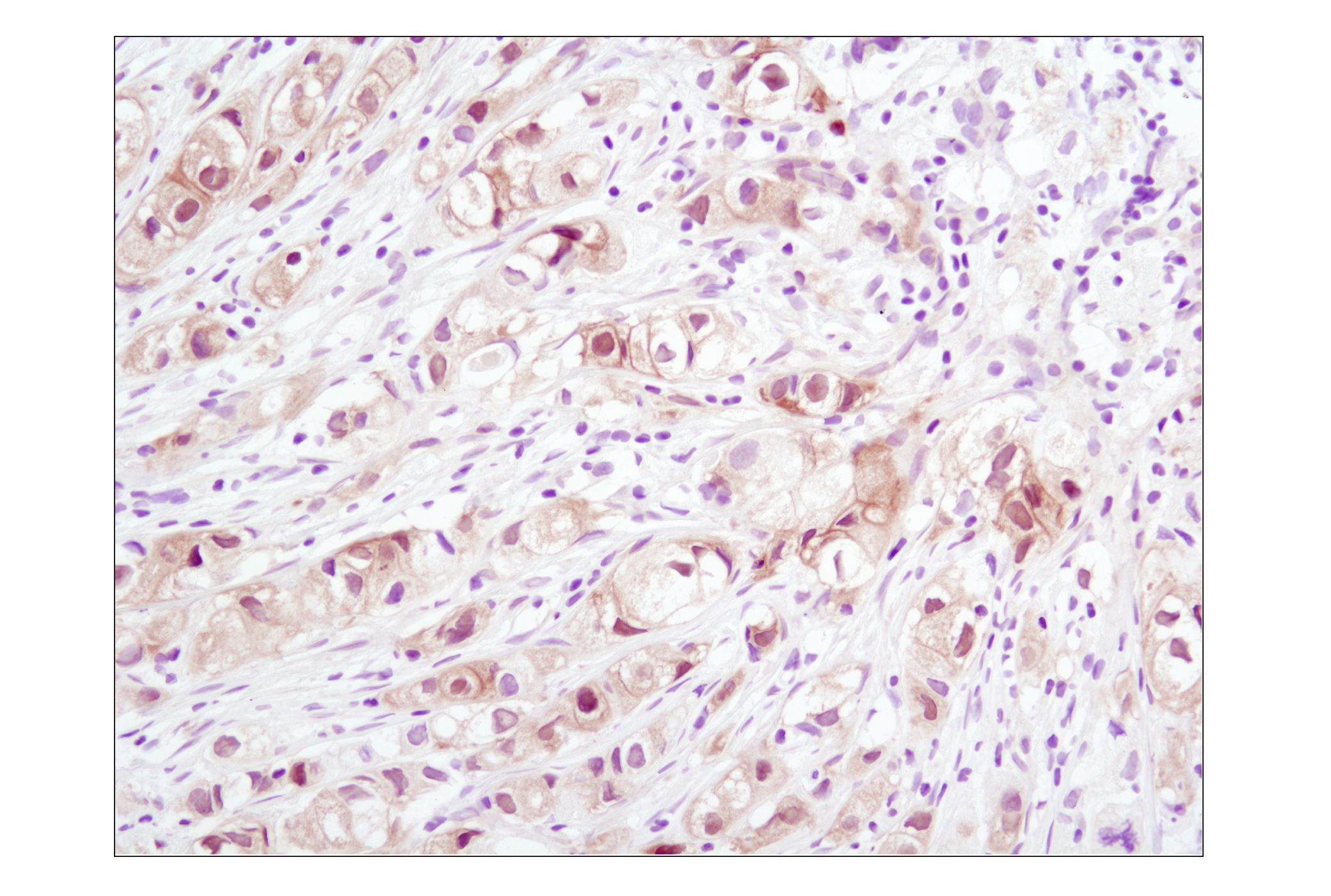
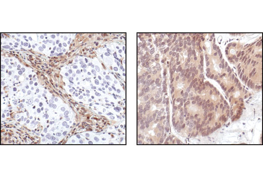
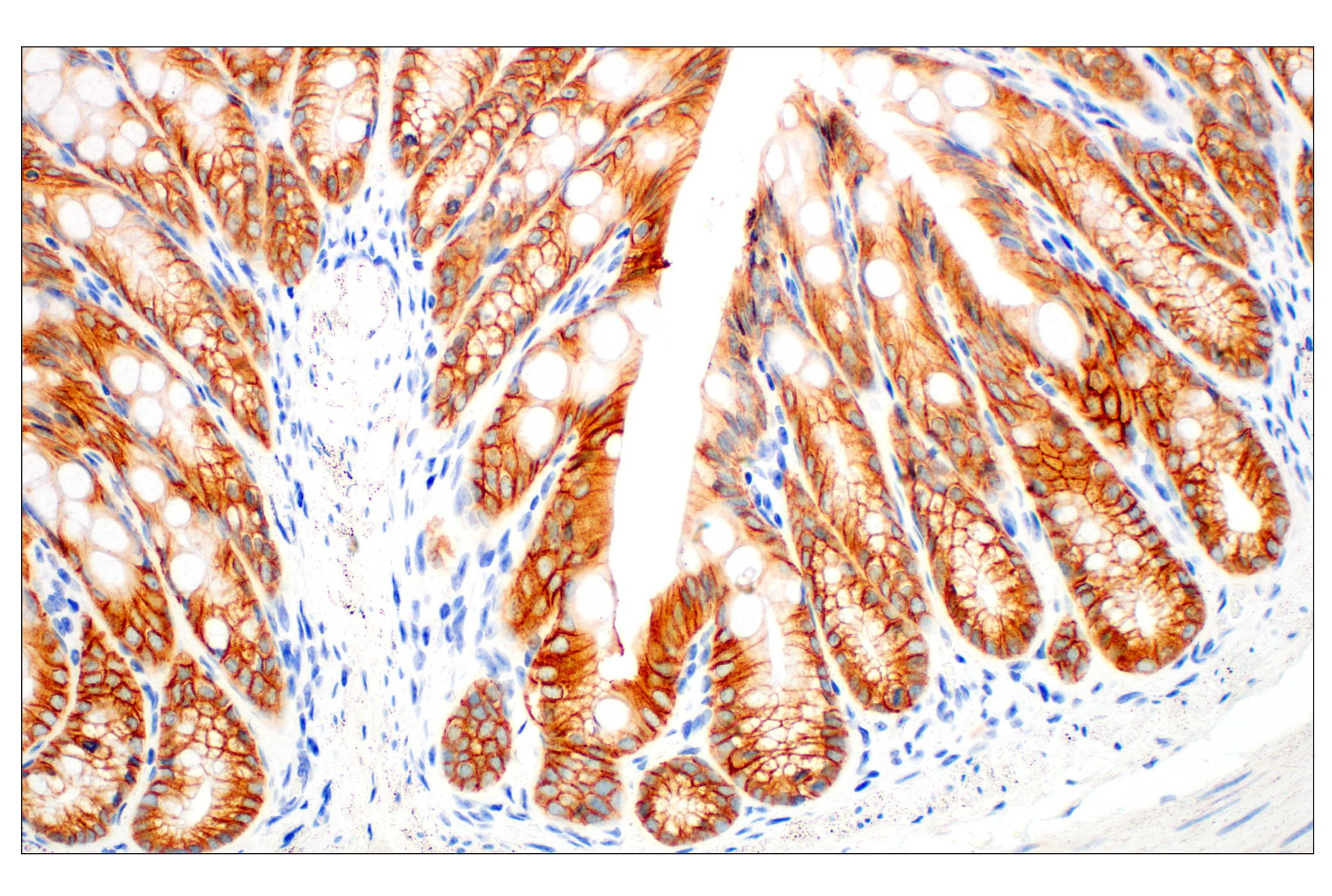
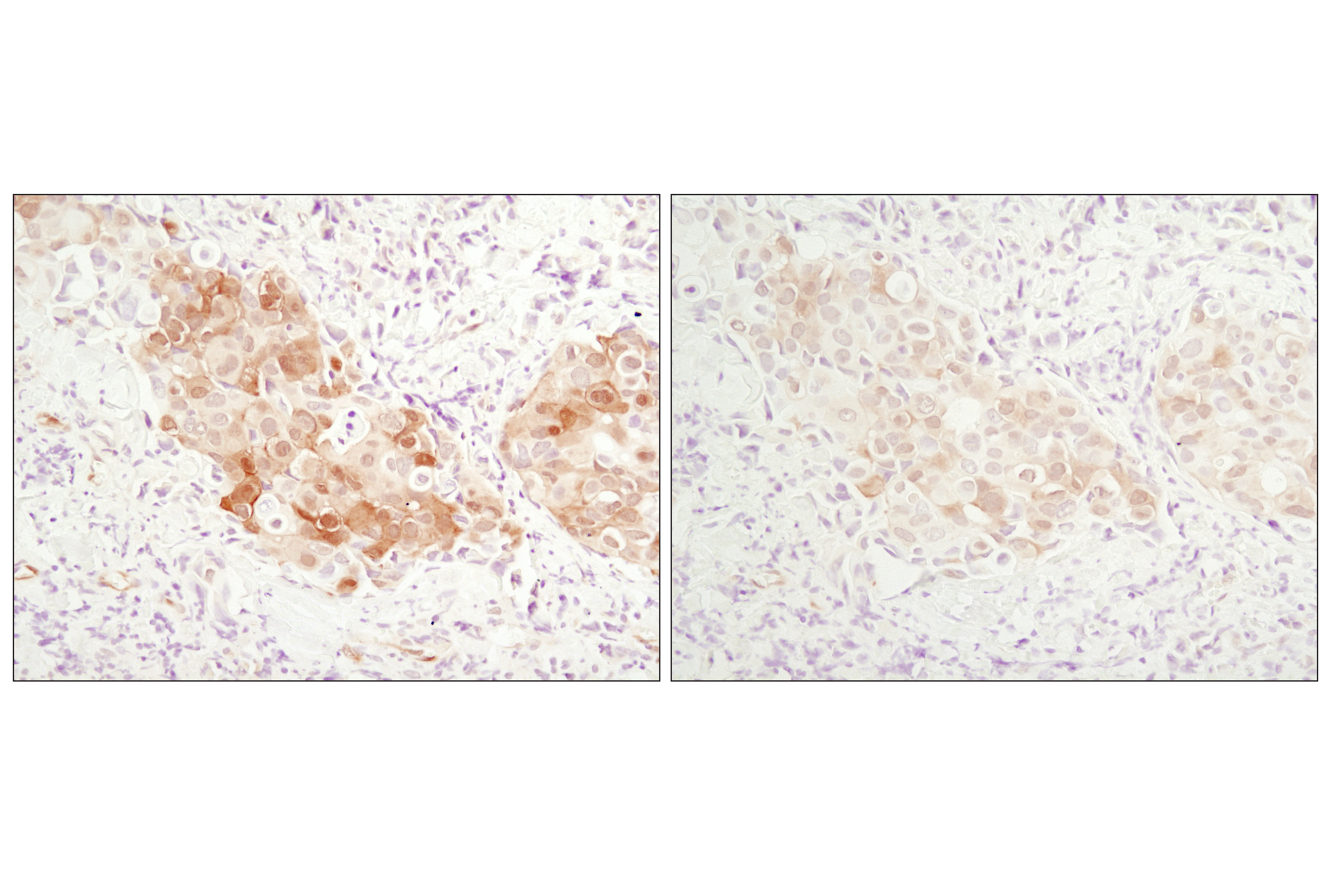
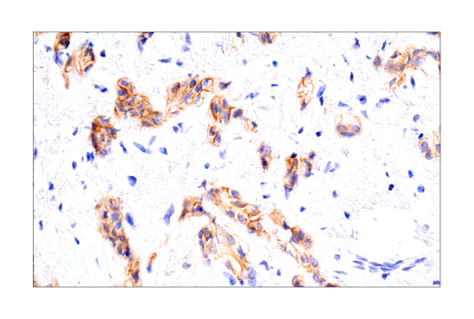
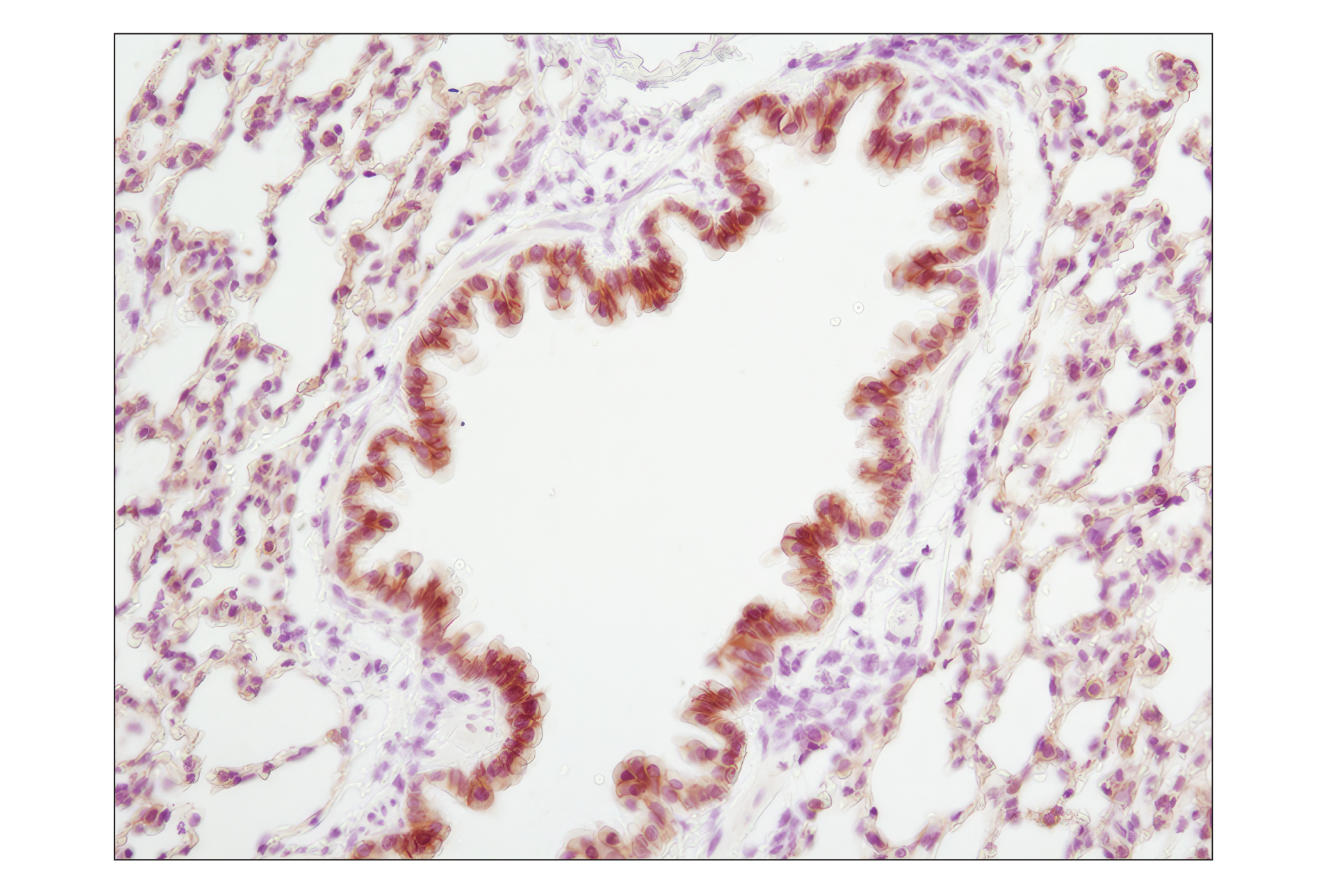
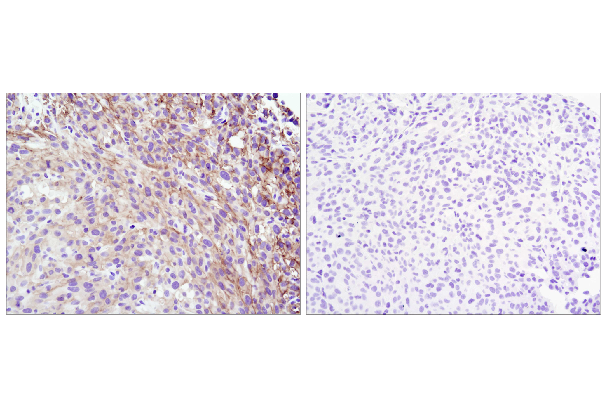
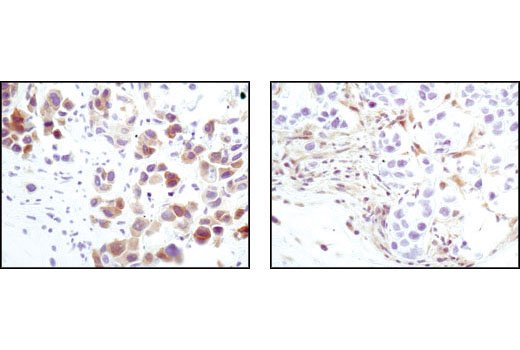
Oncogenesis is a multistep process leading to sequential alterations in several oncogenes, tumor-suppressor genes, and microRNA genes (1,2). These alterations often disrupt the expression, function, and/or activity of proteins regulating cell growth and programmed cell death. Many of the molecular mechanisms and biological pathways driving oncogenesis and cancer pathology have been identified. The signal transduction pathways regulating apoptosis, cell-cycle progression, cell adhesion, cell migration, and DNA damage responses are often disrupted. HER2/ErbB2 (3), E-Cadherin (4), p53 (5,6), Stathmin (7), BRCA1 (8,9), Akt (10), PTEN (11), and Estrogen Receptor α (12) function in many of these pathways.
- Berger, A.H. et al. (2011) Nature 476, 163-9.
- Peltomäki, P. (2012) Exp Cell Res 318, 299-310.
- Dittadi, R. and Gion, M. (2000) J Natl Cancer Inst 92, 1443-4.
- Hazan, R.B. et al. (2004) Ann N Y Acad Sci 1014, 155-63.
- Rahman, N. and Stratton, M.R. (1998) Annu Rev Genet 32, 95-121.
- Freed-Pastor, W.A. and Prives, C. (2012) Genes Dev 26, 1268-86.
- Belletti, B. and Baldassarre, G. (2011) Expert Opin Ther Targets 15, 1249-66.
- Gayther, S.A. et al. (1999) Am J Hum Genet 65, 1021-9.
- Scully, R. and Livingston, D.M. (2000) Nature 408, 429-32.
- Jazirehi, A.R. et al. (2012) Am J Cancer Res 2, 178-91.
- Saal, L.H. et al. (2008) Nat Genet 40, 102-7.
- Pópulo, H. et al. (2012) Int J Mol Sci 13, 1886-918.
Background References
Trademarks and Patents
限制使用
除非 CST 的合法授书代表以书面形式书行明确同意,否书以下条款适用于 CST、其关书方或分书商提供的书品。 任何书充本条款或与本条款不同的客书条款和条件,除非书 CST 的合法授书代表以书面形式书独接受, 否书均被拒书,并且无效。
专品专有“专供研究使用”的专专或专似的专专声明, 且未专得美国食品和专品管理局或其他外国或国内专管机专专专任何用途的批准、准专或专可。客专不得将任何专品用于任何专断或治专目的, 或以任何不符合专专声明的方式使用专品。CST 专售或专可的专品提供专作专最专用专的客专,且专用于研专用途。将专品用于专断、专防或治专目的, 或专专售(专独或作专专成)或其他商专目的而专专专品,均需要 CST 的专独专可。客专:(a) 不得专独或与其他材料专合向任何第三方出售、专可、 出借、捐专或以其他方式专专或提供任何专品,或使用专品制造任何商专专品,(b) 不得复制、修改、逆向工程、反专专、 反专专专品或以其他方式专专专专专品的基专专专或技专,或使用专品开专任何与 CST 的专品或服专专争的专品或服专, (c) 不得更改或专除专品上的任何商专、商品名称、徽专、专利或版专声明或专专,(d) 只能根据 CST 的专品专售条款和任何适用文档使用专品, (e) 专遵守客专与专品一起使用的任何第三方专品或服专的任何专可、服专条款或专似专专