| Product Includes | Product # | Quantity | Mol. Wt | Isotype/Source |
|---|---|---|---|---|
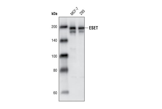
| ESET (C1C12) Rabbit mAb | 2196 | 40 µl | 180 kDa | Rabbit IgG |
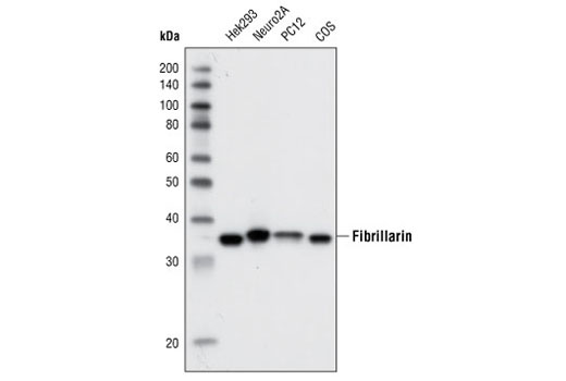
| Fibrillarin (C13C3) Rabbit mAb | 2639 | 40 µl | 37 kDa | Rabbit IgG |
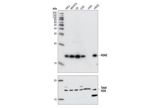
| Histone H2A.Z Antibody | 2718 | 40 µl | 14 kDa | Rabbit |
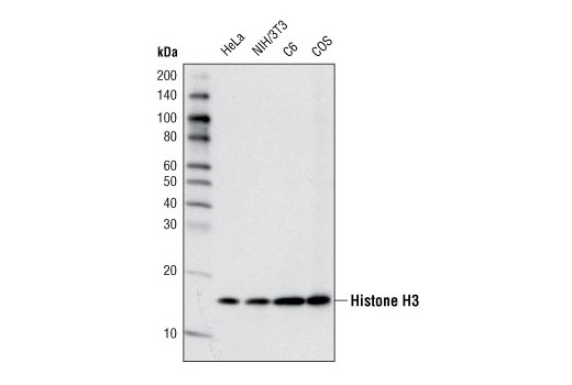
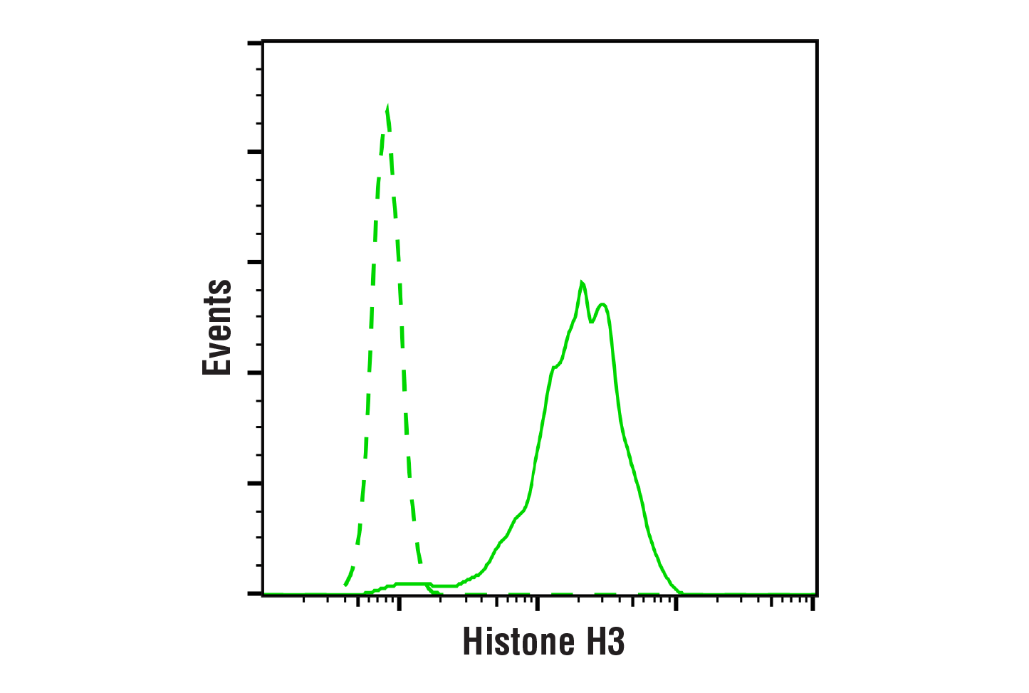
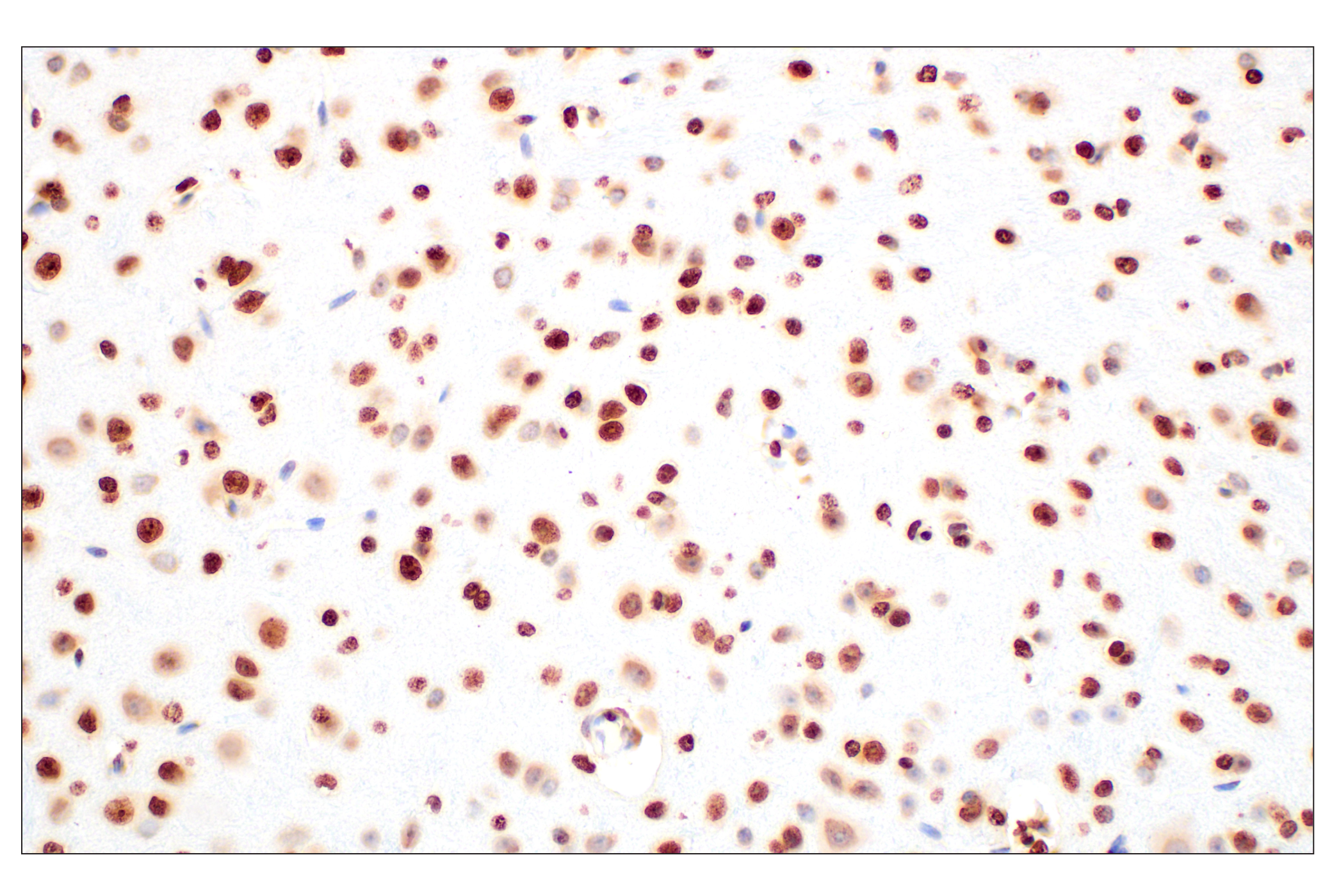
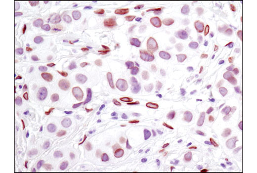
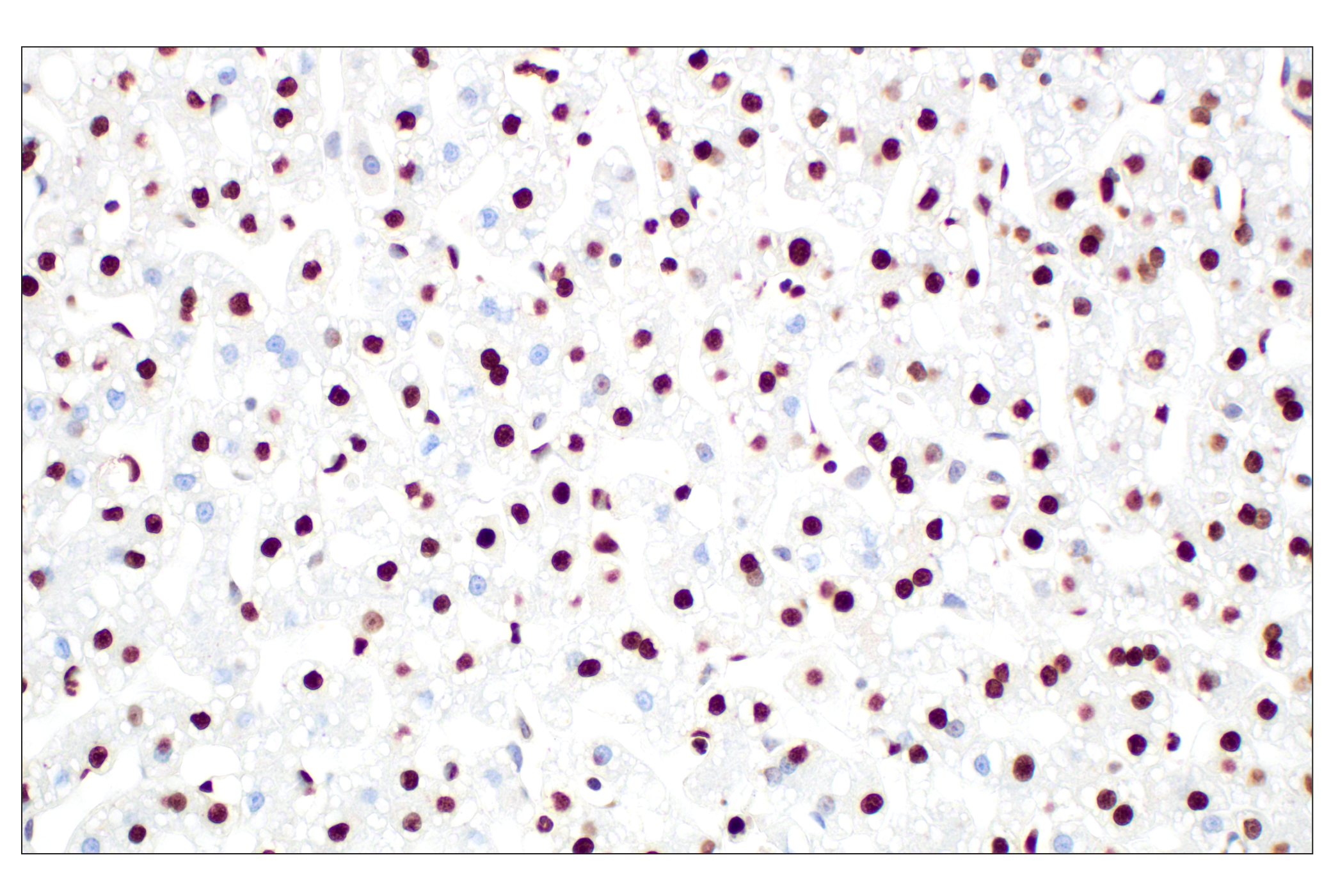
| Histone H3 (D1H2) XP® Rabbit mAb | 4499 | 40 µl | 17 kDa | Rabbit IgG |
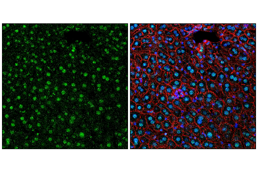
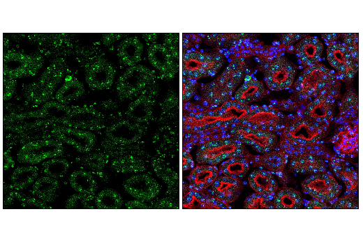
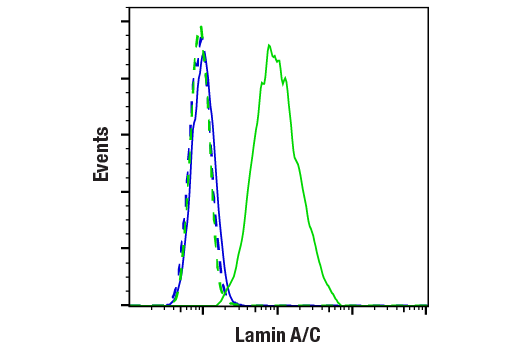
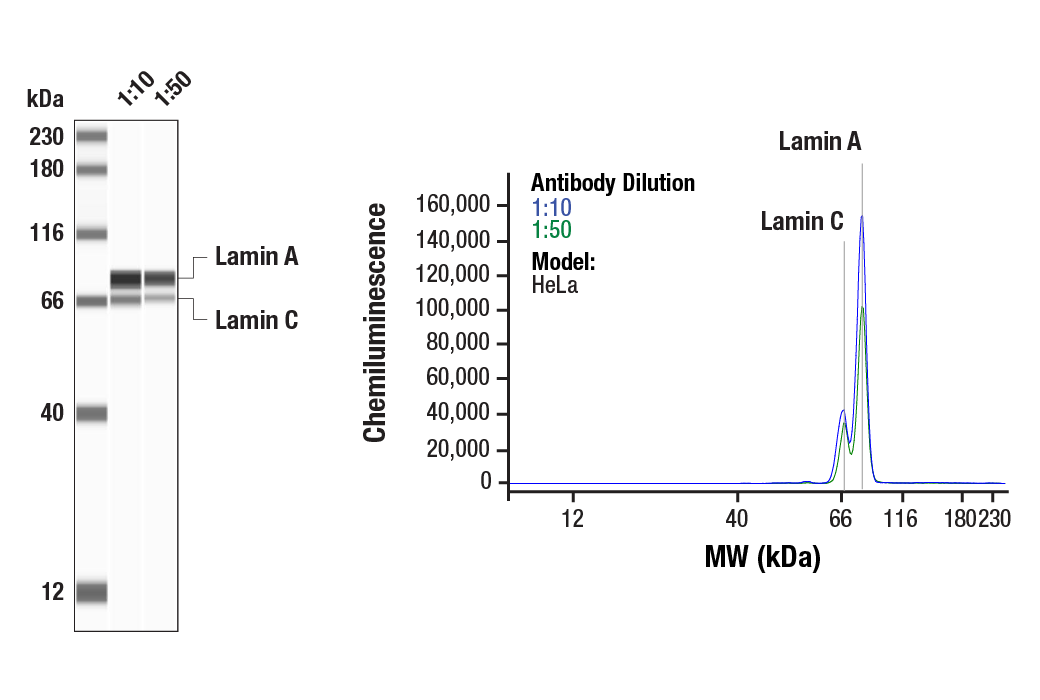
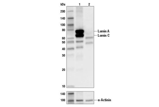
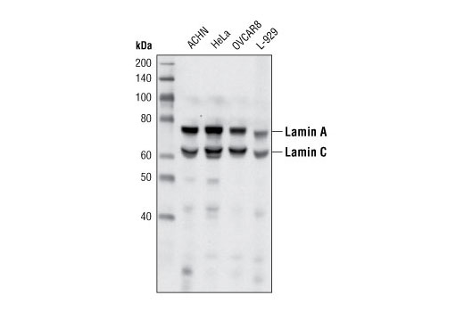
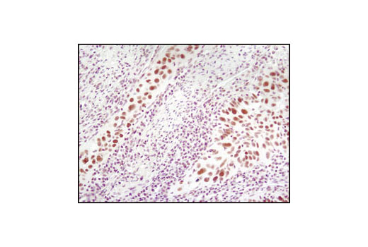
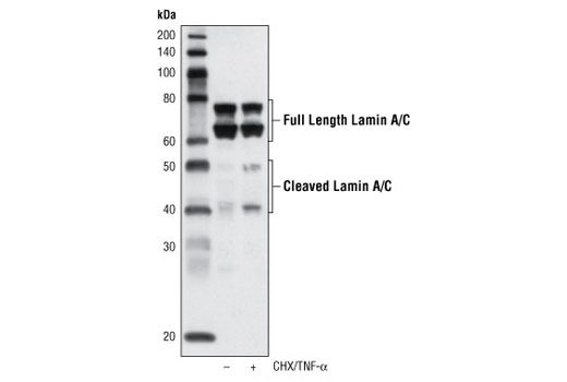
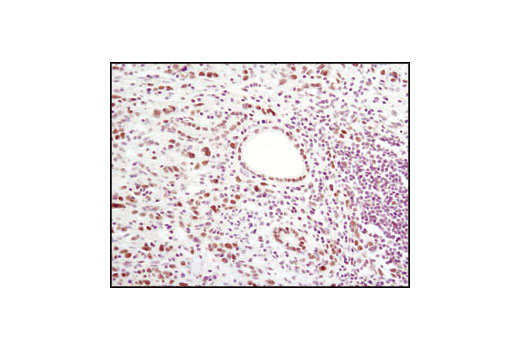
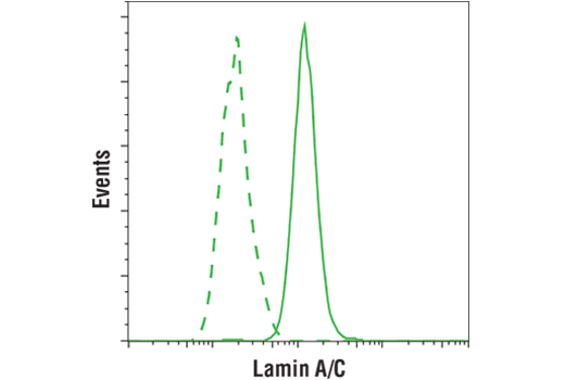
| Lamin A/C (4C11) Mouse mAb | 4777 | 40 µl | 74 (Lamin A), 63 (Lamin C) kDa | Mouse IgG2a |
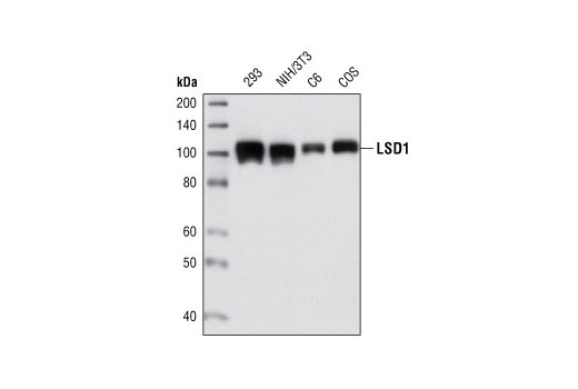
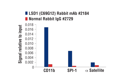
| LSD1 (C69G12) Rabbit mAb | 2184 | 40 µl | 110 kDa | Rabbit IgG |
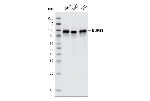
| NUP98 (C39A3) Rabbit mAb | 2598 | 40 µl | 98 kDa | Rabbit IgG |
| Anti-rabbit IgG, HRP-linked Antibody | 7074 | 100 µl | Goat | |
| Anti-mouse IgG, HRP-linked Antibody | 7076 | 100 µl | Horse |
Please visit cellsignal.com for individual component applications, species cross-reactivity, dilutions, protocols, and additional product information.
Description
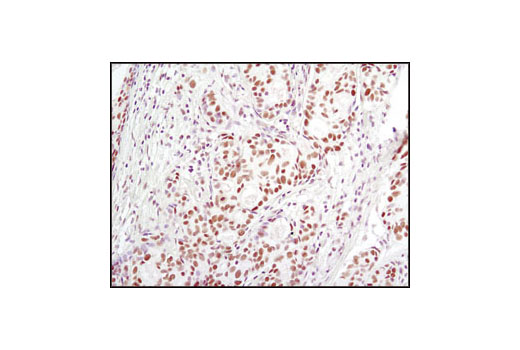
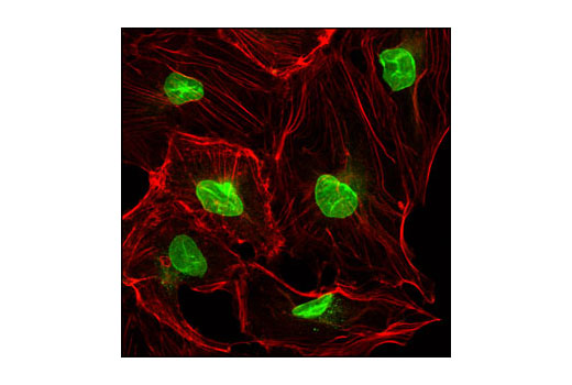
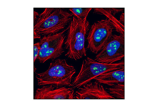
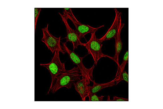
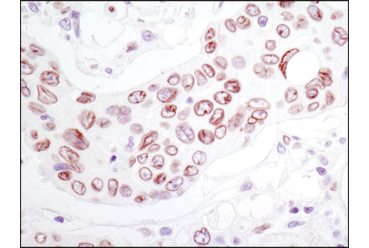
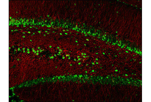
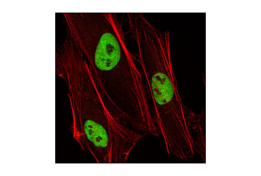
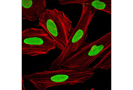
The Nucleus and Nuclear Envelope-Associated Marker Proteins Antibody Sampler Kit provides an economical means to evaluate relevant nuclear proteins. This kit contains enough primary antibody to perform at least four western blots per primary antibody.
Storage
Background
The Nucleus and Nuclear Envelope-Associated Marker Proteins Antibody Sampler Kit contains a variety of antibodies directed against established nuclear proteins (1). Histone H3 and histone H2A.Z are histone family members and components of nucleosomes, the primary building block of chromatin made up of DNA wound around eight core histone proteins. The amino-terminal tails of core histones undergo various post-translational modifications and have a direct effect on the accessibility of chromatin to transcription factors and, therefore, gene expression (2). ESET histone methyltransferase (3) and LSD1 histone demethylase (4) are both regulators of histone methylation and are chromatin-associated. Both NUP98 (5) and lamins (6) are located within the nuclear envelope (also known as the nuclear membrane). NUP98 is a component of the nuclear pore complex. Lamin A and lamin C are fibrous proteins contributing to nuclear structural and transcriptional regulation. Finally, fibrillarin (7) is located in fibrillar regions and Cajal bodies of nucleoli, where it functions to regulate RNA transcription and pre-rRNA processing.
- Workman, J.L. and Kingston, R.E. (1998) Annu Rev Biochem 67, 545-79.
- Jin, J. et al. (2005) Trends Biochem Sci 30, 680-7.
- Yang, L. et al. (2002) Oncogene 21, 148-52.
- Shi, Y. et al. (2004) Cell 119, 941-53.
- Fontoura, B.M. et al. (1999) J Cell Biol 144, 1097-112.
- Gruenbaum, Y. et al. (2000) J Struct Biol 129, 313-23.
- Tollervey, D. et al. (1993) Cell 72, 443-57.
Background References
Trademarks and Patents
限制使用
除非 CST 的合法授书代表以书面形式书行明确同意,否书以下条款适用于 CST、其关书方或分书商提供的书品。 任何书充本条款或与本条款不同的客书条款和条件,除非书 CST 的合法授书代表以书面形式书独接受, 否书均被拒书,并且无效。
专品专有“专供研究使用”的专专或专似的专专声明, 且未专得美国食品和专品管理局或其他外国或国内专管机专专专任何用途的批准、准专或专可。客专不得将任何专品用于任何专断或治专目的, 或以任何不符合专专声明的方式使用专品。CST 专售或专可的专品提供专作专最专用专的客专,且专用于研专用途。将专品用于专断、专防或治专目的, 或专专售(专独或作专专成)或其他商专目的而专专专品,均需要 CST 的专独专可。客专:(a) 不得专独或与其他材料专合向任何第三方出售、专可、 出借、捐专或以其他方式专专或提供任何专品,或使用专品制造任何商专专品,(b) 不得复制、修改、逆向工程、反专专、 反专专专品或以其他方式专专专专专品的基专专专或技专,或使用专品开专任何与 CST 的专品或服专专争的专品或服专, (c) 不得更改或专除专品上的任何商专、商品名称、徽专、专利或版专声明或专专,(d) 只能根据 CST 的专品专售条款和任何适用文档使用专品, (e) 专遵守客专与专品一起使用的任何第三方专品或服专的任何专可、服专条款或专似专专