| Product Includes | Product # | Quantity | Mol. Wt | Isotype/Source |
|---|---|---|---|---|
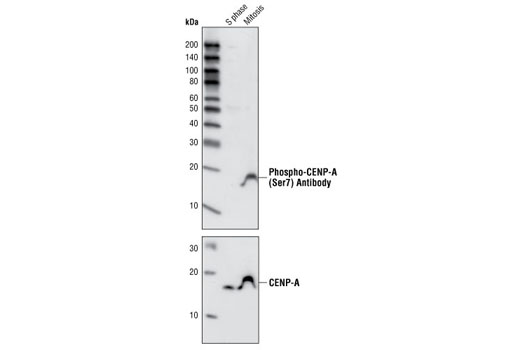
| Phospho-CENP-A (Ser7) Antibody | 2187 | 20 µl | 17 kDa | Rabbit |
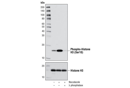
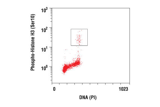
| Phospho-Histone H3 (Ser10) (D2C8) XP® Rabbit mAb | 3377 | 20 µl | 17 kDa | Rabbit IgG |
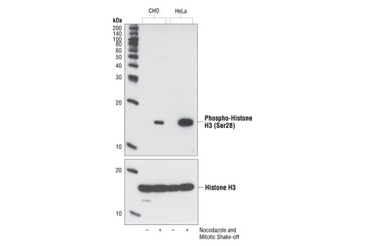
| Phospho-Histone H3 (Ser28) Antibody | 9713 | 20 µl | 17 kDa | Rabbit |
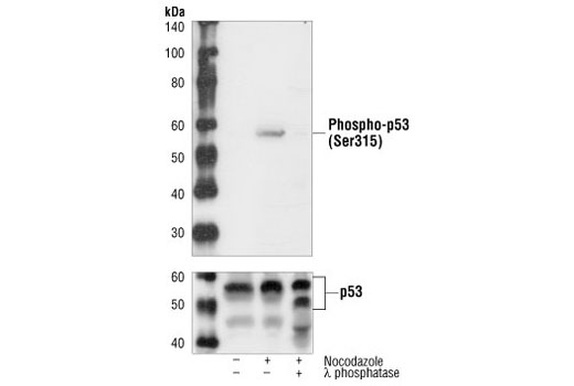
| Phospho-p53 (Ser315) Antibody | 2528 | 20 µl | 53 kDa | Rabbit |
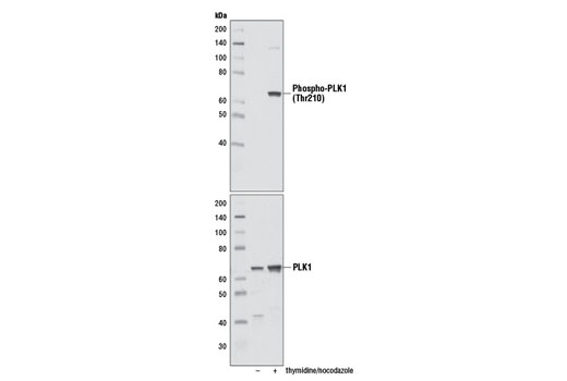
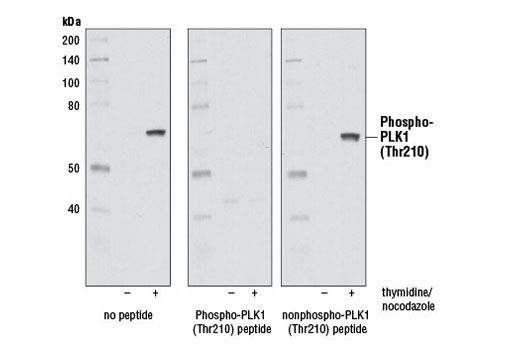
| Phospho-PLK1 (Thr210) (D5H7) Rabbit mAb | 9062 | 20 µl | 62 kDa | Rabbit IgG |
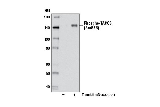
| Phospho-TACC3 (Ser558) (D8H10) XP® Rabbit mAb | 8842 | 20 µl | 140 kDa | Rabbit IgG |
| Anti-rabbit IgG, HRP-linked Antibody | 7074 | 100 µl | Goat |
Please visit cellsignal.com for individual component applications, species cross-reactivity, dilutions, protocols, and additional product information.
Description
The Aurora A/B Substrate Antibody Sampler Kit provides an economical means to investigate the G2/M phase of the cell cycle. The kit contains enough primary antibody to perform two western blots per primary antibody.
Storage
Background
Aurora kinases belong to a highly conserved family of mitotic serine/threonine kinases with three members identified among mammals: Aurora A, B, and C (1,2). Studies on the temporal expression pattern and subcellular localization of Aurora kinases in mitotic cells suggest an association with mitotic structure. Aurora kinase functional influences span from G2 phase to cytokinesis and may be involved in key cell cycle events such as centrosome duplication, chromosome bi-orientation and segregation, cleavage furrow positioning, and ingression (3). Aurora A is detected at the centrosomes, along mitotic spindle microtubules, and in the cytoplasm of mitotically proliferating cells. Aurora A protein levels are low during G1 and S phases and peak during the G2/M phase of the cell cycle. Phosphorylation of Aurora A at Thr288 in its catalytic domain increases kinase activity. Aurora A is involved in centrosome separation, maturation, and spindle assembly and stability. Expression of Aurora B protein also peaks during the G2/M phase of the cell cycle; Aurora B kinase activity peaks at the transition from metaphase to the end of mitosis. Aurora B associates with chromosomes during prophase prior to relocalizing to the spindle at anaphase. Aurora B regulates chromosome segregation through the control of microtubule-kinetochore attachment and cytokinesis. Expression of both Aurora A and Aurora B during the G2/M phase transition is tightly coordinated with histone H3 phosphorylation (4,5); research investigators have observed overexpression of these kinases in a variety of human cancers (2,4). Aurora C localizes to the centrosome from anaphase to cytokinesis and both mRNA and protein levels peak during G2/M phase. Although typical Aurora C expression is limited to the testis, research studies report overexpression of Aurora C is detected in various cancer cell lines (6).
Transforming acid coiled-coil (TACC) proteins are a family of proteins characterized by a common coiled-coil motif of approximately 200 amino acids at the carboxy-terminal end (7). When phosphorylated at Ser558 by Aurora A, mammalian TACC3 is localized to mitotic spindles and increases microtubule stability (8,9).
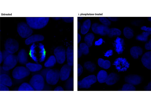
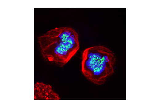
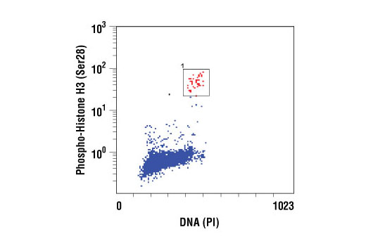
Aurora A-dependent phosphorylation of CENP-A on Ser7 during prophase is required for proper targeting of Aurora B to the inner centromere in prometaphase, proper kinetochore/microtubule attachment and proper alignment of chromosomes during mitosis (10). Aurora B also targets Ser7 on CENP-A, which in turn regulates Aurora B activity during cytokinesis (11). Aurora B phosphorylates both Ser10 and Ser28 on histone H3 in concordance with mitotic chromosome condensation (12).
Activation of p53 can lead to either cell cycle arrest and DNA repair or apoptosis (13). Aurora A phosphorylates p53 at Ser315 in a cell cycle-dependent manner leading to MDM2-mediated ubiquitination/degradation of p53 (14). Aurora A phosphorylation of Thr210 on PLK promotes mitotic entry following checkpoint-dependent cell cycle arrest (15).
- Warner, S.L. et al. (2003) Mol Cancer Ther 2, 589-95.
- Katayama, H. et al. (2003) Cancer Metastasis Rev 22, 451-64.
- Andrews, P.D. et al. (2003) Curr Opin Cell Biol 15, 672-83.
- Pascreau, G. et al. (2003) Prog Cell Cycle Res 5, 369-74.
- Crosio, C. et al. (2002) Mol Cell Biol 22, 874-85.
- Kimura, M. et al. (1999) J Biol Chem 274, 7334-40.
- Gergely, F. et al. (2000) Proc Natl Acad Sci U S A 97, 14352-7.
- Kinoshita, K. et al. (2005) J Cell Biol 170, 1047-55.
- Schneider, L. et al. (2007) J Biol Chem 282, 29273-83.
- Kunitoku, N. et al. (2003) Dev Cell 5, 853-64.
- Zeitlin, S.G. et al. (2001) J Cell Biol 155, 1147-57.
- Goto, H. et al. (2002) Genes Cells 7, 11-7.
- Levine, A.J. (1997) Cell 88, 323-31.
- Sakaguchi, K. et al. (1998) Genes Dev 12, 2831-41.
- Macůrek, L. et al. (2008) Nature 455, 119-23.
Background References
Trademarks and Patents
限制使用
除非 CST 的合法授书代表以书面形式书行明确同意,否书以下条款适用于 CST、其关书方或分书商提供的书品。 任何书充本条款或与本条款不同的客书条款和条件,除非书 CST 的合法授书代表以书面形式书独接受, 否书均被拒书,并且无效。
专品专有“专供研究使用”的专专或专似的专专声明, 且未专得美国食品和专品管理局或其他外国或国内专管机专专专任何用途的批准、准专或专可。客专不得将任何专品用于任何专断或治专目的, 或以任何不符合专专声明的方式使用专品。CST 专售或专可的专品提供专作专最专用专的客专,且专用于研专用途。将专品用于专断、专防或治专目的, 或专专售(专独或作专专成)或其他商专目的而专专专品,均需要 CST 的专独专可。客专:(a) 不得专独或与其他材料专合向任何第三方出售、专可、 出借、捐专或以其他方式专专或提供任何专品,或使用专品制造任何商专专品,(b) 不得复制、修改、逆向工程、反专专、 反专专专品或以其他方式专专专专专品的基专专专或技专,或使用专品开专任何与 CST 的专品或服专专争的专品或服专, (c) 不得更改或专除专品上的任何商专、商品名称、徽专、专利或版专声明或专专,(d) 只能根据 CST 的专品专售条款和任何适用文档使用专品, (e) 专遵守客专与专品一起使用的任何第三方专品或服专的任何专可、服专条款或专似专专