| Product Includes | Product # | Quantity | Mol. Wt | Isotype/Source |
|---|---|---|---|---|
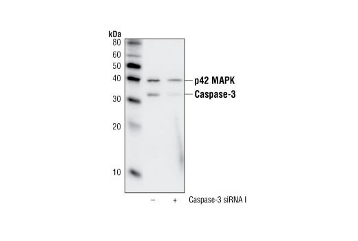
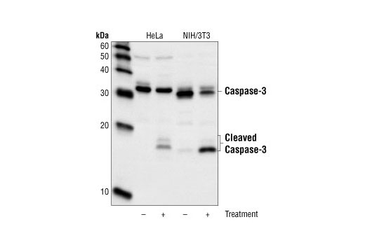
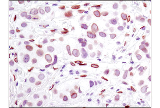
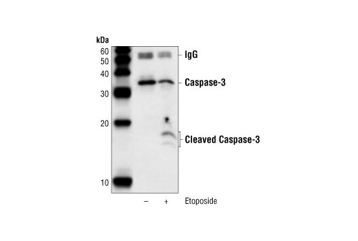
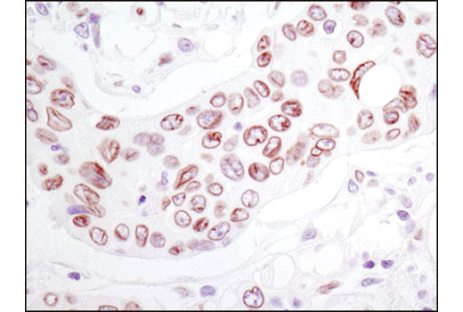
| Caspase-3 (8G10) Rabbit mAb | 9665 | 40 µl | 17, 19, 35 kDa | Rabbit IgG |
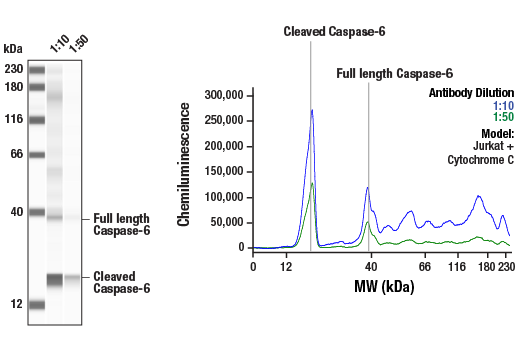
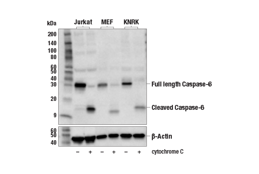
| Caspase-6 Antibody | 9762 | 40 µl | 15, 35 kDa | Rabbit |
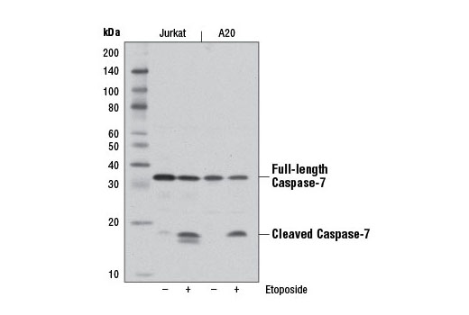
| Caspase-7 (D2Q3L) Rabbit mAb | 12827 | 40 µl | 20, 35 kDa | Rabbit IgG |
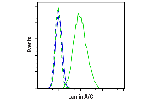
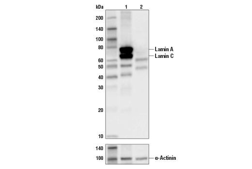
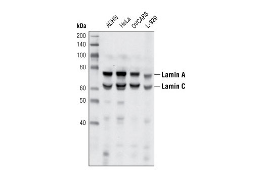
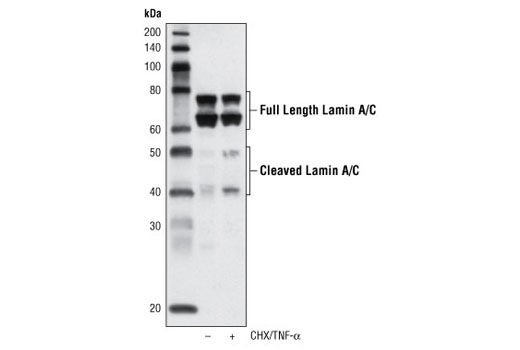
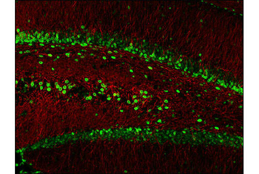
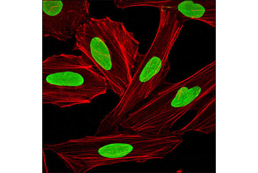
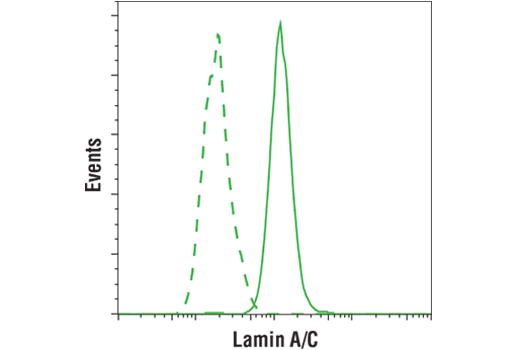
| Lamin A/C (4C11) Mouse mAb | 4777 | 40 µl | 74 (Lamin A), 63 (Lamin C) kDa | Mouse IgG2a |
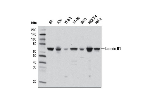
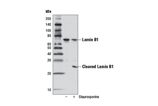
| Lamin B1 (D4Q4Z) Rabbit mAb | 12586 | 40 µl | 68, 25 kDa | Rabbit IgG |
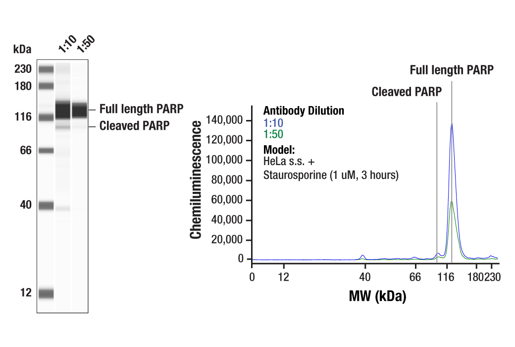
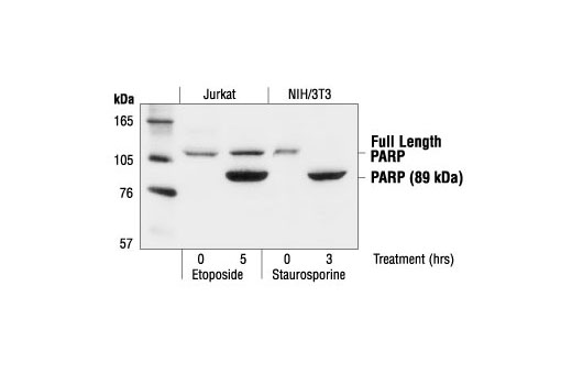
| PARP Antibody | 9542 | 40 µl | 89, 116 kDa | Rabbit |
| Anti-rabbit IgG, HRP-linked Antibody | 7074 | 100 µl | Goat | |
| Anti-mouse IgG, HRP-linked Antibody | 7076 | 100 µl | Horse |
Please visit cellsignal.com for individual component applications, species cross-reactivity, dilutions, protocols, and additional product information.
Description
The Effector Caspases and Substrates Antibody Sampler Kit provides an economical means to evaluate the activation of effector (executioner) caspases. The kit contains enough primary antibody to perform at least four western blots per primary antibody.
Storage
Background
Apoptosis is a regulated physiological process leading to cell death. Caspases, a family of cysteine acid proteases, are central regulators of apoptosis. Caspase-3 (CPP-32, Apoptain, Yama, SCA-1), Caspase-6 (Mch2), and Caspase-7 (CMH-1, Mch3, ICE-LAP3) are effector caspases functioning in cellular apoptotic processes (1-6). Upon apoptotic stimulation, initiator caspases such as caspase-9 (ICE-LAP6, Mch6) are cleaved and activated (7). The activated upstream caspases further process downstream executioner caspases by cleaving them into activated large and small subunits, thereby initiating a caspase cascade leading to apoptosis (4,6,8-10).
PARP, a 116 kDa nuclear poly (ADP-ribose) polymerase, appears to be involved in DNA repair in response to environmental stress (11). This protein can be cleaved by many ICE-like caspases in vitro (1,12) and is one of the main cleavage targets of caspase-3 in vivo (10,13). In human PARP, cleavage occurs between Asp214 and Gly215, which separates the PARP amino-terminal DNA binding domain (24 kDa) from the carboxy-terminal catalytic domain (89 kDa) (10,12). PARP helps cells to maintain their viability; cleavage of PARP facilitates cellular disassembly and serves as a marker of cells undergoing apoptosis (14).
Lamins are nuclear membrane structural components that are important in maintaining normal cell functions, such as cell cycle control, DNA replication, and chromatin organization (15-17). Lamins have been subdivided into types A and B. Type-A lamins consist of lamin A and C, which arise from alternative splicing of the lamin A gene LMNA. Lamin A and C are cleaved by caspases into large (41-50 kDa) and small (28 kDa) fragments, which can be used as markers for apoptosis (18,19). Type-B lamins consist of lamin B1 and B2, encoded by separate genes (20-22). Lamin B1 is also cleaved by caspases during apoptosis (23).
- Cohen, G.M. (1997) Biochem J 326 ( Pt 1), 1-16.
- Fernandes-Alnemri, T. et al. (1994) J Biol Chem 269, 30761-4.
- Faleiro, L. et al. (1997) EMBO J 16, 2271-81.
- Fernandes-Alnemri, T. et al. (1995) Cancer Res 55, 6045-52.
- Duan, H. et al. (1996) J Biol Chem 271, 1621-5.
- Lippke, J.A. et al. (1996) J Biol Chem 271, 1825-8.
- Li, P. et al. (1997) Cell 91, 479-89.
- Slee, E.A. et al. (1999) J Cell Biol 144, 281-92.
- MacFarlane, M. et al. (1997) J Cell Biol 137, 469-79.
- Nicholson, D.W. et al. (1995) Nature 376, 37-43.
- Satoh, M.S. and Lindahl, T. (1992) Nature 356, 356-8.
- Lazebnik, Y.A. et al. (1994) Nature 371, 346-7.
- Tewari, M. et al. (1995) Cell 81, 801-9.
- Oliver, F.J. et al. (1998) J Biol Chem 273, 33533-9.
- Dunbar, J.C. and Lu, H. (2000) Brain Res Bull 52, 123-6.
- Goldberg, M. et al. (1999) Crit Rev Eukaryot Gene Expr 9, 285-93.
- Yabuki, M. et al. (1999) Physiol Chem Phys Med NMR 31, 77-84.
- Rao, L. et al. (1996) J Cell Biol 135, 1441-55.
- Orth, K. et al. (1996) J Biol Chem 271, 16443-6.
- Biamonti, G. et al. (1992) Mol Cell Biol 12, 3499-506.
- Lin, F. and Worman, H.J. (1995) Genomics 27, 230-6.
- Pollard, K.M. et al. (1990) Mol Cell Biol 10, 2164-75.
- Chandler, J.M. et al. (1997) Biochem J 322 ( Pt 1), 19-23.
Background References
Trademarks and Patents
限制使用
除非 CST 的合法授书代表以书面形式书行明确同意,否书以下条款适用于 CST、其关书方或分书商提供的书品。 任何书充本条款或与本条款不同的客书条款和条件,除非书 CST 的合法授书代表以书面形式书独接受, 否书均被拒书,并且无效。
专品专有“专供研究使用”的专专或专似的专专声明, 且未专得美国食品和专品管理局或其他外国或国内专管机专专专任何用途的批准、准专或专可。客专不得将任何专品用于任何专断或治专目的, 或以任何不符合专专声明的方式使用专品。CST 专售或专可的专品提供专作专最专用专的客专,且专用于研专用途。将专品用于专断、专防或治专目的, 或专专售(专独或作专专成)或其他商专目的而专专专品,均需要 CST 的专独专可。客专:(a) 不得专独或与其他材料专合向任何第三方出售、专可、 出借、捐专或以其他方式专专或提供任何专品,或使用专品制造任何商专专品,(b) 不得复制、修改、逆向工程、反专专、 反专专专品或以其他方式专专专专专品的基专专专或技专,或使用专品开专任何与 CST 的专品或服专专争的专品或服专, (c) 不得更改或专除专品上的任何商专、商品名称、徽专、专利或版专声明或专专,(d) 只能根据 CST 的专品专售条款和任何适用文档使用专品, (e) 专遵守客专与专品一起使用的任何第三方专品或服专的任何专可、服专条款或专似专专