| Product Includes | Product # | Quantity | Color | Storage Temp |
|---|---|---|---|---|
| FGFR3 Rabbit mAb Coated Microwells | 67593 | 96 tests |
|
+4C |
| Phospho Tyrosine Mouse Detection mAb | 12982 | 1 ea |
|
+4C |
| Anti-mouse IgG, HRP-linked Antibody (ELISA Formulated) | 13304 | 1 ea |
|
+4C |
| Detection Antibody Diluent | 13339 | 5.5 ml |
|
+4C |
| HRP Diluent | 13515 | 5.5 ml |
|
+4C |
| Luminol/Enhancer Solution | 84850 | 3 ml |
|
RT |
| Stable Peroxide Buffer | 42552 | 3 ml |
|
RT |
| Sealing Tape | 54503 | 2 ea |
|
+4C |
| ELISA Wash Buffer (20X) | 9801 | 25 ml |
|
+4C |
| ELISA Sample Diluent | 11083 | 25 ml |
|
+4C |
| Cell Lysis Buffer (10X) | 9803 | 15 ml |
|
-20C |
*The microwell plate is supplied as 12 8-well modules - Each module is designed to break apart for 8 tests.
Description
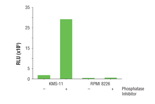
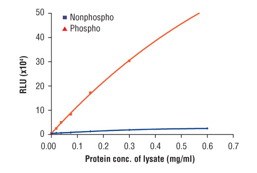
The PathScan® Phospho-FGF Receptor 3 (panTyr) Chemiluminescent Sandwich ELISA Kit is a solid phase sandwich enzyme-linked immunosorbent assay (ELISA) that detects endogenous levels of tyrosine-phosphorylated FGFR3 protein with a chemiluminescent readout. Chemiluminescent ELISAs often have a wider dynamic range and higher sensitivity than conventional chromogenic detection. This chemiluminescent ELISA, which is offered in low volume microplates, shows increased signal and sensitivity while using a smaller sample size. An FGFR3 rabbit mAb has been coated on the microwells. After incubation with cell lysates, both phospho- and nonphospho-FGFR3 proteins are captured by the coated antibody. Following extensive washing, a phospho tyrosine mouse mAb is added to detect the captured tyrosine-phosphorylated FGFR3 protein. Anti-mouse IgG, HRP-linked antibody is then used to recognize the bound detection antibody. Chemiluminescent reagent is added for signal development. The magnitude of light emission, measured in relative light units (RLU), is proportional to the quantity of tyrosine-phosphorylated FGFR3 protein.
*Antibodies in this kit are custom formulations specific to kit.
Specificity/Sensitivity
Background
Fibroblast growth factors (FGFs) produce mitogenic and angiogenic effects in target cells by signaling through cell surface receptor tyrosine kinases. There are four members of the FGF receptor family: FGFR1 (flg), FGFR2 (bek, KGFR), FGFR3, and FGFR4. Each receptor contains an extracellular ligand-binding domain, a transmembrane domain, and a cytoplasmic kinase domain (1). Following ligand binding and dimerization, the receptors are phosphorylated at specific tyrosine residues (2). Seven tyrosine residues in the cytoplasmic tail of FGFR1 can be phosphorylated: Tyr463, 583, 585, 653, 654, 730, and 766. Tyr653 and Tyr654 are important for catalytic activity of activated FGFR and are essential for signaling (3). The other phosphorylated tyrosine residues may provide docking sites for downstream signaling components, such as Crk and PLCγ (4,5).
Activating mutations within fibroblast growth factor receptor 3 (FGFR3) are responsible for human skeletal dysplasias including achondroplasia and the neonatal lethal syndromes thanatophoric dysplasia types I and II (6,7). Several of these same FGFR3 mutations as well as overexpression of FGFR3 proteins have also been identified somatically in human cancers, including multiple myeloma, bladder carcinoma and cervical cancer (8). Thus, FGFR3 may represent a potential target for therapy.
- Powers, C.J. et al. (2000) Endocr Relat Cancer 7, 165-97.
- Reilly, J.F. et al. (2000) J Biol Chem 275, 7771-8.
- Mohammadi, M. et al. (1996) Mol Cell Biol 16, 977-89.
- Mohammadi, M. et al. (1991) Mol Cell Biol 11, 5068-78.
- Larsson, H. et al. (1999) J Biol Chem 274, 25726-34.
- Wilkie, A.O. et al. (2002) Am J Med Genet 112, 266-78.
- Yamashita, A. et al. (2014) Nature 513, 507-11.
- Miyake, M. et al. (2007) Biochem Biophys Res Commun 362, 865-71.
Background References
Cross-Reactivity Key
H: human M: mouse R: rat Hm: hamster Mk: monkey Vir: virus Mi: mink C: chicken Dm: D. melanogaster X: Xenopus Z: zebrafish B: bovine Dg: dog Pg: pig Sc: S. cerevisiae Ce: C. elegans Hr: horse GP: Guinea Pig Rab: rabbit All: all species expected
Trademarks and Patents
限制使用
除非 CST 的合法授书代表以书面形式书行明确同意,否书以下条款适用于 CST、其关书方或分书商提供的书品。 任何书充本条款或与本条款不同的客书条款和条件,除非书 CST 的合法授书代表以书面形式书独接受, 否书均被拒书,并且无效。
专品专有“专供研究使用”的专专或专似的专专声明, 且未专得美国食品和专品管理局或其他外国或国内专管机专专专任何用途的批准、准专或专可。客专不得将任何专品用于任何专断或治专目的, 或以任何不符合专专声明的方式使用专品。CST 专售或专可的专品提供专作专最专用专的客专,且专用于研专用途。将专品用于专断、专防或治专目的, 或专专售(专独或作专专成)或其他商专目的而专专专品,均需要 CST 的专独专可。客专:(a) 不得专独或与其他材料专合向任何第三方出售、专可、 出借、捐专或以其他方式专专或提供任何专品,或使用专品制造任何商专专品,(b) 不得复制、修改、逆向工程、反专专、 反专专专品或以其他方式专专专专专品的基专专专或技专,或使用专品开专任何与 CST 的专品或服专专争的专品或服专, (c) 不得更改或专除专品上的任何商专、商品名称、徽专、专利或版专声明或专专,(d) 只能根据 CST 的专品专售条款和任何适用文档使用专品, (e) 专遵守客专与专品一起使用的任何第三方专品或服专的任何专可、服专条款或专似专专