Revision 1
Cell Signaling Technology

Orders: 877-616-CELL (2355) [email protected]

Support: 877-678-TECH (8324)

Web: [email protected] cellsignal.com

3 Trask LaneDanversMassachusetts01923USA
For Research Use Only. Not for Use in Diagnostic Procedures.
:

:

:

:

UniProt ID:

#P21709, #P40763, #P27361, #P29317, #P16234, #P35568, #P54753, #Q06418, #P04629, #P21860, #P31751, #Q9Y243, #P43403, #P07947, #P07948, #Q02763, #P30530, #P07333, #Q9UM73, #P62753, #P22455, #P28482, #P06239, #P00533, #P29320, #P31749, #P08581, #P11362, #P04626, #P10721, #P06213, #P54760, #P08631, #P07949, #P42224, #P08069, #Q16620, #P12931, #Q04912, #P35968, #P06241, #P00519

Entrez-Gene Id:

2041, 6774, 5595, 1969, 5156, 3667, 2049, 7301, 4914, 2065, 208, 10000, 7535, 7525, 4067, 7010, 558, 1436, 238, 6194, 2264, 5594, 3932, 1956, 2042, 207, 4233, 2260, 2064, 3815, 3643, 2050, 3055, 5979, 6772, 3480, 4915, 6714, 4486, 3791, 2534, 25

Product Information

Storage

Kit should be stored at 4°C with the exception of Lysis Buffer, which is stored at –20°C (packaged separately).

Specificity / Sensitivity

Cell Signaling Technology's PathScan® RTK Signaling Antibody Array Kit detects the indicated RTKs and signaling nodes only when phosphorylated at tyrosine or specified residues (see Array Target Map). No significant crossreactivity has been observed between targets, with the exception of some crossreactivity of the FLT3 antibody with phosphorylated EphB3. In addition, Stat1 (Tyr701) and Stat3 (Tyr705) may be detected when phosphorylated at other tyrosine sites within the proteins. This kit is optimized for cell lysates diluted to a total protein concentration between 0.2 and 1 mg/ml (see Figure 4). All capture antibodies have been validated for human targets. Although this kit has not been tested with mouse lysates, it is expected than many capture antibodies will crossreact in murine systems.

Species Reactivity:

Human

Product Description

The PathScan® RTK Signaling Antibody Array Kit (Chemiluminescent Readout) is a slide-based antibody array founded upon the sandwich immunoassay principle. The array kit allows for the simultaneous detection of 28 receptor tyrosine kinases and 11 important signaling nodes, when phosphorylated at tyrosine or other residues. Target-specific capture antibodies have been spotted in duplicate onto nitrocellulose-coated glass slides. Each kit contains two 8-pad slides, allowing the user to test up to 16 samples. Cell lysate is incubated on the slide followed by a biotinylated detection antibody cocktail. Streptavidin-conjugated HRP and LumiGLO® Reagent are then used to visualize the bound detection antibody by chemiluminescence. An image of the slide can be captured with either a digital imaging system or standard chemiluminescent film. The image can be analyzed visually or the spot intensities quantified using array analysis software.

Background

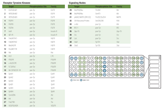
Receptor Tyrosine Kinases (RTKs) are a family of cell surface receptors that signal primarily through tyrosine phosphorylation events (1). RTKs trigger a wide range of downstream signaling cascades, including the PI3K/Akt, MAPK and Jak/Stat pathways. These pathways control basic cellular functions such as division, growth, metabolism, differentiation, migration and survival. Dysregulation of RTK signaling has been implicated in a large number of cancers (2), making RTKs popular targets for pharmaceutical intervention.

  1. Schlessinger, J. (2000) Cell 103, 211-25.
  2. Blume-Jensen, P. and Hunter, T. (2001) Nature 411, 355-65.

Species Reactivity

Species reactivity is determined by testing in at least one approved application (e.g., western blot).

Cross-Reactivity Key

H: human M: mouse R: rat Hm: hamster Mk: monkey Vir: virus Mi: mink C: chicken Dm: D. melanogaster X: Xenopus Z: zebrafish B: bovine Dg: dog Pg: pig Sc: S. cerevisiae Ce: C. elegans Hr: horse GP: Guinea Pig Rab: rabbit All: all species expected

Trademarks and Patents

Cell Signaling Technology is a trademark of Cell Signaling Technology, Inc.
PathScan is a registered trademark of Cell Signaling Technology, Inc.
All other trademarks are the property of their respective owners. Visit cellsignal.com/trademarks for more information.

限制使用

除非 CST 的合法授书代表以书面形式书行明确同意,否书以下条款适用于 CST、其关书方或分书商提供的书品。 任何书充本条款或与本条款不同的客书条款和条件,除非书 CST 的合法授书代表以书面形式书独接受, 否书均被拒书,并且无效。

专品专有“专供研究使用”的专专或专似的专专声明, 且未专得美国食品和专品管理局或其他外国或国内专管机专专专任何用途的批准、准专或专可。客专不得将任何专品用于任何专断或治专目的, 或以任何不符合专专声明的方式使用专品。CST 专售或专可的专品提供专作专最专用专的客专,且专用于研专用途。将专品用于专断、专防或治专目的, 或专专售(专独或作专专成)或其他商专目的而专专专品,均需要 CST 的专独专可。客专:(a) 不得专独或与其他材料专合向任何第三方出售、专可、 出借、捐专或以其他方式专专或提供任何专品,或使用专品制造任何商专专品,(b) 不得复制、修改、逆向工程、反专专、 反专专专品或以其他方式专专专专专品的基专专专或技专,或使用专品开专任何与 CST 的专品或服专专争的专品或服专, (c) 不得更改或专除专品上的任何商专、商品名称、徽专、专利或版专声明或专专,(d) 只能根据 CST 的专品专售条款和任何适用文档使用专品, (e) 专遵守客专与专品一起使用的任何第三方专品或服专的任何专可、服专条款或专似专专

Revision 1
#7982

PathScan® RTK Signaling Antibody Array Kit (Chemiluminescent Readout)

PathScan® RTK Signaling Antibody Array Kit (Chemiluminescent Readout): Image 1 Expand Image
图 1. 使用 PathScan® RTK Signaling Antibody Array Kit (Chemiluminescent Readout) #7982 对一组细胞系进行的筛选显示了各种磷酸化 RTK 和信号转导节点。A431 细胞饥饿 24 小时,随后在 37ºC 下用 100 ng/ml EGF #8916 处理 3 分钟。NIH/3T3 细胞饥饿 24 小时,随后在 37ºC 下用 100 ng/ml PDGF #9909 处理 5 分钟。Karpas-299 和 K562 细胞裂解无需饥饿或处理。使用化学发光膜捕捉芯片图像,曝光时间为 2-5 秒。
PathScan® RTK Signaling Antibody Array Kit (Chemiluminescent Readout): Image 2 Expand Image
图 2. Treatment of MCF-7 cells with IGF-I stimulates phosphorylation of IGF-IR at tyrosine residues, Akt at Ser473 and p44/42 MAPK at Thr202/Tyr204 as detected by the PathScan® RTK Signaling Antibody Array Kit (Chemiluminescent Readout) #7982. MCF-7 细胞饥饿 24 小时,随后在 37ºC 下用 100 ng/ml IGF-I #3093 处理 5 分钟。化学发光膜图像(下小图)和该图像的定量分析(上小图)如图所示。膜曝光 2-5 秒后,捕捉化学发光芯片图像。
PathScan® RTK Signaling Antibody Array Kit (Chemiluminescent Readout): Image 3 Expand Image
图 3. 使用 PathScan® RTK Signaling Antibody Array Kit (Chemiluminescent Readout) #7982 检测发现,A431 细胞用 EGF 处理会刺激 EGFR、Akt、p44/42 MAPK 和 Stat3 磷酸化。A431 细胞饥饿 24 小时,并用 100 ng/ml EGF #8916 处理 5 或 40 分钟。在某些情况下,细胞用 1 μM 渥曼青霉素 #9951 处理 1 小时,或用 1 μM 吉非替尼处理 2 小时后再用 EGF 刺激。从数字显影仪上获得的芯片图像像素强度如顶部小图所示,蛋白质印迹如底部小图所示。
PathScan® RTK Signaling Antibody Array Kit (Chemiluminescent Readout): Image 4 Expand Image
图 4. 未经处理和已经 IGF-I 处理的 MCF-7 细胞的裂解物蛋白浓度与磷酸化 IGF-IR (pan-Tyr)、磷酸化 Akt (Ser473) 和磷酸化 p44/42 (Thr202/Tyr204) 的像素强度之间的关系如图所示。MCF-7 细胞饥饿 24 小时,随后在 37ºC 下用 100 ng/ml IGF-I #3093 处理 5 分钟。
PathScan® RTK Signaling Antibody Array Kit (Chemiluminescent Readout): Image 5 Expand Image
图 5. PathScan® RTK Signaling Antibody Array Kit (Chemiluminscent Readout) 的靶标图