| Product Includes | Product # | Quantity | Color | Storage Temp |
|---|---|---|---|---|
| Phospho Tyrosine Mouse Detection mAb (Biotinylated) | 13770 | 1 ea |
|
+4C |
| HRP-Linked Streptavidin (ELISA Formulated) | 11805 | 1 ea |
|
+4C |
| Detection Antibody Diluent | 13339 | 11 ml |
|
+4C |
| HRP Diluent | 13515 | 11 ml |
|
+4C |
| TMB Substrate | 7004 | 11 ml |
|
+4C |
| STOP Solution | 7002 | 11 ml |
|
+4C |
| ELISA Wash Buffer (20X) | 9801 | 25 ml |
|
+4C |
| ELISA Sample Diluent | 11083 | 25 ml |
|
+4C |
| Cell Lysis Buffer (10X) | 9803 | 15 ml |
|
-20C |
| Sealing Tape | 54503 | 2 ea |
|
+4C |
*The microwell plate is supplied as 12 8-well modules - Each module is designed to break apart for 8 tests.
Description
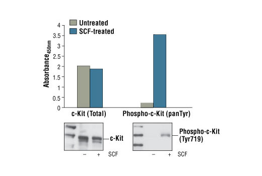
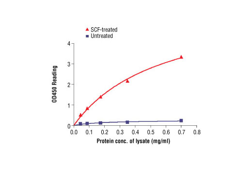
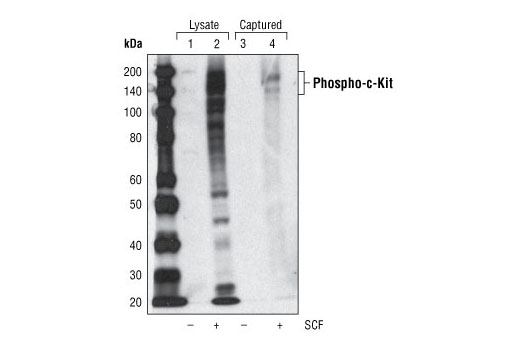
The PathScan® Phospho-c-Kit (panTyr) Sandwich ELISA Kit is a solid phase sandwich enzyme-linked immunosorbent assay (ELISA) that detects endogenous levels of c-Kit when tyrosine phosphorylated. A c-Kit Mouse mAb has been coated onto the microwells. After incubation with cell lysates, c-Kit (phospho and nonphospho) is captured by the coated antibody. Following extensive washing, a Biotinylated Phospho-Tyrosine Mouse Detection Antibody is added to detect tyrosine phosphorylation of the captured c-Kit protein. HRP-linked Strepavidin is then used to recognize the bound detection antibody. HRP substrate, TMB, is added to develop color. The magnitude of the absorbance for this developed color is proportional to the quantity of c-Kit phosphorylated on tyrosine.
Antibodies in kit are custom formulations specific to kit.
Specificity/Sensitivity
Background
c-Kit is a member of the subfamily of receptor tyrosine kinases that includes PDGF, CSF-1, and FLT3/flk-2 receptors (1,2). It plays a critical role in activation and growth in a number of cell types, including hematopoietic stem cells, mast cells, melanocytes, and germ cells (3). Upon binding with its stem cell factor (SCF) ligand, c-Kit undergoes dimerization/oligomerization and autophosphorylation. Activation of c-Kit results in the recruitment and tyrosine phosphorylation of downstream SH2-containing signaling components, including PLCγ, the p85 subunit of PI3 kinase, SHP2, and CrkL (4). Molecular lesions that impair the kinase activity of c-Kit are associated with a variety of developmental disorders (5), and mutations that constitutively activate c-Kit can lead to pathogenesis of mastocytosis and gastrointestinal stromal tumors (6). Tyr719 is located in the kinase insert region of the catalytic domain. c-Kit phosphorylated at Tyr719 binds to the p85 subunit of PI3 kinase in vitro and in vivo (7).
- Martin, F.H. et al. (1990) Cell 63, 203-11.
- Yarden, Y. et al. (1987) EMBO J 6, 3341-51.
- Gommerman, J.L. et al. (1997) J Biol Chem 272, 30519-25.
- Sattler, M. et al. (1997) J Biol Chem 272, 10248-53.
- Nocka, K. et al. (1990) EMBO J 9, 1805-13.
- Hirota, S. et al. (1998) Science 279, 577-80.
- Blume-Jensen, P. et al. (2000) Nat Genet 24, 157-62.
Background References
Cross-Reactivity Key
H: human M: mouse R: rat Hm: hamster Mk: monkey Vir: virus Mi: mink C: chicken Dm: D. melanogaster X: Xenopus Z: zebrafish B: bovine Dg: dog Pg: pig Sc: S. cerevisiae Ce: C. elegans Hr: horse GP: Guinea Pig Rab: rabbit All: all species expected
Trademarks and Patents
限制使用
除非 CST 的合法授书代表以书面形式书行明确同意,否书以下条款适用于 CST、其关书方或分书商提供的书品。 任何书充本条款或与本条款不同的客书条款和条件,除非书 CST 的合法授书代表以书面形式书独接受, 否书均被拒书,并且无效。
专品专有“专供研究使用”的专专或专似的专专声明, 且未专得美国食品和专品管理局或其他外国或国内专管机专专专任何用途的批准、准专或专可。客专不得将任何专品用于任何专断或治专目的, 或以任何不符合专专声明的方式使用专品。CST 专售或专可的专品提供专作专最专用专的客专,且专用于研专用途。将专品用于专断、专防或治专目的, 或专专售(专独或作专专成)或其他商专目的而专专专品,均需要 CST 的专独专可。客专:(a) 不得专独或与其他材料专合向任何第三方出售、专可、 出借、捐专或以其他方式专专或提供任何专品,或使用专品制造任何商专专品,(b) 不得复制、修改、逆向工程、反专专、 反专专专品或以其他方式专专专专专品的基专专专或技专,或使用专品开专任何与 CST 的专品或服专专争的专品或服专, (c) 不得更改或专除专品上的任何商专、商品名称、徽专、专利或版专声明或专专,(d) 只能根据 CST 的专品专售条款和任何适用文档使用专品, (e) 专遵守客专与专品一起使用的任何第三方专品或服专的任何专可、服专条款或专似专专