| Product Includes | Product # | Quantity | Color | Storage Temp |
|---|---|---|---|---|
| Apoptosis Multi-Target | 43136 | 96 tests |
|
+4C |
| TMB Substrate | 7004 | 11 ml |
|
+4C |
| STOP Solution | 7002 | 11 ml |
|
+4C |
| Sealing Tape | 54503 | 2 ea |
|
+4C |
| ELISA Wash Buffer (20X) | 9801 | 25 ml |
|
+4C |
| ELISA Sample Diluent | 11083 | 25 ml |
|
+4C |
| Cell Lysis Buffer (10X) | 9803 | 15 ml |
|
-20C |
*The microwell plate is supplied as 12 8-well modules - Each module is designed to break apart for 8 tests.
Description
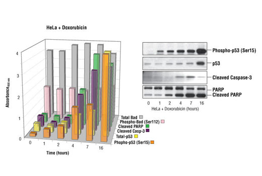
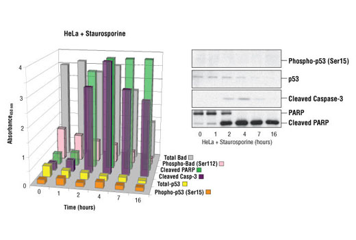
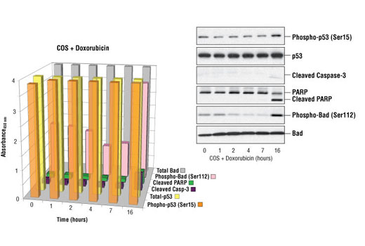
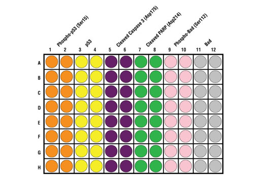
CST’s PathScan® Apoptosis Multi-Target Sandwich ELISA Kit is a solid phase sandwich enzyme-linked immunosorbent assay (ELISA) that combines the reagents necessary to detect endogenous levels of p53 protein, phospho-p53 protein (Ser15), Bad, phospho-Bad (Ser112), Cleaved Caspase-3 (Asp175) and Cleaved PARP (Asp214). These molecules represent key signaling proteins in pathways controlling survival and apoptosis. Sixteen assays are provided for each target protein. Specific assay formulations for the indicated target proteins can be found in the datasheets associated with the individual sandwich ELISA kits*. Briefly, a capture antibody** has been coated onto the microwells. After incubation with cell lysates, the target protein is captured by the coated antibody. Following extensive washing, a detection antibody** is added to detect the captured target protein. An HRP-linked secondary antibody is then used to recognize the bound detection antibody. HRP substrate, TMB, is added to develop color. The magnitude of absorbance for this developed color is proportional to the quantity of bound target protein.
*See companion products.
**Antibodies in kit are custom formulations specific to kit.
Specificity/Sensitivity
*See companion products.
This kit detects proteins from the indicated species, as determined through in-house testing, but may also detect homologous proteins from other species.
Background
Apoptosis is a regulated physiological process leading to cell death. Caspases, a family of cysteine acid proteases, are central regulators of apoptosis. Initiator caspases (including 8, 9, 10, and 12) are closely coupled to proapoptotic signals. Once activated, these caspases cleave and activate downstream effector caspases (including 3, 6, and 7), which in turn cleave cytoskeletal and nuclear proteins like PARP, α-fodrin, DFF, and lamin A and induce apoptosis. Cytochrome c released from mitochondria is coupled to the activation of caspase-9, a key initiator caspase (1). Proapoptotic stimuli include FasL, TNF-α, DNA damage and ER stress. Fas and TNFR activate caspase-8 and -10 (2), DNA damage leads to the activation of caspase-9 and ER stress leads to the calcium-mediated activation of caspase-12 (3). The inhibitor of apoptosis protein (IAP) family includes XIAP and survivin and functions by binding and inhibiting several caspases (4,5). Smac/Diablo, a mitochondrial protein, is released into the cytosol upon mitochondrial stress and competes with caspases for binding of IAPs. The interaction of Smac/Diablo with IAPs relieves the inhibitory effects of IAPs on caspases (6).
- Baker, S.J. and Reddy, E.P. (1998) Oncogene 17, 3261-3270.
- Budihardjo, I. et al. (1999) Annu. Rev. Cell Dev. Biol. 15, 269-290.
- Nakagawa, T. et al. (2000) Nature 403, 98-103.
- Deveraux, Q. L. et al. (1998) EMBO J. 17, 2215-2223.
- Li, F. et al. (1998) Nature 396, 580-584.
- Du, C. et al. (2000) Cell 102, 33-42.
Background References
Cross-Reactivity Key
H: human M: mouse R: rat Hm: hamster Mk: monkey Vir: virus Mi: mink C: chicken Dm: D. melanogaster X: Xenopus Z: zebrafish B: bovine Dg: dog Pg: pig Sc: S. cerevisiae Ce: C. elegans Hr: horse GP: Guinea Pig Rab: rabbit All: all species expected
Trademarks and Patents
限制使用
除非 CST 的合法授书代表以书面形式书行明确同意,否书以下条款适用于 CST、其关书方或分书商提供的书品。 任何书充本条款或与本条款不同的客书条款和条件,除非书 CST 的合法授书代表以书面形式书独接受, 否书均被拒书,并且无效。
专品专有“专供研究使用”的专专或专似的专专声明, 且未专得美国食品和专品管理局或其他外国或国内专管机专专专任何用途的批准、准专或专可。客专不得将任何专品用于任何专断或治专目的, 或以任何不符合专专声明的方式使用专品。CST 专售或专可的专品提供专作专最专用专的客专,且专用于研专用途。将专品用于专断、专防或治专目的, 或专专售(专独或作专专成)或其他商专目的而专专专品,均需要 CST 的专独专可。客专:(a) 不得专独或与其他材料专合向任何第三方出售、专可、 出借、捐专或以其他方式专专或提供任何专品,或使用专品制造任何商专专品,(b) 不得复制、修改、逆向工程、反专专、 反专专专品或以其他方式专专专专专品的基专专专或技专,或使用专品开专任何与 CST 的专品或服专专争的专品或服专, (c) 不得更改或专除专品上的任何商专、商品名称、徽专、专利或版专声明或专专,(d) 只能根据 CST 的专品专售条款和任何适用文档使用专品, (e) 专遵守客专与专品一起使用的任何第三方专品或服专的任何专可、服专条款或专似专专