| Product Includes | Product # | Quantity | Color | Storage Temp |
|---|---|---|---|---|
| Met Mouse mAb Coated Microwells | 36629 | 96 tests |
|
+4C |
| Met Rabbit Detection mAb | 14242 | 1 ea |
|
+4C |
| Anti-rabbit IgG, HRP-linked Antibody (ELISA Formulated) | 13272 | 1 ea |
|
+4C |
| Detection Antibody Diluent | 13339 | 11 ml |
|
+4C |
| HRP Diluent | 13515 | 11 ml |
|
+4C |
| TMB Substrate | 7004 | 11 ml |
|
+4C |
| STOP Solution | 7002 | 11 ml |
|
+4C |
| Sealing Tape | 54503 | 2 ea |
|
+4C |
| ELISA Wash Buffer (20X) | 9801 | 25 ml |
|
+4C |
| ELISA Sample Diluent | 11083 | 25 ml |
|
+4C |
| Cell Lysis Buffer (10X) | 9803 | 15 ml |
|
-20C |
*The microwell plate is supplied as 12 8-well modules - Each module is designed to break apart for 8 tests.
Description
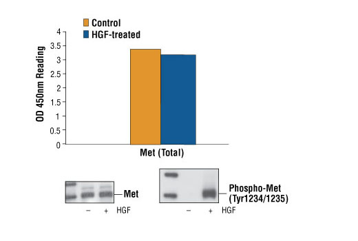
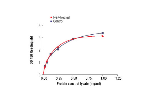
CST's PathScan® Total Met Sandwich ELISA Kit is a solid phase sandwich enzyme-linked immunosorbent assay (ELISA) that detects endogenous levels of total Met protein. A Met Mouse mAb has been coated onto the microwells. After incubation with cell lysates, both phospho- and nonphospho-Met proteins are captured by the coated antibody. Following extensive washing, Met Rabbit Antibody is added to detect both the captured phospho- and nonphospho-Met protein. Anti-rabbit IgG, HRP-linked Antibody is then used to recognize the bound detection antibody. HRP substrate, TMB, is added to develop color. The magnitude of optical density for this developed color is proportional to the quantity of total Met protein.
*Antibodies in kit are custom formulations specific to kit.
Specificity/Sensitivity
Background
Met, a high affinity tyrosine kinase receptor for hepatocyte growth factor (HGF, also known as scatter factor) is a disulfide-linked heterodimer made of 45 kDa α- and 145 kDa β-subunits (1,2). The α-subunit and the amino-terminal region of the β-subunit form the extracellular domain. The remainder of the β-chain spans the plasma membrane and contains a cytoplasmic region with tyrosine kinase activity. Interaction of Met with HGF results in autophosphorylation at multiple tyrosines, which recruit several downstream signaling components, including Gab1, c-Cbl, and PI3 kinase (3). These fundamental events are important for all of the biological functions involving Met kinase activity. The addition of a phosphate at cytoplasmic Tyr1003 is essential for Met protein ubiquitination and degradation (4). Phosphorylation at Tyr1234/1235 in the Met kinase domain is critical for kinase activation. Phosphorylation at Tyr1349 in the Met cytoplasmic domain provides a direct binding site for Gab1 (5). Research studies have shown that altered Met levels and/or tyrosine kinase activities are found in several types of tumors, including renal, colon, and breast. Thus, investigators have concluded that Met is an attractive potential cancer therapeutic and diagnostic target (6,7).
- Cooper, C.S. et al. (1984) Nature 311, 29-33.
- Bottaro, D.P. et al. (1991) Science 251, 802-4.
- Bardelli, A. et al. (1997) Oncogene 15, 3103-11.
- Taher, T.E. et al. (2002) J Immunol 169, 3793-800.
- Schaeper, U. et al. (2000) J Cell Biol 149, 1419-32.
- Eder, J.P. et al. (2009) Clin Cancer Res 15, 2207-14.
- Sattler, M. and Salgia, R. (2009) Update Cancer Ther 3, 109-118.
Background References
Cross-Reactivity Key
H: human M: mouse R: rat Hm: hamster Mk: monkey Vir: virus Mi: mink C: chicken Dm: D. melanogaster X: Xenopus Z: zebrafish B: bovine Dg: dog Pg: pig Sc: S. cerevisiae Ce: C. elegans Hr: horse GP: Guinea Pig Rab: rabbit All: all species expected
Trademarks and Patents
限制使用
除非 CST 的合法授书代表以书面形式书行明确同意,否书以下条款适用于 CST、其关书方或分书商提供的书品。 任何书充本条款或与本条款不同的客书条款和条件,除非书 CST 的合法授书代表以书面形式书独接受, 否书均被拒书,并且无效。
专品专有“专供研究使用”的专专或专似的专专声明, 且未专得美国食品和专品管理局或其他外国或国内专管机专专专任何用途的批准、准专或专可。客专不得将任何专品用于任何专断或治专目的, 或以任何不符合专专声明的方式使用专品。CST 专售或专可的专品提供专作专最专用专的客专,且专用于研专用途。将专品用于专断、专防或治专目的, 或专专售(专独或作专专成)或其他商专目的而专专专品,均需要 CST 的专独专可。客专:(a) 不得专独或与其他材料专合向任何第三方出售、专可、 出借、捐专或以其他方式专专或提供任何专品,或使用专品制造任何商专专品,(b) 不得复制、修改、逆向工程、反专专、 反专专专品或以其他方式专专专专专品的基专专专或技专,或使用专品开专任何与 CST 的专品或服专专争的专品或服专, (c) 不得更改或专除专品上的任何商专、商品名称、徽专、专利或版专声明或专专,(d) 只能根据 CST 的专品专售条款和任何适用文档使用专品, (e) 专遵守客专与专品一起使用的任何第三方专品或服专的任何专可、服专条款或专似专专