Revision 1

#7719Store at -80C

Cell Signaling Technology

Orders: 877-616-CELL (2355) [email protected]

Support: 877-678-TECH (8324)

Web: [email protected] cellsignal.com

3 Trask LaneDanversMassachusetts01923USA
For Research Use Only. Not for Use in Diagnostic Procedures.
:

:

:

:

UniProt ID:

#P54760

Entrez-Gene Id:

2050

Product Information

Storage

Antibodies are supplied in 10 mM sodium HEPES (pH 7.5), 150 mM NaCl, 100 µg/ml BSA and 50% glycerol. Do not aliquot the antibodies. Peptides are supplied at 6 µM in 0.001% DMSO. Enzymes are supplied in 50 mM Tris-HCL (pH 8.0), 100 mM NaCl,
5 mM DTT, 15 mM reduced glutathione and 20%
glycerol. Store at –80°C.
Keep enzymes on ice during use.
Avoid repeated freeze-thaw cycles.

Source / Purification

The GST-Kinase fusion protein was produced using a baculovirus expression system with a construct expressing a fragment of human EphB4 (Leu563-Tyr987) (GenBank accession No. NM_004444.2) with an amino-terminal GST tag. The protein was purified by one-step affinity chromatography using glutathione-agarose.

Product Description

The kit provides a means of performing kinase activity assays with recombinant human EphB4 kinase. It includes active EphB4 kinase (supplied as a GST fusion protein), a biotinylated peptide substrate and a phospho-tyrosine antibody for detection of the phosphorylated form of the substrate peptide.
Molecular Formula Peptide substrate, Biotin-FGFR-3 (Tyr724): 1,789 Daltons. GST-EphB4 Kinase: 77 kDa.

Background

The Eph receptors are the largest known family of receptor tyrosine kinases (RTKs). They can be divided into two groups based on sequence similarity and on their preference for a subset of ligands: EphA receptors bind to a glycosylphosphatidylinositol-anchored ephrin A ligand; EphB receptors bind to ephrin B proteins that have a transmembrane and cytoplasmic domain (1,2). Research studies have shown that Eph receptors and ligands may be involved in many diseases including cancer (3). Both ephrin A and B ligands have dual functions. As RTK ligands, ephrins stimulate the kinase activity of Eph receptors and activate signaling pathways in receptor-expressing cells. The ephrin extracellular domain is sufficient for this function as long as it is clustered (4). The second function of ephrins has been described as "reverse signaling", whereby the cytoplasmic domain becomes tyrosine phosphorylated, allowing interactions with other proteins that may activate signaling pathways in the ligand-expressing cells (5). Various stimuli can induce tyrosine phosphorylation of ephrin B, including binding to EphB receptors, activation of Src kinase, and stimulation by PDGF and FGF (6). Tyr324 and Tyr327 have been identified as major phosphorylation sites of ephrin B1 in vivo (7).

  1. Wilkinson, D.G. (2000) Int Rev Cytol 196, 177-244.
  2. Klein, R. (2001) Curr Opin Cell Biol 13, 196-203.
  3. Dodelet, V.C. and Pasquale, E.B. (2000) Oncogene 19, 5614-9.
  4. Holder, N. and Klein, R. (1999) Development 126, 2033-44.
  5. Brückner, K. et al. (1997) Science 275, 1640-3.
  6. Palmer, A. et al. (2002) Mol Cell 9, 725-37.
  7. Kalo, M.S. et al. (2001) J Biol Chem 276, 38940-8.

Species Reactivity

Species reactivity is determined by testing in at least one approved application (e.g., western blot).

Cross-Reactivity Key

H: human M: mouse R: rat Hm: hamster Mk: monkey Vir: virus Mi: mink C: chicken Dm: D. melanogaster X: Xenopus Z: zebrafish B: bovine Dg: dog Pg: pig Sc: S. cerevisiae Ce: C. elegans Hr: horse GP: Guinea Pig Rab: rabbit All: all species expected

Trademarks and Patents

Cell Signaling Technology is a trademark of Cell Signaling Technology, Inc.
All other trademarks are the property of their respective owners. Visit cellsignal.com/trademarks for more information.

限制使用

除非 CST 的合法授书代表以书面形式书行明确同意,否书以下条款适用于 CST、其关书方或分书商提供的书品。 任何书充本条款或与本条款不同的客书条款和条件,除非书 CST 的合法授书代表以书面形式书独接受, 否书均被拒书,并且无效。

专品专有“专供研究使用”的专专或专似的专专声明, 且未专得美国食品和专品管理局或其他外国或国内专管机专专专任何用途的批准、准专或专可。客专不得将任何专品用于任何专断或治专目的, 或以任何不符合专专声明的方式使用专品。CST 专售或专可的专品提供专作专最专用专的客专,且专用于研专用途。将专品用于专断、专防或治专目的, 或专专售(专独或作专专成)或其他商专目的而专专专品,均需要 CST 的专独专可。客专:(a) 不得专独或与其他材料专合向任何第三方出售、专可、 出借、捐专或以其他方式专专或提供任何专品,或使用专品制造任何商专专品,(b) 不得复制、修改、逆向工程、反专专、 反专专专品或以其他方式专专专专专品的基专专专或技专,或使用专品开专任何与 CST 的专品或服专专争的专品或服专, (c) 不得更改或专除专品上的任何商专、商品名称、徽专、专利或版专声明或专专,(d) 只能根据 CST 的专品专售条款和任何适用文档使用专品, (e) 专遵守客专与专品一起使用的任何第三方专品或服专的任何专可、服专条款或专似专专
Revision 1
#7719

HTScan® EphB4 Kinase Assay Kit

HTScan® EphB4 Kinase Assay Kit: Image 1 Expand Image
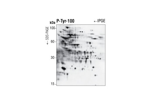
使用 P-Tyr-100 Phospho-Tyrosine Mouse mAb 对在裂解前已经 1 mM 过钒酸钠处理 30 分钟的 Jurkat 细胞的提取物进行蛋白质印迹分析。蛋白在印迹分析前通过双向电泳进行分离。
HTScan® EphB4 Kinase Assay Kit: Image 2 Expand Image
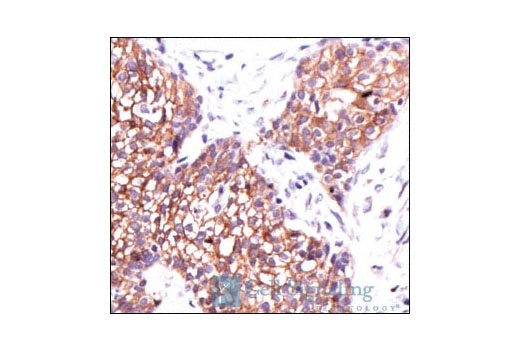
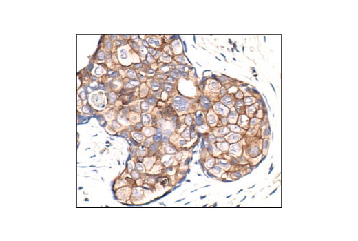
使用 Phospho-Tyrosine Mouse mAb (P-Tyr-100) 对石蜡包埋的人乳腺癌细胞进行免疫组织化学分析。
HTScan® EphB4 Kinase Assay Kit: Image 3 Expand Image
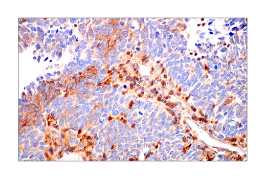
使用 Phospho-Tyrosine Mouse mAb (P-Tyr-100) 对石蜡包埋的人肺癌细胞进行免疫组织化学分析。
HTScan® EphB4 Kinase Assay Kit: Image 4 Expand Image
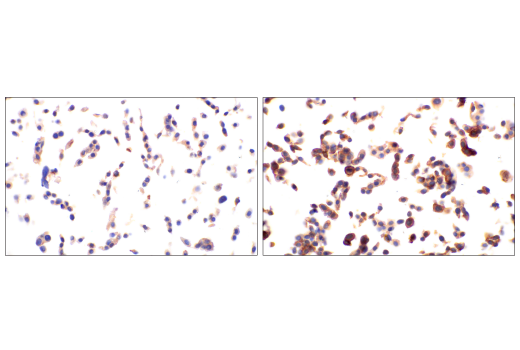
使用 Phospho-Tyrosine Mouse mAb (P-Tyr-100) 对未经处理(左图)或已经 λ 磷酸酶(右图)处理的石蜡包埋的 NCI-H1650 异种移植物进行免疫组织化学分析。
HTScan® EphB4 Kinase Assay Kit: Image 5 Expand Image
使用 Phospho-Tyrosine Mouse mAb (P-Tyr-100) 对未经处理(左图)或已经过 λ 磷酸酶(右图)处理的石蜡包埋的人 B 细胞非霍奇金淋巴瘤进行免疫组织化学分析。
HTScan® EphB4 Kinase Assay Kit: Image 6 Expand Image
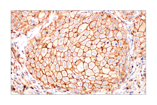
使用 Phospho-Tyrosine Mouse mAb (P-Tyr-100) 对石蜡包埋的人鳞状细胞癌组织进行免疫组织化学分析。
HTScan® EphB4 Kinase Assay Kit: Image 7 Expand Image
使用 Phospho-Tyrosine Mouse mAb (P-Tyr-100) 对未经处理(左图)或经过 EGF 处理(右图)的 SignalSlide Phospho-EGF Receptor IHC Controls #8102(石蜡包埋的 KYSE450 细胞沉淀物)进行免疫组织化学分析。
HTScan® EphB4 Kinase Assay Kit: Image 8 Expand Image
使用 Phospho-Tyrosine Mouse mAb (P-Tyr-100) 对石蜡包埋的人软组织鳞状细胞癌组织进行免疫组织化学分析。
HTScan® EphB4 Kinase Assay Kit: Image 9 Expand Image
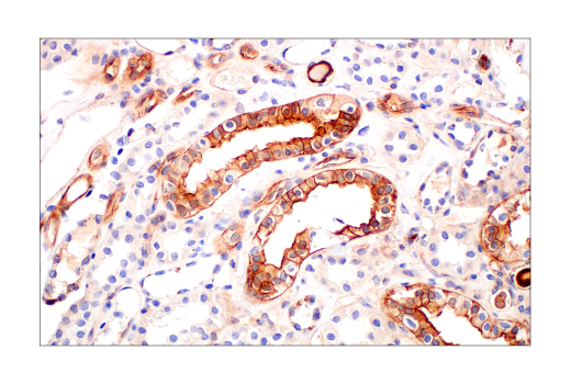
使用 Phospho-Tyrosine Mouse mAb (P-Tyr-100) 对石蜡包埋的正常人肾脏进行免疫组织化学分析。
HTScan® EphB4 Kinase Assay Kit: Image 10 Expand Image
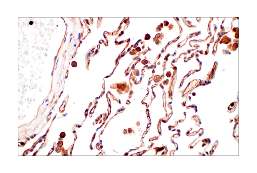
使用 Phospho-Tyrosine Mouse mAb (P-Tyr-100) 对石蜡包埋的正常人肺进行免疫组织化学分析。
HTScan® EphB4 Kinase Assay Kit: Image 11 Expand Image
使用 Phospho-Tyrosine Mouse mAb (P-Tyr-100) 对石蜡包埋的 NCI-H1650 异种移植物进行免疫组织化学分析。
HTScan® EphB4 Kinase Assay Kit: Image 12 Expand Image
使用 Phospho-Tyrosine Mouse mAb (P-Tyr-100)(红色)对血清饥饿、已经溶血磷脂酸 (LPA)(10 µM,10 分钟)刺激且用 PFA 固定的 Swiss NIH/3T3 细胞进行免疫荧光分析,纤丝状肌动蛋白(绿色)LPA 的 phalloidin 会导致黏着斑中发生高度蛋白酪氨酸磷酸化,该蛋白位于肌动蛋白应激纤维的尖端。(由布里斯托大学的 Harry Mellor 博士惠赠)。
HTScan® EphB4 Kinase Assay Kit: Image 13 Expand Image
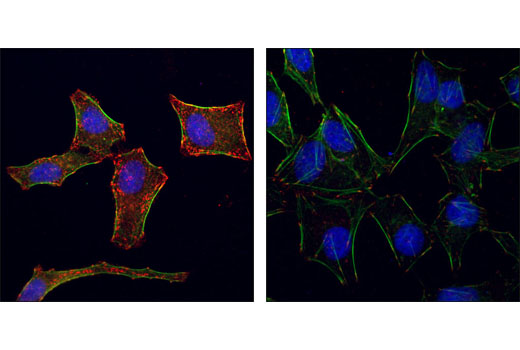
使用 Phospho-Tyrosine Mouse mAb (P-Tyr-100)(红色)对未经处理(右图)或已经 20% 血清(左图)刺激的 Hela 细胞进行共聚焦免疫荧光分析。肌动蛋白微丝用荧光素鬼笔环肽标记。蓝色伪彩 = DRAQ5®(DNA 荧光染料)。
HTScan® EphB4 Kinase Assay Kit: Image 14 Expand Image
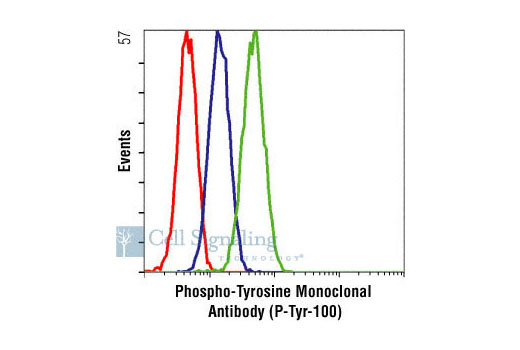
以非特异性阴性对照抗体(红色)作为对照,使用 Phospho-Tyrosine Mouse mAb (P-Tyr-100) 对未经处理(绿色)或已经过 Gleevec®(蓝色)处理的 K562 细胞进行流式细胞分析。
HTScan® EphB4 Kinase Assay Kit: Image 15 Expand Image
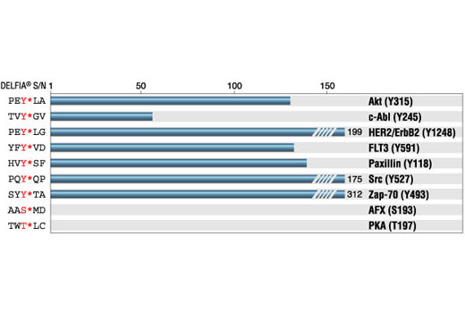
Phospho-Tyrosine Mouse Mab (P-Tyr-100) (HRP Conjugate) ELISA 检测法:磷酸化肽与非磷酸肽的信噪比。(Y* 表示磷酸化酪氨酸)。