Revision 1
Cell Signaling Technology

Orders: 877-616-CELL (2355) [email protected]

Support: 877-678-TECH (8324)

Web: [email protected] cellsignal.com

3 Trask LaneDanversMassachusetts01923USA
For Research Use Only. Not for Use in Diagnostic Procedures.
Product Includes Product # Quantity Mol. Wt Isotype/Source
COX IV (3E11) Rabbit mAb (Alexa Fluor® 647 Conjugate) 7561 40 µl Rabbit IgG
PDI (C81H6) Rabbit mAb (Alexa Fluor® 647 Conjugate) 8463 40 µl Rabbit 
β-Tubulin (9F3) Rabbit mAb (Alexa Fluor® 647 Conjugate) 3624 40 µl Rabbit IgG
β-Actin (13E5) Rabbit mAb (Alexa Fluor® 647 Conjugate) 8584 40 µl Rabbit IgG
Rabbit (DA1E) mAb IgG XP® Isotype Control (Alexa Fluor® 647 Conjugate) 2985 40 µl Rabbit IgG

Please visit cellsignal.com for individual component applications, species cross-reactivity, dilutions, protocols, and additional product information.

Description

The Cellular Localization Alexa Fluor® 647 Conjugated Antibody Sampler Kit provides an economical means for identification of cellular organelles without the need for a fluorescent secondary antibody. This kit includes enough primary antibody to perform at least 20 IF-IC tests per antibody.

Storage

Supplied in 10 mM sodium HEPES (pH 7.5), 150 mM NaCl, 100 µg/ml BSA, 50% glycerol and less than 0.02% sodium azide. Store at –20°C. Do not aliquot the antibody.

Background

Knowledge of the subcellular location of a protein may reveal the potential role it plays in the cell. The subcellular location of a protein of interest may be confirmed by colocalization with one of the organelle-specific antibodies in this kit.
β-Actin and β-tubulin are ubiquitous eukaryotic proteins that form the major component of the cellular cytoskeleton (1,2). Cytochrome c oxidase (COX) IV is part of the hetero-oligomeric enzyme found in the inner mitochondrial membrane (3). Protein disulfide isomerase (PDI) catalyzes the formation and isomerization of disulfide bonds in the endoplasmic reticulum (4).

  1. Herman, I.M. (1993) Curr Opin Cell Biol 5, 48-55.
  2. Westermann, S. and Weber, K. (2003) Nat Rev Mol Cell Biol 4, 938-47.
  3. Ostermeier, C. et al. (1996) Curr Opin Struct Biol 6, 460-6.
  4. Ellgaard, L. and Ruddock, L.W. (2005) EMBO Rep 6, 28-32.

Background References

    Trademarks and Patents

    Cell Signaling Technology is a trademark of Cell Signaling Technology, Inc.
    All other trademarks are the property of their respective owners. Visit cellsignal.com/trademarks for more information.

    限制使用

    除非 CST 的合法授书代表以书面形式书行明确同意,否书以下条款适用于 CST、其关书方或分书商提供的书品。 任何书充本条款或与本条款不同的客书条款和条件,除非书 CST 的合法授书代表以书面形式书独接受, 否书均被拒书,并且无效。

    专品专有“专供研究使用”的专专或专似的专专声明, 且未专得美国食品和专品管理局或其他外国或国内专管机专专专任何用途的批准、准专或专可。客专不得将任何专品用于任何专断或治专目的, 或以任何不符合专专声明的方式使用专品。CST 专售或专可的专品提供专作专最专用专的客专,且专用于研专用途。将专品用于专断、专防或治专目的, 或专专售(专独或作专专成)或其他商专目的而专专专品,均需要 CST 的专独专可。客专:(a) 不得专独或与其他材料专合向任何第三方出售、专可、 出借、捐专或以其他方式专专或提供任何专品,或使用专品制造任何商专专品,(b) 不得复制、修改、逆向工程、反专专、 反专专专品或以其他方式专专专专专品的基专专专或技专,或使用专品开专任何与 CST 的专品或服专专争的专品或服专, (c) 不得更改或专除专品上的任何商专、商品名称、徽专、专利或版专声明或专专,(d) 只能根据 CST 的专品专售条款和任何适用文档使用专品, (e) 专遵守客专与专品一起使用的任何第三方专品或服专的任何专可、服专条款或专似专专

    Revision 1
    #8663

    Cellular Localization Alexa Fluor® 647 Conjugated Antibody Sampler Kit

    Cellular Localization Alexa Fluor® 647 Conjugated Antibody Sampler Kit: Image 1 Expand Image
    以浓度匹配的 Rabbit (DA1E) mAb IgG XP® Isotype Control (Alexa Fluor® 647 Conjugate(蓝色伪彩,右图)作为对照,使用 GAPDH (14C10) Rabbit mAb (Alexa Fluor® 647 Conjugate) #3907(蓝色伪彩,左图)对 C2C12 细胞进行共聚焦免疫荧光分析。红色 = Propidium Iodide (PI)/RNase Staining Solution #4087(DNA 荧光染料)。
    Cellular Localization Alexa Fluor® 647 Conjugated Antibody Sampler Kit: Image 2 Expand Image
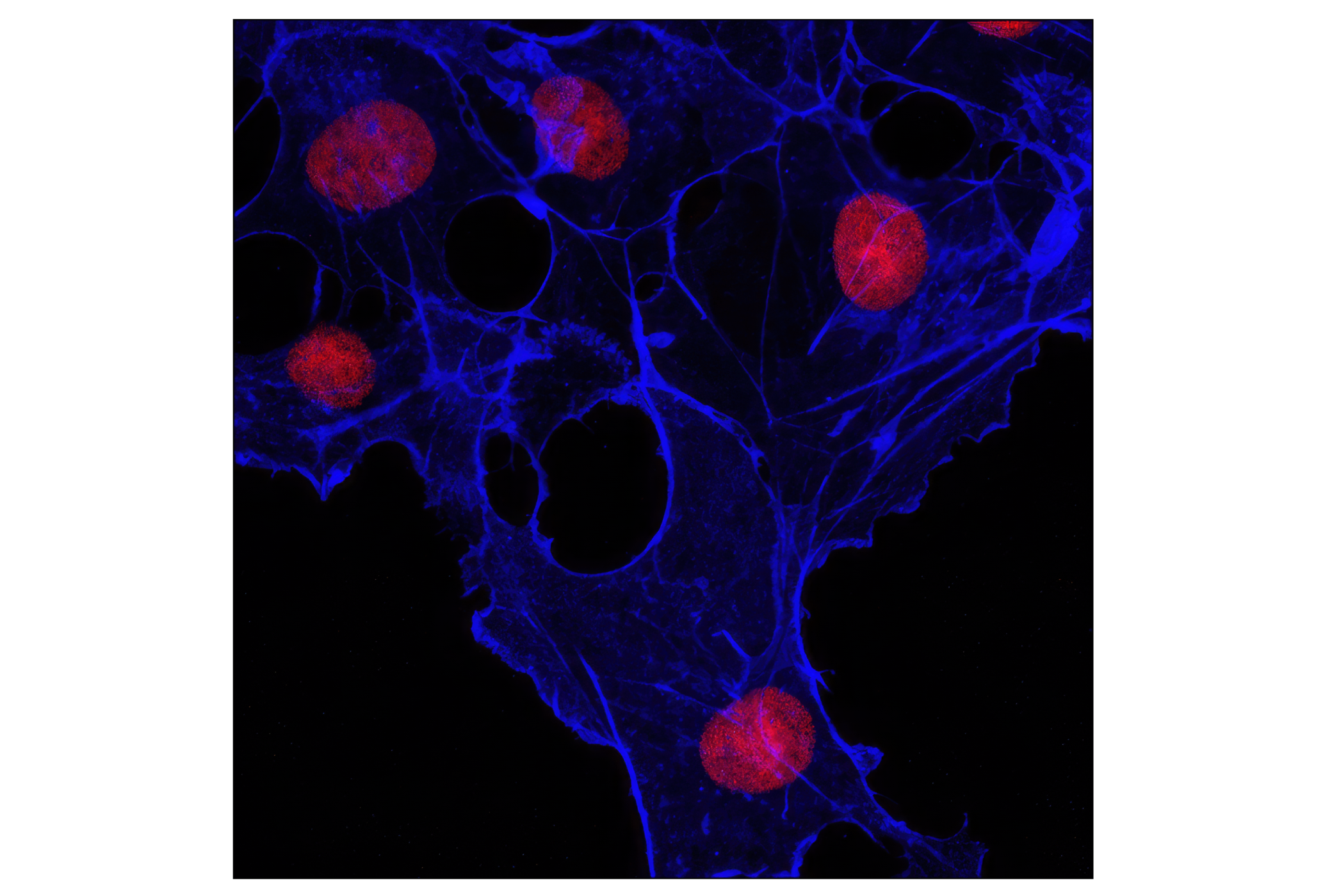
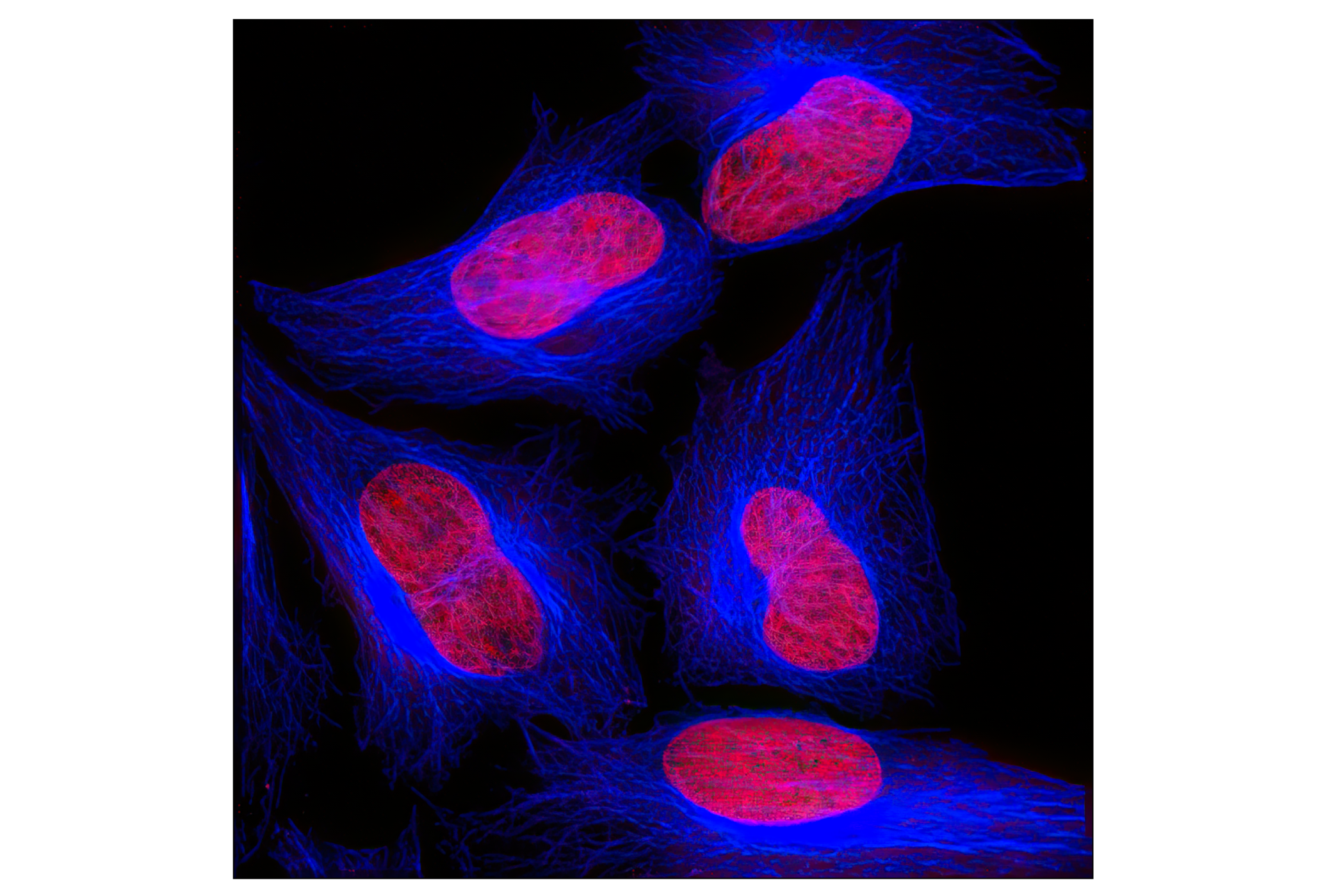
    使用 β-Tubulin (9F3) Rabbit mAb (Alexa Fluor® 647 Conjugate)(蓝色伪彩)对 HeLa 细胞进行共聚焦免疫荧光分析。红色 = 碘化丙啶(DNA 荧光染料)。
    Cellular Localization Alexa Fluor® 647 Conjugated Antibody Sampler Kit: Image 3 Expand Image
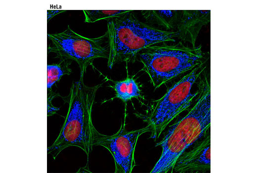
    使用 COX IV (3E11) Rabbit mAb (Alexa Fluor® 647 Conjugate)(蓝色伪彩)对 HeLa 细胞进行共聚焦免疫荧光分析。肌动蛋白丝用 Alexa Fluor® 488 phalloidin(绿色)标记。红色 = Propidium Iodide (PI)/RNase Staining Solution #4087。
    Cellular Localization Alexa Fluor® 647 Conjugated Antibody Sampler Kit: Image 4 Expand Image
    使用 PDI (C81H6) Rabbit mAb (Alexa Fluor® 647 Conjugate)(伪彩蓝色)和 Alexa Fluor® 488 phalloidin(绿色)对 NIH/3T3 细胞进行共聚焦免疫荧光分析。红色 = Propidium Iodide (PI)/RNase Staining Solution #4087(DNA 荧光染料)。
    Cellular Localization Alexa Fluor® 647 Conjugated Antibody Sampler Kit: Image 5 Expand Image
    使用 β-Actin (13E5) Rabbit mAb (Alexa Fluor® 647 Conjugate)(蓝色假色)对 COS-7 细胞进行共聚焦免疫荧光分析。红色 = Propidium Iodide (PI)/RNase Staining Solution #4087。
    Cellular Localization Alexa Fluor® 647 Conjugated Antibody Sampler Kit: Image 6 Expand Image
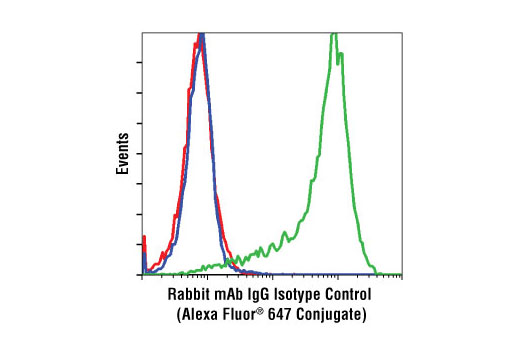
    以未经处理(绿色)或已经 LY294002 和 wortmannin(蓝色)处理的 Phospho-Akt (Ser473) (D9E) XP® Rabbit mAb (Alexa Fluor® 647 Conjugate) #4075 作为对照,使用 Rabbit (DA1E) mAb IgG XP® Isotype Control (Alexa Fluor® 647 Conjugate)(红色)对 Jurkat 细胞进行流式细胞分析。
    Cellular Localization Alexa Fluor® 647 Conjugated Antibody Sampler Kit: Image 7 Expand Image
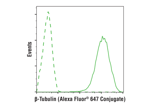
    使用 β-Tubulin (9F3) Rabbit mAb (Alexa Fluor® 647 Conjugate) (solid line)(实线)或浓度匹配的 Rabbit (DA1E) mAb IgG XP® Isotype Control (Alexa Fluor® 647 Conjugate) #2985(虚线)对 Hela 细胞进行流式细胞分析。
    Cellular Localization Alexa Fluor® 647 Conjugated Antibody Sampler Kit: Image 8 Expand Image
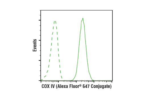
    以浓度匹配的 Rabbit (DA1E) mAb IgG XP® Isotype Control (Alexa Fluor® 647 Conjugate) #2985(虚线)作为对照,使用 COX IV (3E11) Rabbit mAb (Alexa Fluor® 647 Conjugate)(实线)对 HeLa 细胞进行流式细胞分析。
    Cellular Localization Alexa Fluor® 647 Conjugated Antibody Sampler Kit: Image 9 Expand Image
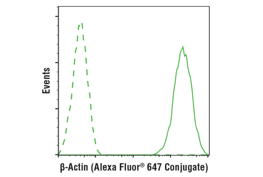
    以浓度匹配的 Rabbit (DA1E) mAb IgG XP® Isotype Control (Alexa Fluor® 647 Conjugate) #2985(虚线)作为对照,使用 β-Actin (13E5) Rabbit mAb (Alexa Fluor® 647 Conjugate)(实线)对 Hela 细胞进行流式细胞分析。
    Cellular Localization Alexa Fluor® 647 Conjugated Antibody Sampler Kit: Image 10 Expand Image
    以未经处理(蓝色)或已经 IFN-α(绿色)处理的 Phospho-S6 Ribosomal Protein (Ser235/236) (D57.2.2E) XP® Rabbit mAb (Alexa Fluor® 647 Conjugate) #4851 作为对照,使用 Rabbit (DA1E) mAb IgG XP® Isotype Control (Alexa Fluor® 647 Conjugate)(红色)对 Jurkat 细胞进行流式细胞分析。