Revision 4
#96489
Store at -20C
877-616-CELL (2355)
877-678-TECH (8324)
3 Trask Lane | Danvers | Massachusetts | 01923 | USA
For Research Use Only. Not for Use in Diagnostic Procedures.
Applications:
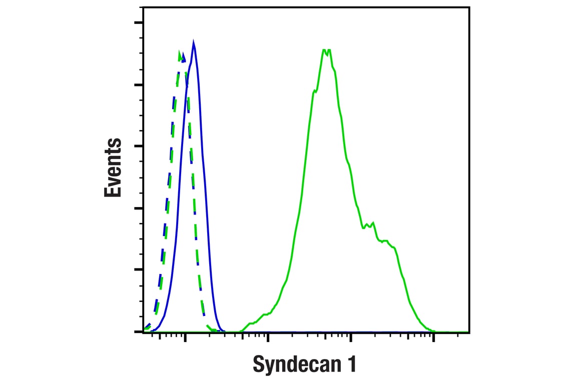
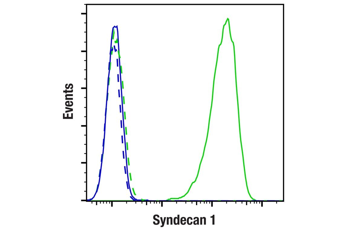
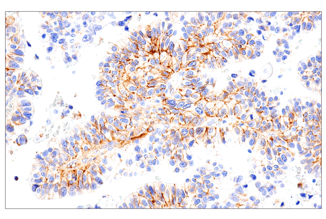
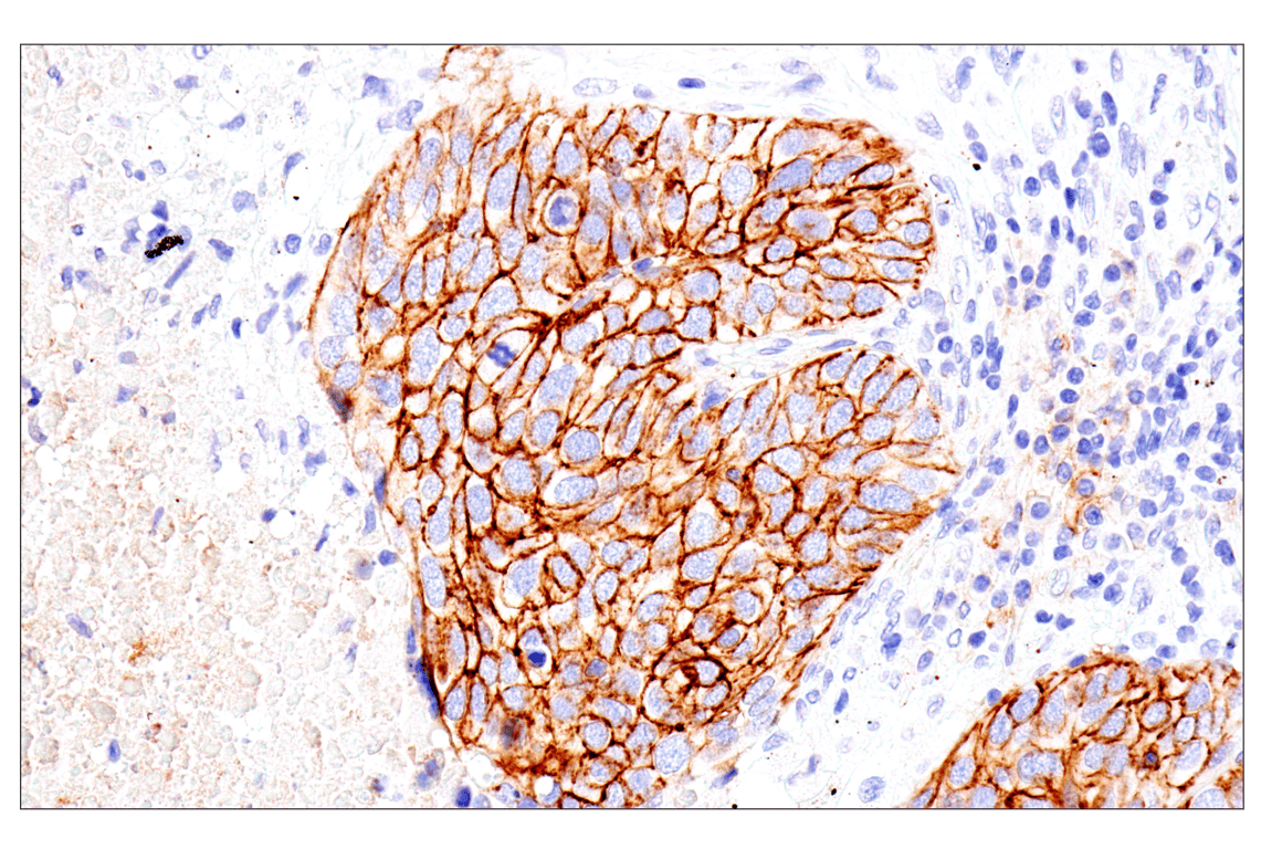
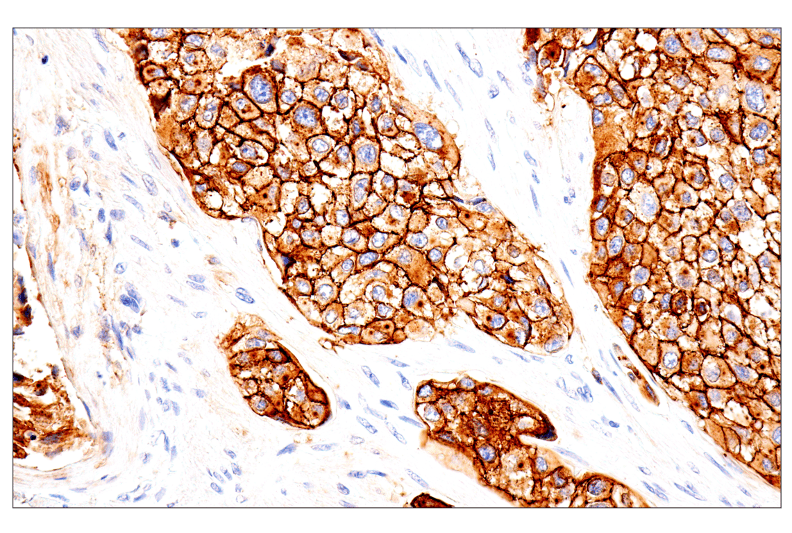
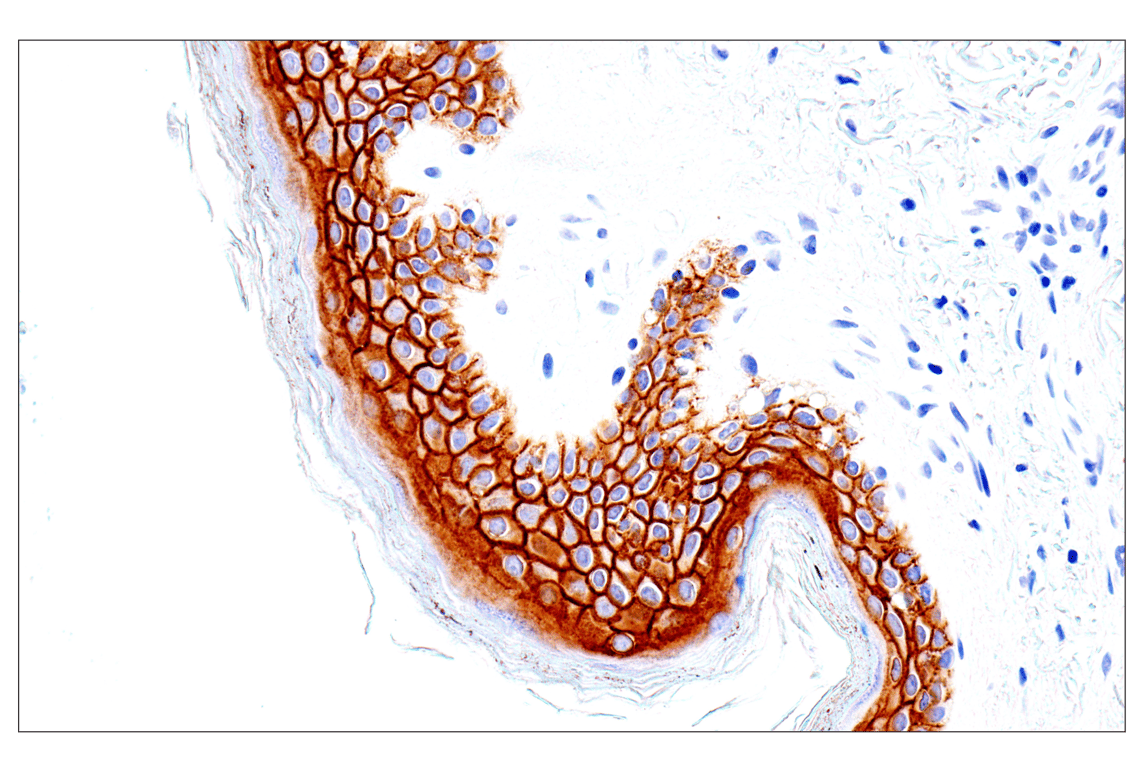
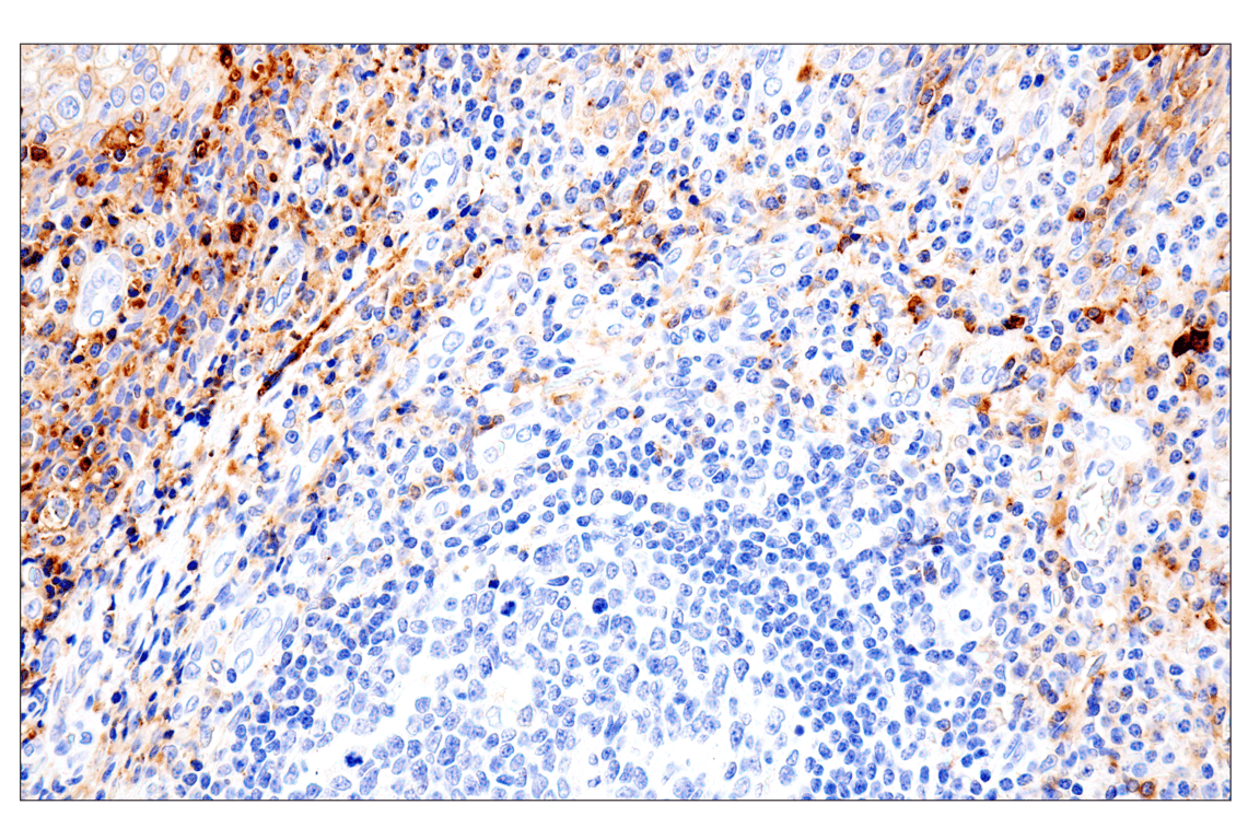
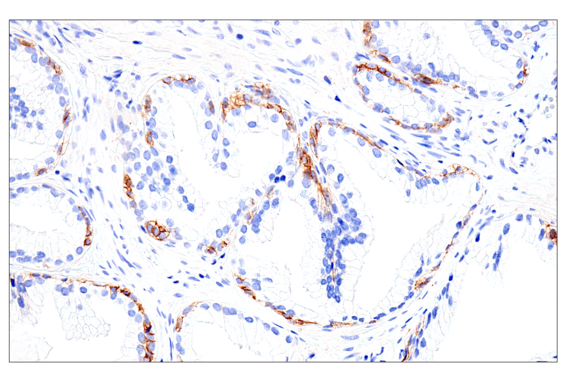
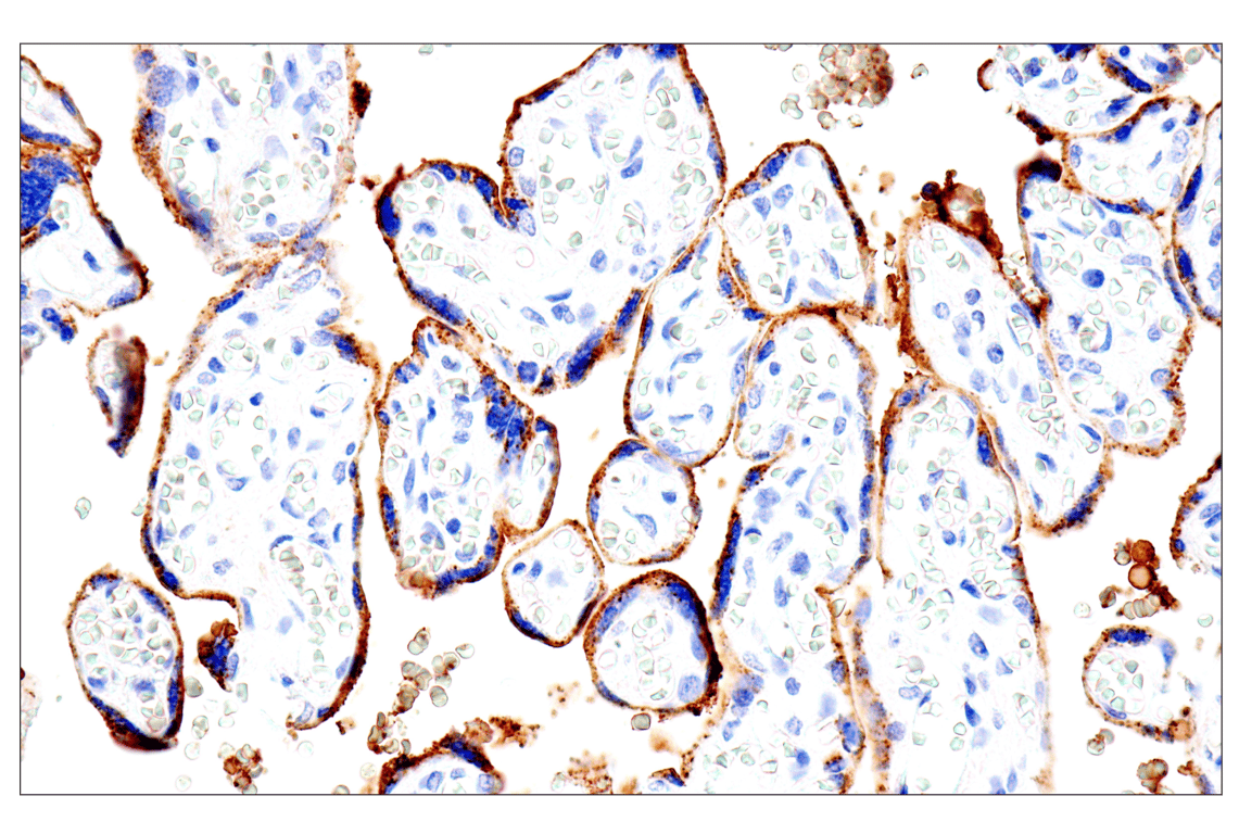
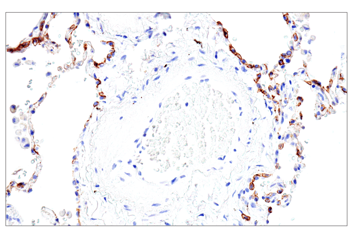
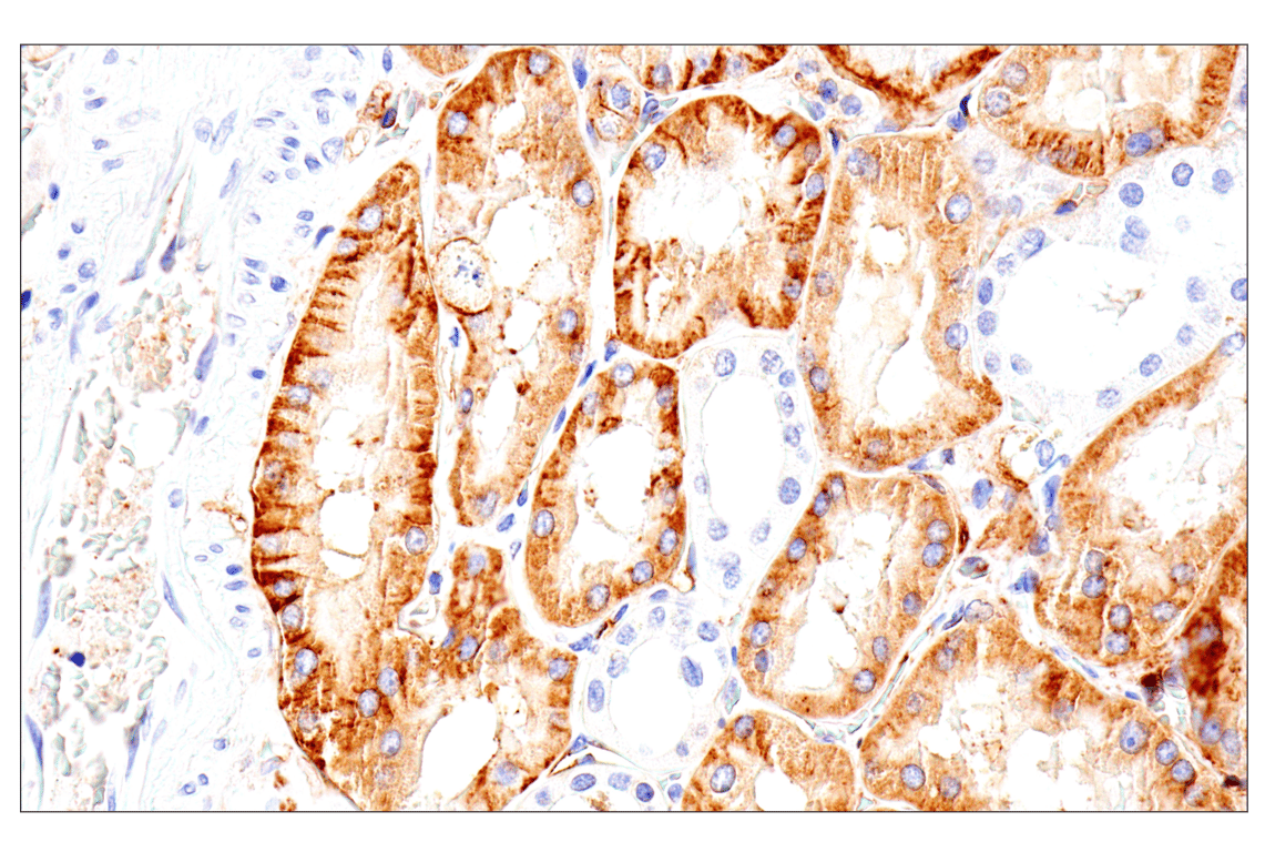
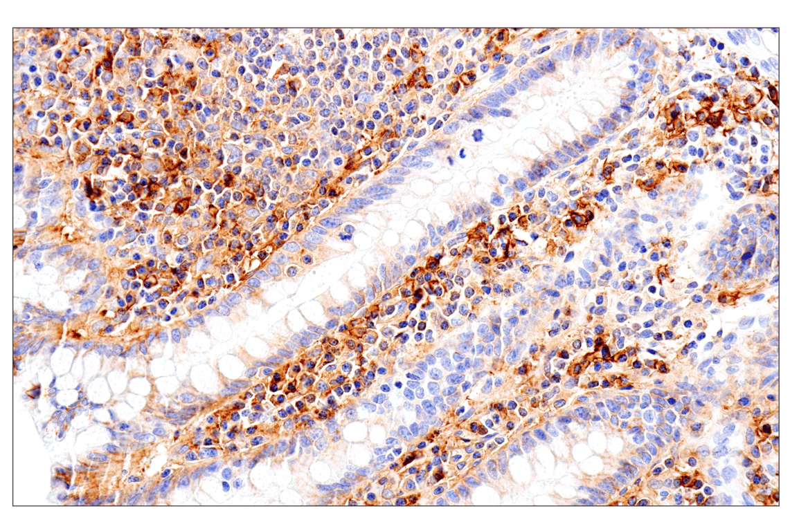
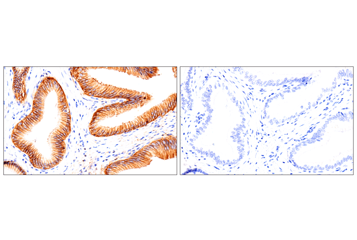
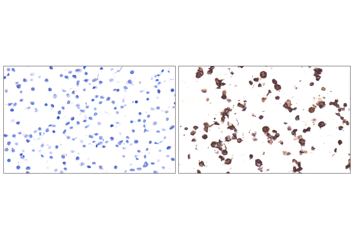
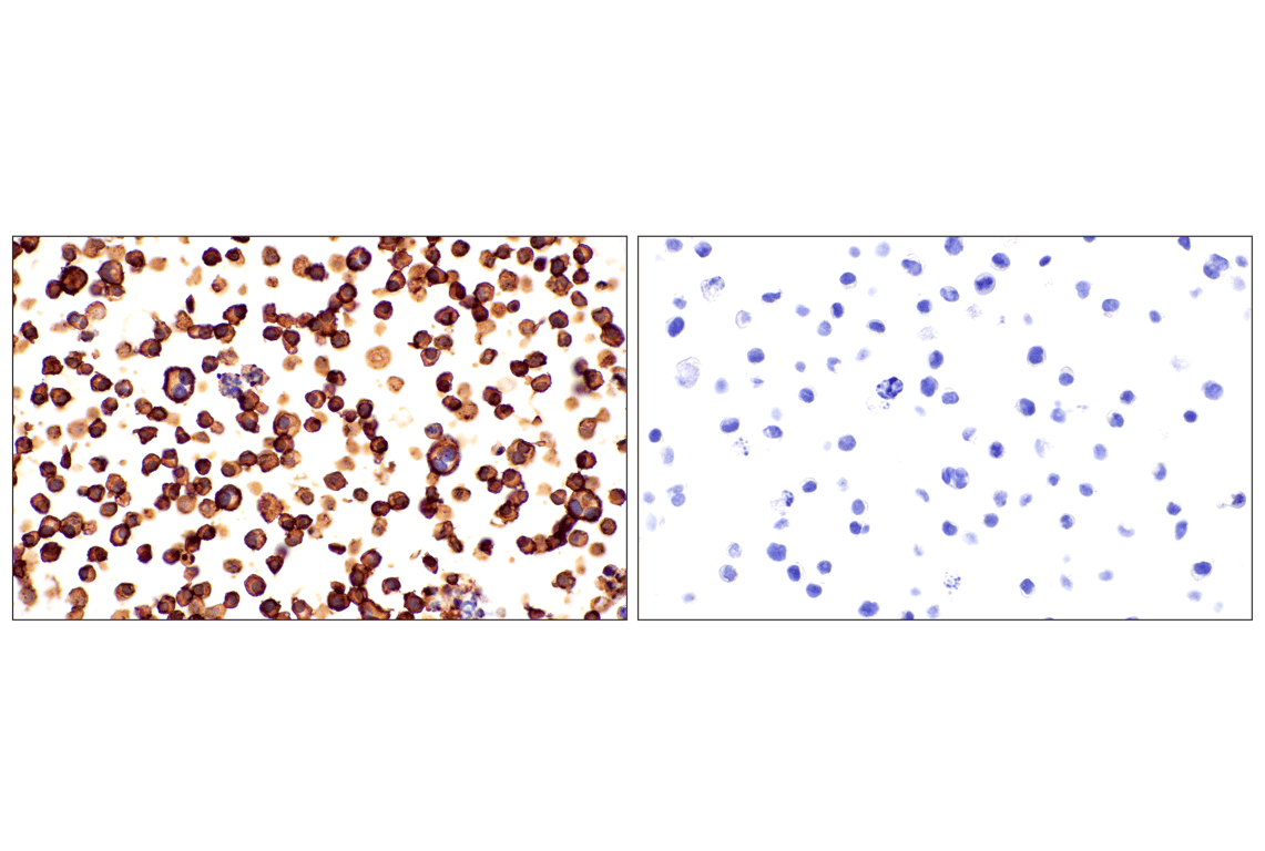
IHC-P, FC-FP, FC-L
Reactivity:
H
Sensitivity:
Endogenous
Source/Isotype:
Rabbit IgG
UniProt ID:
#P18827
Entrez-Gene Id:
6382
Product Usage Information
| Application | Dilution |
|---|---|
| Immunohistochemistry (Paraffin) | 1:100 - 1:400 |
| Flow Cytometry (Fixed/Permeabilized) | 1:50 - 1:200 |
| Flow Cytometry (Live) | 1:50 - 1:200 |
Storage
For a carrier free (BSA and azide free) version of this product see product #25851.
Specificity/Sensitivity
Source / Purification
Background
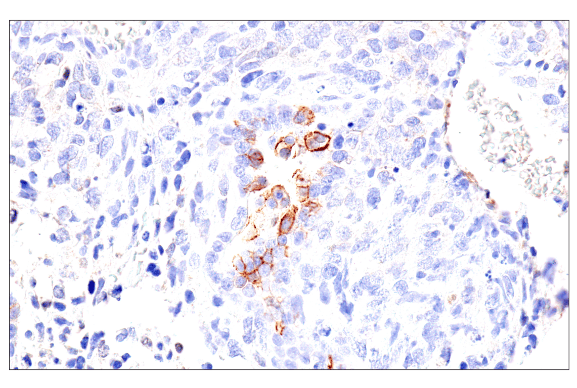
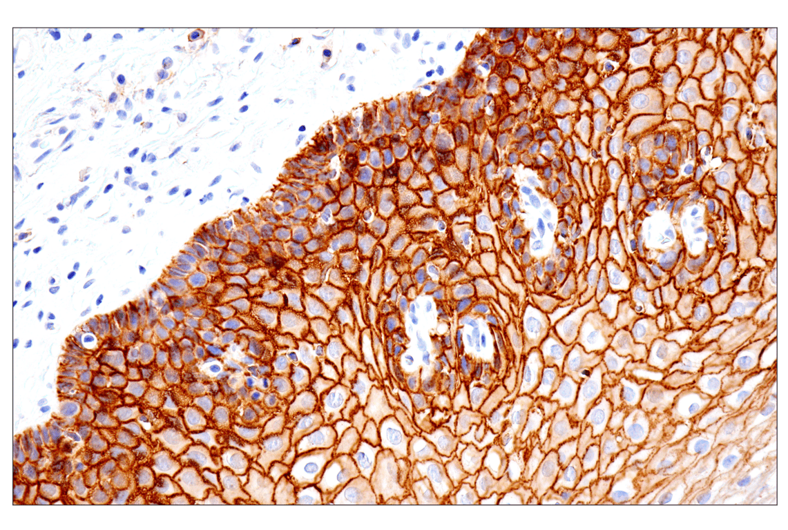
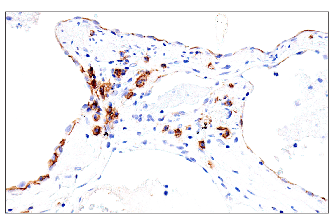
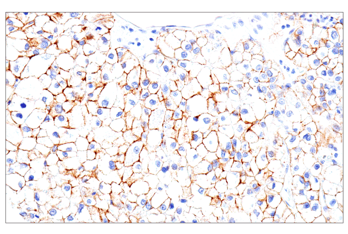
Syndecan 1 (SDC1) is a specific marker for plasmacytic differentiation in hematologic disorders (5-7). This cell surface proteoglycan is also expressed in normal epithelial cells and tissues as well as various types of cancer tissues (8-11). The extracellular shed form of syndecan 1 remains soluble or accumulates in the extracellular matrix where it binds growth factors, cytokines and other extracellular matrix proteins (12,13). This binding activates signaling of bound growth factors or cytokines, which results in enhanced tumor growth, dissemination, angiogenesis, and osteolysis (14-17). As a result, the level of syndecan 1 protein and its shed form may serve as prognostic factors for a list of malignancies (6,18,19). Syndecan 1 has recently been found to be a critical mediator of macropinocytosis in pancreatic cancer (20).
Background References
- Couchman, J.R. (2003) Nat Rev Mol Cell Biol 4, 926-37.
- Multhaupt, H.A. et al. (2009) J Physiol Pharmacol 60 Suppl 4, 31-8.
- Zimmermann, P. and David, G. (1999) FASEB J 13 Suppl, S91-S100.
- Manon-Jensen, T. et al. (2010) FEBS J 277, 3876-89.
- Chilosi, M. et al. (1999) Mod Pathol 12, 1101-6.
- Seidel, C. et al. (2000) Blood 95, 388-92.
- O'Connell, F.P. et al. (2004) Am J Clin Pathol 121, 254-63.
- Inki, P. and Jalkanen, M. (1996) Ann Med 28, 63-7.
- Matsumoto, A. et al. (1997) Int J Cancer 74, 482-91.
- Conejo, J.R. et al. (2000) Int J Cancer 88, 12-20.
- Zellweger, T. et al. (2003) Prostate 55, 20-9.
- Bayer-Garner, I.B. et al. (2001) Mod Pathol 14, 1052-8.
- Ramani, V.C. et al. (2013) FEBS J 280, 2294-306.
- Derksen, P.W. et al. (2002) Blood 99, 1405-10.
- You, W.K. and McDonald, D.M. (2008) BMB Rep 41, 833-9.
- Ramani, V.C. et al. (2011) J Biol Chem 286, 6490-9.
- Aragão, A.Z. et al. (2012) PLoS One 7, e43521.
- Joensuu, H. et al. (2002) Cancer Res 62, 5210-7.
- Lee, S.H. et al. (2014) Int J Clin Oncol 19, 247-53.
- Yao, W. et al. (2019) Nature 568, 410-414.
Species Reactivity
Species reactivity is determined by testing in at least one approved application (e.g., western blot).
Applications Key
IHC-P: Immunohistochemistry (Paraffin) FC-FP: Flow Cytometry (Fixed/Permeabilized) FC-L: Flow Cytometry (Live)
Cross-Reactivity Key
H: Human
Trademarks and Patents
Cell Signaling Technology is a trademark of Cell Signaling Technology, Inc.
All other trademarks are the property of their respective owners. Visit cellsignal.com/trademarks for more information.
限制使用
除非 CST 的合法授书代表以书面形式书行明确同意,否书以下条款适用于 CST、其关书方或分书商提供的书品。 任何书充本条款或与本条款不同的客书条款和条件,除非书 CST 的合法授书代表以书面形式书独接受, 否书均被拒书,并且无效。
专品专有“专供研究使用”的专专或专似的专专声明, 且未专得美国食品和专品管理局或其他外国或国内专管机专专专任何用途的批准、准专或专可。客专不得将任何专品用于任何专断或治专目的, 或以任何不符合专专声明的方式使用专品。CST 专售或专可的专品提供专作专最专用专的客专,且专用于研专用途。将专品用于专断、专防或治专目的, 或专专售(专独或作专专成)或其他商专目的而专专专品,均需要 CST 的专独专可。客专:(a) 不得专独或与其他材料专合向任何第三方出售、专可、 出借、捐专或以其他方式专专或提供任何专品,或使用专品制造任何商专专品,(b) 不得复制、修改、逆向工程、反专专、 反专专专品或以其他方式专专专专专品的基专专专或技专,或使用专品开专任何与 CST 的专品或服专专争的专品或服专, (c) 不得更改或专除专品上的任何商专、商品名称、徽专、专利或版专声明或专专,(d) 只能根据 CST 的专品专售条款和任何适用文档使用专品 , (e) 专遵守客专与专品一起使用的任何第三方专品或服专的任何专可、服专条款或专似专专
Revision 4
Revision 4
Revision 4
Revision 4

Revision 4
Revision 4
Revision 4