Revision 1
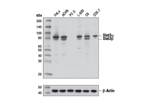
Western blot analysis of extracts from various cell lines using Stat3 (D3Z2G) Rabbit mAb (upper), or β-Actin (D6A8) Rabbit mAb #8457 (lower). PC-3 cells are negative for Stat3.
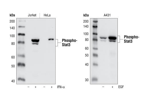
Western blot analysis of extracts from IFN-alpha treated Jurkat cells and HeLa cells (left), as well as EGF treated A431 cells (right), using Phospho-Stat3 (Tyr705) (D3A7) XP® Rabbit mAb. Note that the basal phospho-Stat3 in A431 is detected by the antibody.
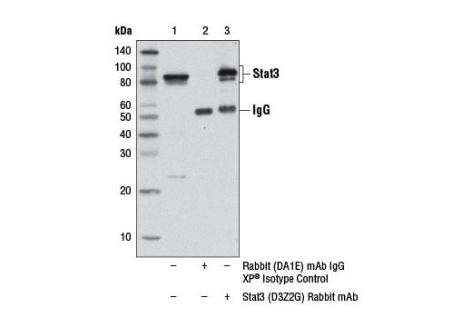
Immunoprecipitation of Stat3 from HeLa cell extracts using Rabbit (DA1E) mAb IgG XP® Isotype Control #3900 (lane 2) or Stat3 (D3Z2G) Rabbit mAb (lane 3). Lane 1 is 10% input.
Orders: 877-616-CELL (2355) • [email protected] • Support: 877-678-TECH (8324) • [email protected] •
Web:
cellsignal.com For Research Use Only. Not for Use in Diagnostic Procedures.
Revision 1
Immunoprecipitation of phospho-Stat3 (Tyr705) from U266 extracts treated with human IFN-α (50 ng/ml, 15 min). Lane 1 is 10% input, lane 2 is Rabbit (DA1E) mAb IgG XP® Isotype Control #3900, and lane 3 is Phospho-Stat3 (Tyr705) (D3A7) XP® Rabbit mAb. Western blot analysis was performed using Phospho-Stat3 (Tyr705) (3E2) Mouse mAb #9138. Anti-mouse IgG, HRP-linked Antibody #7076 was used as a secondary antibody.
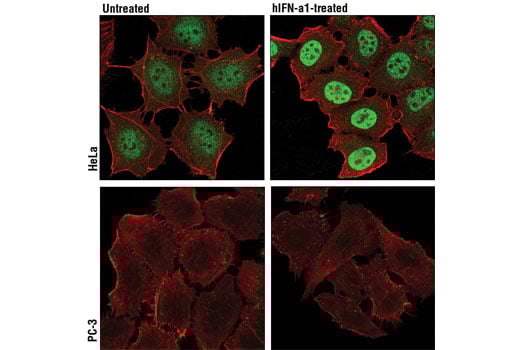
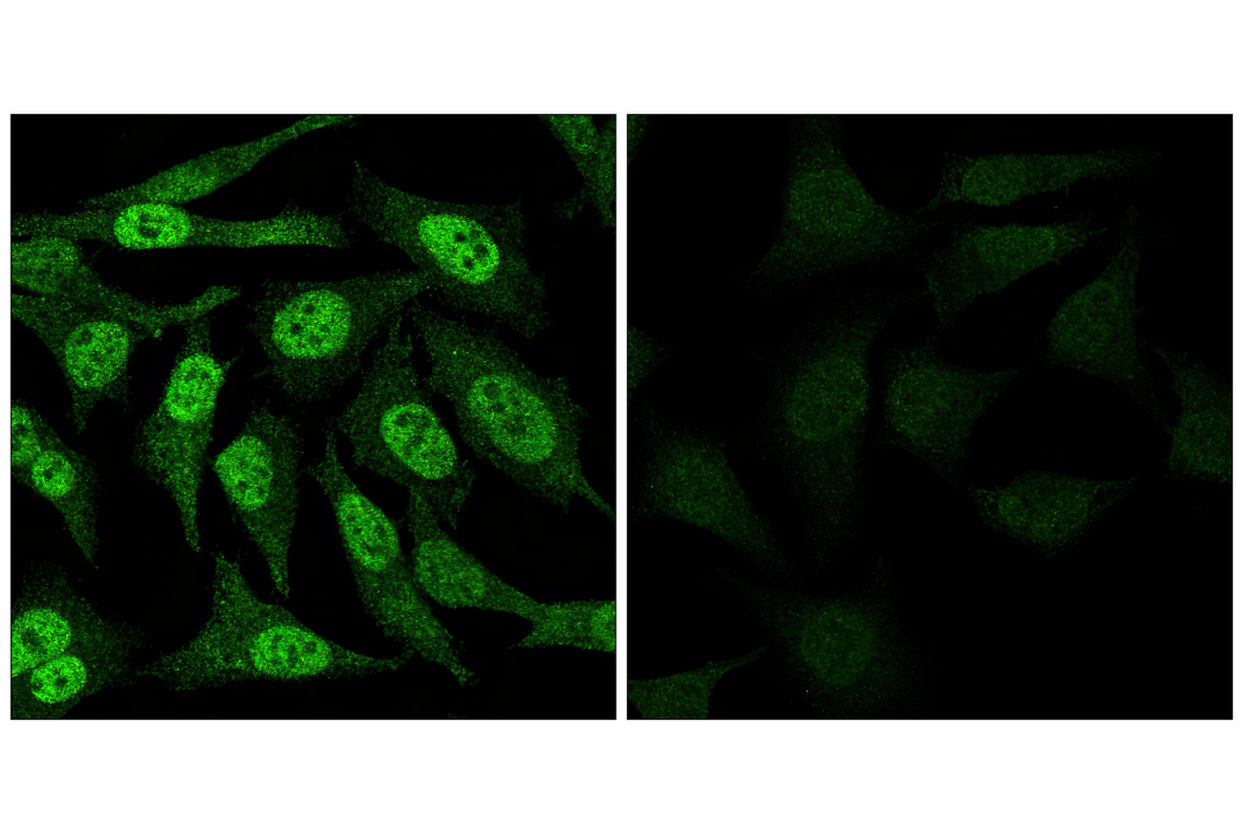
Confocal immunofluorescent analysis of serum-starved HeLa cells, untreated (upper left) or treated with Human Interferon-α1 (hIFN-α1) #8927 (100 ng/ml, 30 min; upper right), and serum-starved PC-3 cells, untreated (lower right) or treated with Human Interferon-α1 (hIFN-α1) #8927 (100 ng/ml, 30 min; lower right), using Stat3 (D3Z2G) Rabbit mAb (green) and β-Actin (8H10D10) Mouse mAb #3700 (red).
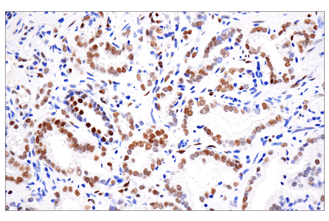
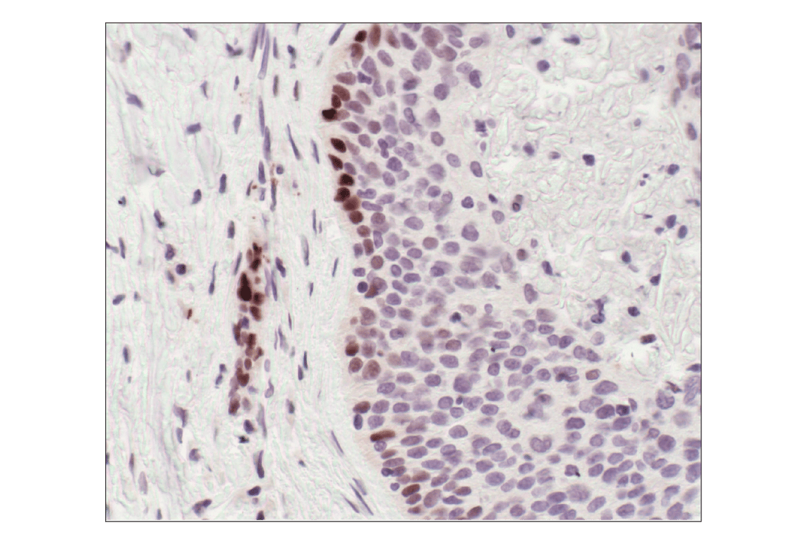
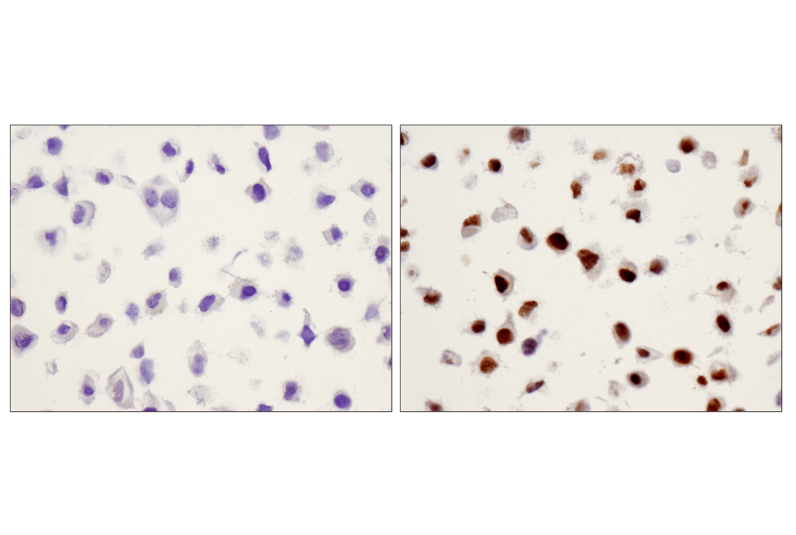
Immunohistochemical analysis of paraffin-embedded human prostate adenocarcinoma using Phospho-Stat3 (Tyr705) (D3A7) XP® Rabbit mAb performed on the Leica® BOND™ Rx.
Orders: 877-616-CELL (2355) • [email protected] • Support: 877-678-TECH (8324) • [email protected] •
Web:
cellsignal.com For Research Use Only. Not for Use in Diagnostic Procedures.
Revision 1
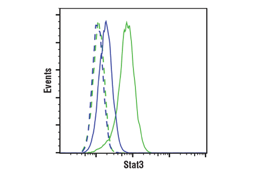
Flow cytometric analysis of PC-3 cells (blue) and HeLa cells (green) using Stat3 (D3Z2G) Rabbit mAb or a concentration-matched Rabbit (DA1E) mAb IgG XP® Isotype Control #3900 (dashed lines). Anti-rabbit IgG (H+L), F(ab')2 Fragment (Alexa Fluor® 488 Conjugate) was used as a secondary antibody.
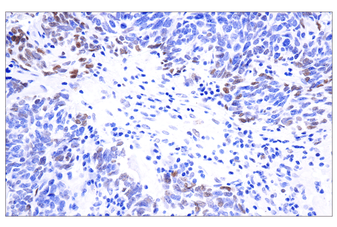
Immunohistochemical analysis of paraffin-embedded human neuroendocrine lung carcinoma using Phospho-Stat3 (Tyr705) (D3A7) XP® Rabbit mAb performed on the Leica® BOND™ Rx.
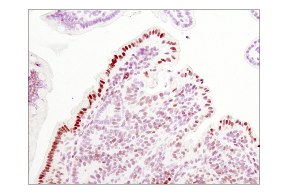
Immunohistochemical analysis of paraffin-embedded Apc (min/+) mouse intestine, using Phosho-Stat3 (Tyr705) (D3A7) XP® Rabbit mAb.
Orders: 877-616-CELL (2355) • [email protected] • Support: 877-678-TECH (8324) • [email protected] •
Web:
cellsignal.com For Research Use Only. Not for Use in Diagnostic Procedures.
Revision 1
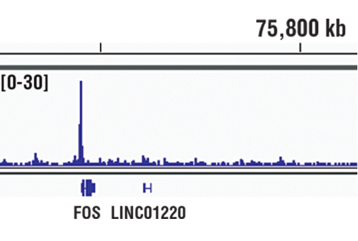
Chromatin immunoprecipitations were performed with cross-linked chromatin from Hep G2 cells starved overnight and treated with Human Interleukin-6 (hIL-6) #8904 (100 ng/ml) for 30 minutes and Stat3 (D3Z2G) Rabbit mAb, using SimpleChIP® Plus Enzymatic Chromatin IP Kit (Magnetic Beads) #9005. DNA Libraries were prepared using DNA Library Prep Kit for Illumina® (ChIP-seq, CUT&RUN) #56795. The figure shows binding across Fos, a known target gene of Stat3 (see additional figure containing ChIP-qPCR data). For additional ChIP-seq tracks, please download the product datasheet.
Immunnohistochemical analysis of paraffin-embedded human lung carcinoma, showing nuclear localization, using Phospho-Stat3 (Tyr705) (D3A7) XP® Rabbit mAb.
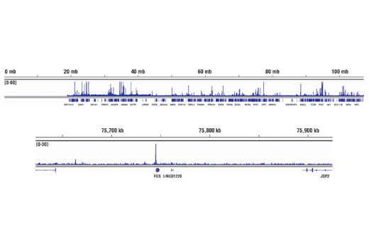
Chromatin immunoprecipitations were performed with cross-linked chromatin from Hep G2 cells starved overnight and treated with Human Interleukin-6 (hIL-6) #8904 (100 ng/ml) for 30 minutes and Stat3 (D3Z2G) Rabbit mAb, using SimpleChIP® Plus Enzymatic Chromatin IP Kit (Magnetic Beads) #9005. DNA Libraries were prepared using DNA Library Prep Kit for Illumina® (ChIP-seq, CUT&RUN) #56795. The figure shows binding across chromosome 14 (upper), including Fos (lower), a known target gene of Stat3 (see additional figure containing ChIP-qPCR data).
Orders: 877-616-CELL (2355) • [email protected] • Support: 877-678-TECH (8324) • [email protected] •
Web:
cellsignal.com For Research Use Only. Not for Use in Diagnostic Procedures.
Revision 1
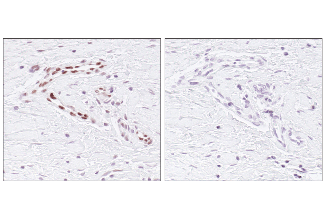
Immunohistochemical analysis of paraffin embedded human breast carcinoma, specifically endothelial cells, untreated (left) or lambda phosphatase treated (right), using Phospho-Stat3 (Tyr705) (D3A7) XP® Rabbit mAb.
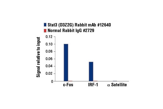
Chromatin immunoprecipitations were performed with cross-linked chromatin from Hep G2 cells starved overnight and treated with Human Interleukin-6 (hIL-6) #8904 (100 ng/ml) for 30 minutes, and either Stat3 (D3Z2G) Rabbit mAb or Normal Rabbit IgG #2729 using SimpleChIP® Enzymatic Chromatin IP Kit (Magnetic Beads) #9003. The enriched DNA was quantified by real-time PCR using human IRF-1 promoter primers, SimpleChIP® Human c-Fos Promoter Primers #4663, and SimpleChIP® Human α Satellite Repeat Primers #4486. The amount of immunoprecipitated DNA in each sample is represented as signal relative to the total amount of input chromatin, which is equivalent to one.
Immunohistochemical analysis using Phospho-Stat3 (Tyr705) (D3A7) XP® Rabbit mAb on SignalSlide® HeLa -/+ IFNa IHC Controls #55861 (paraffin-embedded HeLa cell pellets, untreated (left) or treated with Human Interferon-α1 (hIFN-α1) #8927 (right)).
Orders: 877-616-CELL (2355) • [email protected] • Support: 877-678-TECH (8324) • [email protected] •
Web:
cellsignal.com For Research Use Only. Not for Use in Diagnostic Procedures.
Revision 1
Confocal immunofluorescent analysis of HeLa cells, IFN-alpha treated (left) or untreated (right), labeled with Phospho-Stat3 (Tyr705) (D3A7) XP® Rabbit mAb (green).
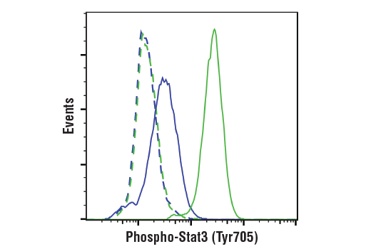
Flow cytometric analysis of U266 cells, untreated (blue) or treated with IFNalpha (50 ng/ml, 15 min; green) using Phospho-Stat3 (Tyr705) (D3A7) XP® Rabbit mAb (solid lines) or concentration matched Rabbit (DA1E) mAb IgG XP® Isotype Control #3900 (dashed lines). Anti-rabbit IgG (H+L), F(ab')2 Fragment (Alexa Fluor® 488 Conjugate) #4412 was used as a secondary antibody.
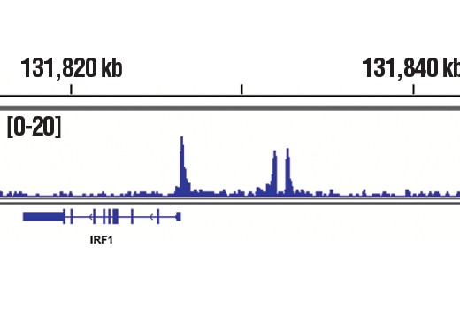
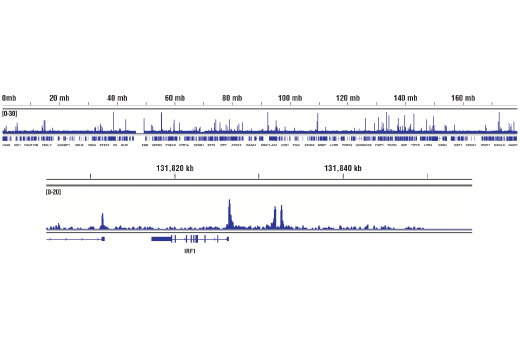
Chromatin immunoprecipitations were performed with cross-linked chromatin from Hep G2 cells starved overnight and treated with IL-6 (100 ng/ml) for 30 minutes and Phospho-Stat3 (Tyr705) (D3A7) XP® Rabbit mAb, using SimpleChIP® Plus Enzymatic Chromatin IP Kit (Magnetic Beads) #9005. DNA Libraries were prepared using DNA Library Prep Kit for Illumina® (ChIP-seq, CUT&RUN) #56795. The figure shows binding across IRF1, a known target gene of Phospho-Sata3 (see additional figure containing ChIP-qPCR data). For additional ChIP-seq tracks, please download the product datasheet.
Orders: 877-616-CELL (2355) • [email protected] • Support: 877-678-TECH (8324) • [email protected] •
Web:
cellsignal.com For Research Use Only. Not for Use in Diagnostic Procedures.
Revision 1
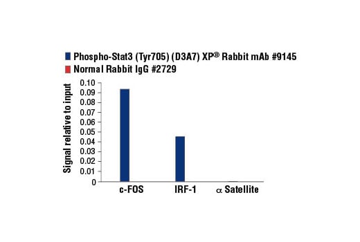
Chromatin immunoprecipitations were performed with cross-linked chromatin from HaCaT cells Hep G2 cells starved overnight and treated with IL-6 (100 ng/ml) for 30 minutes and either Phospho-Stat3 (Tyr705) (D3A7) XP® Rabbit mAb or Normal Rabbit IgG #2729 using SimpleChIP® Plus Sonication Chromatin IP Kit #56383. The enriched DNA was quantified by real-time PCR using SimpleChIP® Human c-Fos Promoter Primers #4663, human IRF1 promoter primers, and SimpleChIP® Human α Satellite Repeat Primers #4486. The amount of immunoprecipitated DNA in each sample is represented as signal relative to the total amount of input chromatin, which is equivalent to one.
Chromatin immunoprecipitations were performed with cross-linked chromatin from Hep G2 cells starved overnight and treated with IL-6 (100 ng/ml) for 30 minutes, and either Phospho-Stat3 (Tyr705) (D3A7) XP® Rabbit mAb or of Normal Rabbit IgG #2729 using SimpleChIP® Enzymatic Chromatin IP Kit (Magnetic Beads) #9003. The enriched DNA was quantified by real-time PCR using human IRF-1 promoter primers, SimpleChIP® Human c-Fos Promoter Primers #4663, and SimpleChIP® Human α Satellite Repeat Primers #4486. The amount of immunoprecipitated DNA in each sample is represented as signal relative to the total amount of input chromatin, which is equivalent to one.
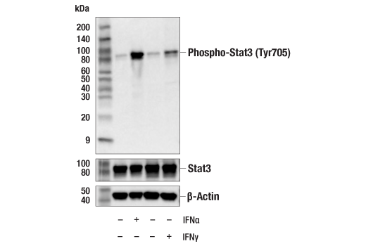
Western blot analysis of extracts from HeLa cells, untreated or treated with IFNa (#36000, 100 ng/mL, 5 min) or IFNg (#80385, 100 ng/mL, 30 min); using Phospho-Stat3 (Tyr705) (D3A7) XP® Rabbit mAb #9145 (upper), Stat3 (D3Z2G) Rabbit mAb #12640 (middle), and β-Actin (D6A8) Rabbit mAb #8457 (lower).
Orders: 877-616-CELL (2355) • [email protected] • Support: 877-678-TECH (8324) • [email protected] •
Web:
cellsignal.com For Research Use Only. Not for Use in Diagnostic Procedures.
Revision 1
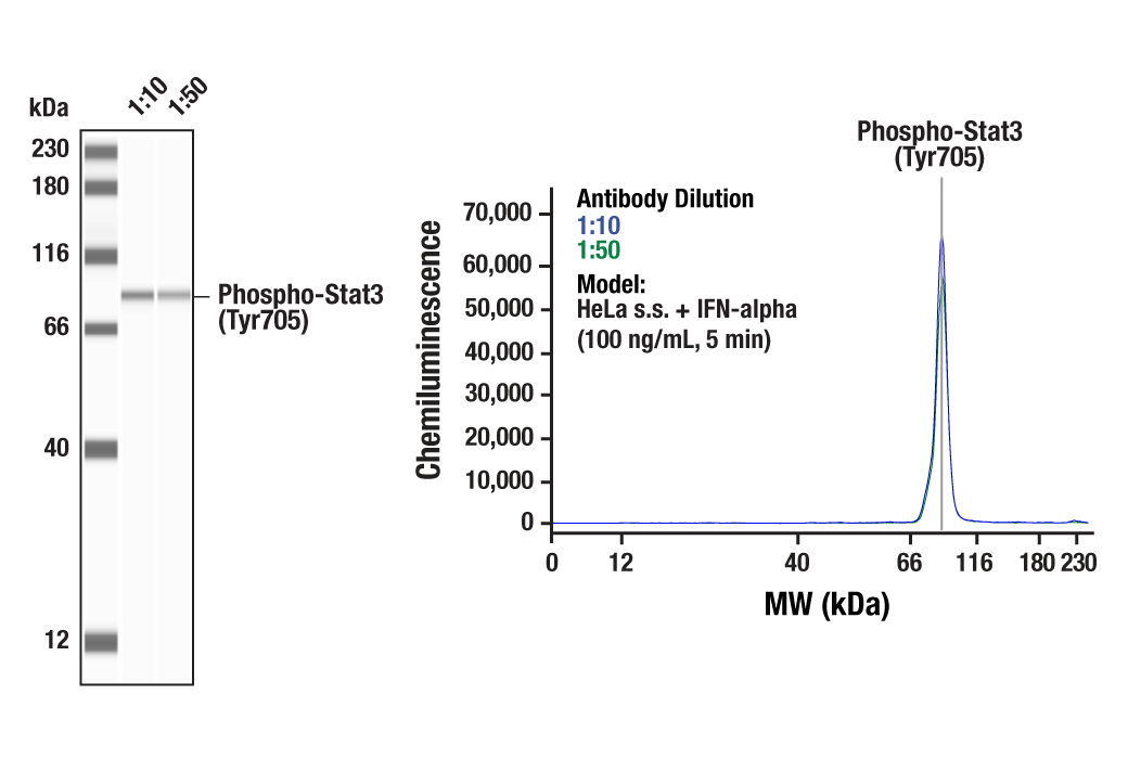
Simple Western™ analysis of lysates (0.1 mg/mL) from serum-starved HeLa cells treated with IFN-alpha (100 ng/mL, 5 min) using Phospho-Stat3 (Tyr705) (D3A7) XP® Rabbit mAb #9145. The virtual lane view (left) shows a single target band (as indicated) at 1:10 and 1:50 dilutions of primary antibody. The corresponding electropherogram view (right) plots chemiluminescence by molecular weight along the capillary at 1:10 (blue line) and 1:50 (green line) dilutions of primary antibody. This experiment was performed under reducing conditions on the Jess™ Simple Western instrument from ProteinSimple, a BioTechne brand, using the 12-230 kDa separation module.
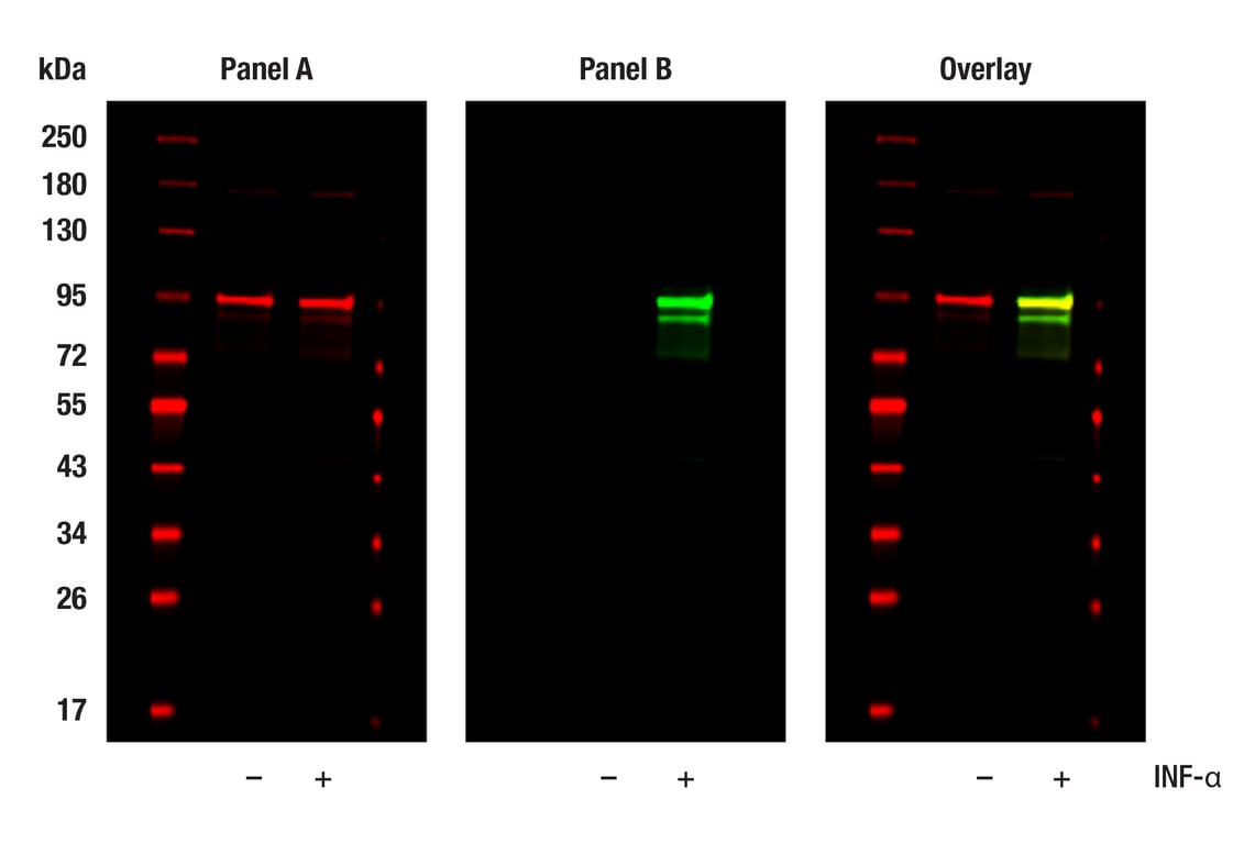
Western blot analysis of extracts from HeLa cells, untreated (-) or treated with INF-⍶ (100 ng/ml, 5 min; +), using Stat3 (124H6) Mouse mAb #9139 (Panel A) and Phospho-Stat3 (Tyr705) (D3A7) XP® Rabbit mAb #9145 (Panel B). Anti-mouse IgG (H+L) (DyLight 680 Conjugate) #5470 (red) and Anti-rabbit IgG (H+L) (DyLight 800 4X PEG Conjugate) #5151 (green) were used as secondary antibodies.
Orders: 877-616-CELL (2355) • [email protected] • Support: 877-678-TECH (8324) • [email protected] •
Web:
cellsignal.com For Research Use Only. Not for Use in Diagnostic Procedures.