Revision 1
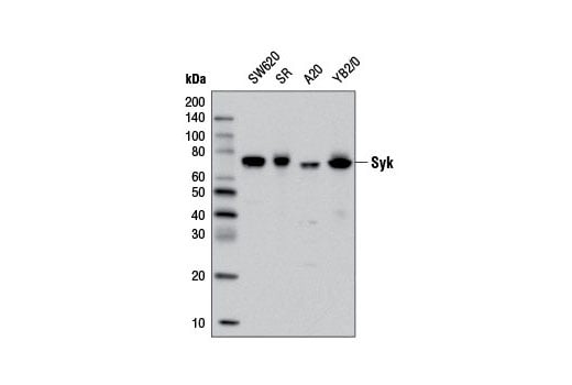
Western blot analysis of extracts from various cell lines using Syk (D3Z1E) XP® Rabbit mAb.
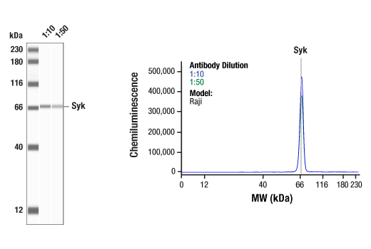
Simple Western™ analysis of lysates (0.1 mg/mL) from Raji cells using Syk (D3Z1E) XP® Rabbit mAb #13198. The virtual lane view (left) shows the target band (as indicated) at 1:10 and 1:50 dilutions of primary antibody. The corresponding electropherogram view (right) plots chemiluminescence by molecular weight along the capillary at 1:10 (blue line) and 1:50 (green line) dilutions of primary antibody. This experiment was performed under reducing conditions on the Jess™ Simple Western instrument from ProteinSimple, a BioTechne brand, using the 12-230 kDa separation module.
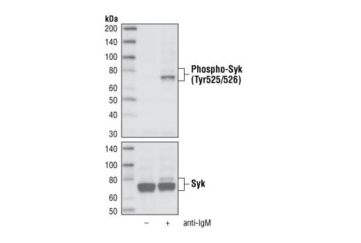
Western blot analysis of extracts from Ramos cells, untreated or treated with anti-IgM, using Phospho-Syk (Tyr525/526) (C87C1) Rabbit mAb (upper) or Syk Antibody #2712 (lower).
Orders: 877-616-CELL (2355) • [email protected] • Support: 877-678-TECH (8324) • [email protected] •
Web:
cellsignal.com For Research Use Only. Not for Use in Diagnostic Procedures.
Revision 1
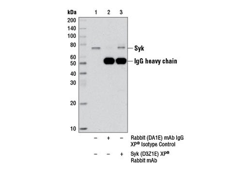
Immunoprecipitation of Syk protein from SR cell extracts, using Rabbit (DA1E) mAb IgG XP® Isotype Control #3900 (lane 2) or Syk(D3Z1E) XP® Rabbit mAb (lane 3). Lane 1 is 10% input. Western blot analysis was performed using Syk (D3Z1E) XP® Rabbit mAb
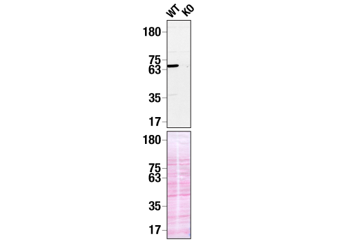
Western blot analysis of extracts from THP-1 WT (left) or SYK KO (right) using Syk (D3Z1E) XP® Rabbit mAb (upper). Membranes stained with Ponceau S for total protein normalization (lower). These data were provided by YCharOS Inc., an open science company with the mission of characterizing commercially available antibodies, as a companion to validation data generated by CST scientists.
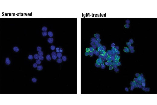
Confocal immunofluorescent analysis of Ramos cells, serum-starved (overnight; left) or IgM-treated (12 ug/ml, 2 minutes; right), using Phospho-Syk (Tyr525/526) (C87C1) Rabbit mAb (green). Blue pseudocolor = DRAQ5® #4084 (fluorescent DNA dye).
Orders: 877-616-CELL (2355) • [email protected] • Support: 877-678-TECH (8324) • [email protected] •
Web:
cellsignal.com For Research Use Only. Not for Use in Diagnostic Procedures.
Revision 1
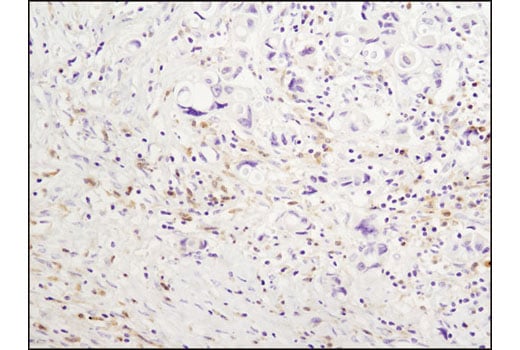
Immunohistochemical analysis of paraffin-embedded human breast carcinoma using Syk (D3Z1E) XP® Rabbit mAb.
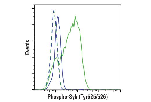
Flow cytometric analysis of Ramos cells, untreated (blue) or treated with anti-IgM (green), using Phospho-Syk(Tyr525/526) (C87C1) Rabbit mAb, or a concentration-matched Rabbit (DA1E) mAb IgG XP® Isotype Control #3900 (dashed lines). Anti-rabbit IgG (H+L), F(ab')2 Fragment (PE Conjugate) #8885 was used as a secondary antibody.
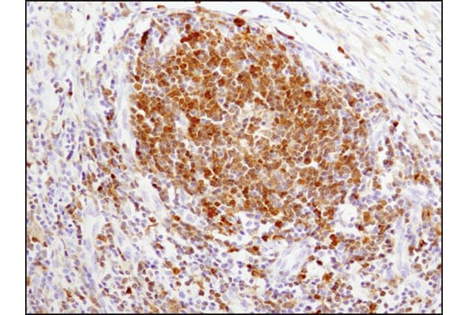
Immunohistochemical analysis of paraffin-embedded human lymph node using Syk (D3Z1E) XP® Rabbit mAb.
Orders: 877-616-CELL (2355) • [email protected] • Support: 877-678-TECH (8324) • [email protected] •
Web:
cellsignal.com For Research Use Only. Not for Use in Diagnostic Procedures.
Revision 1
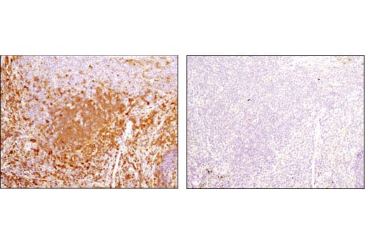
Immunohistochemical analysis of paraffin-embedded mouse spleen using Syk (D3Z1E) XP® Rabbit mAb in the presence of control peptide (left) or antigen-specific peptide (right).
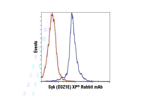
Flow cytometric analysis of RL cells using Syk (D3Z1E) XP® Rabbit mAb (blue) compared to Rabbit (DA1E) mAb IgG XP® Isotype Control #3900 (red). Anti-rabbit IgG (H+L), F(ab')2 Fragment (Alexa Fluor® 647 Conjugate) #4414 was used as a secondary antibody.
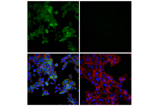
Confocal immunofluorescent analysis of SW620 cells (left, positive) and ACHN cells (right, negative) using Syk (D3Z1E) XP® Rabbit mAb (green), DyLight™ 650 Phalloidin #12956 (red), and DAPI #4083 (blue).
Orders: 877-616-CELL (2355) • [email protected] • Support: 877-678-TECH (8324) • [email protected] •
Web:
cellsignal.com For Research Use Only. Not for Use in Diagnostic Procedures.
Revision 1
Simple Western™ analysis of lysates (0.1 mg/mL) from Ramos cells treated with Anti-Human IgM (12 ug/mL, 10”) using Phospho-Syk (Tyr525/526) (C87C1) Rabbit mAb #2710. The virtual lane view (left) shows the target band (as indicated) at 1:10 and 1:50 dilutions of primary antibody. The corresponding electropherogram view (right) plots chemiluminescence by molecular weight along the capillary at 1:10 (blue line) and 1:50 (green line) dilutions of primary antibody. This experiment was performed under reducing conditions on the Jess™ Simple Western instrument from ProteinSimple, a BioTechne brand, using the 12-230 kDa separation module.
Orders: 877-616-CELL (2355) • [email protected] • Support: 877-678-TECH (8324) • [email protected] •
Web:
cellsignal.com For Research Use Only. Not for Use in Diagnostic Procedures.