WB, W-S, IP, IHC-P, IF-IC, FC-FP
H M R Mk Hm Dm Z B
Endogenous
60
Rabbit IgG
#P31751, #Q9Y243, #P31749
208, 10000, 207
Product Information
Product Usage Information
| Application | Dilution |
|---|---|
| Western Blotting | 1:2000 |
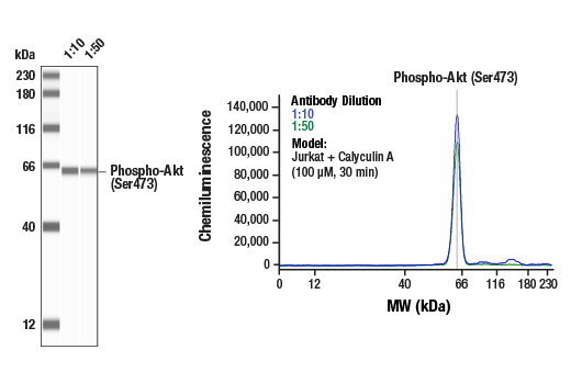
| Simple Western™ | 1:10 - 1:50 |
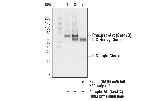
| Immunoprecipitation | 1:50 |
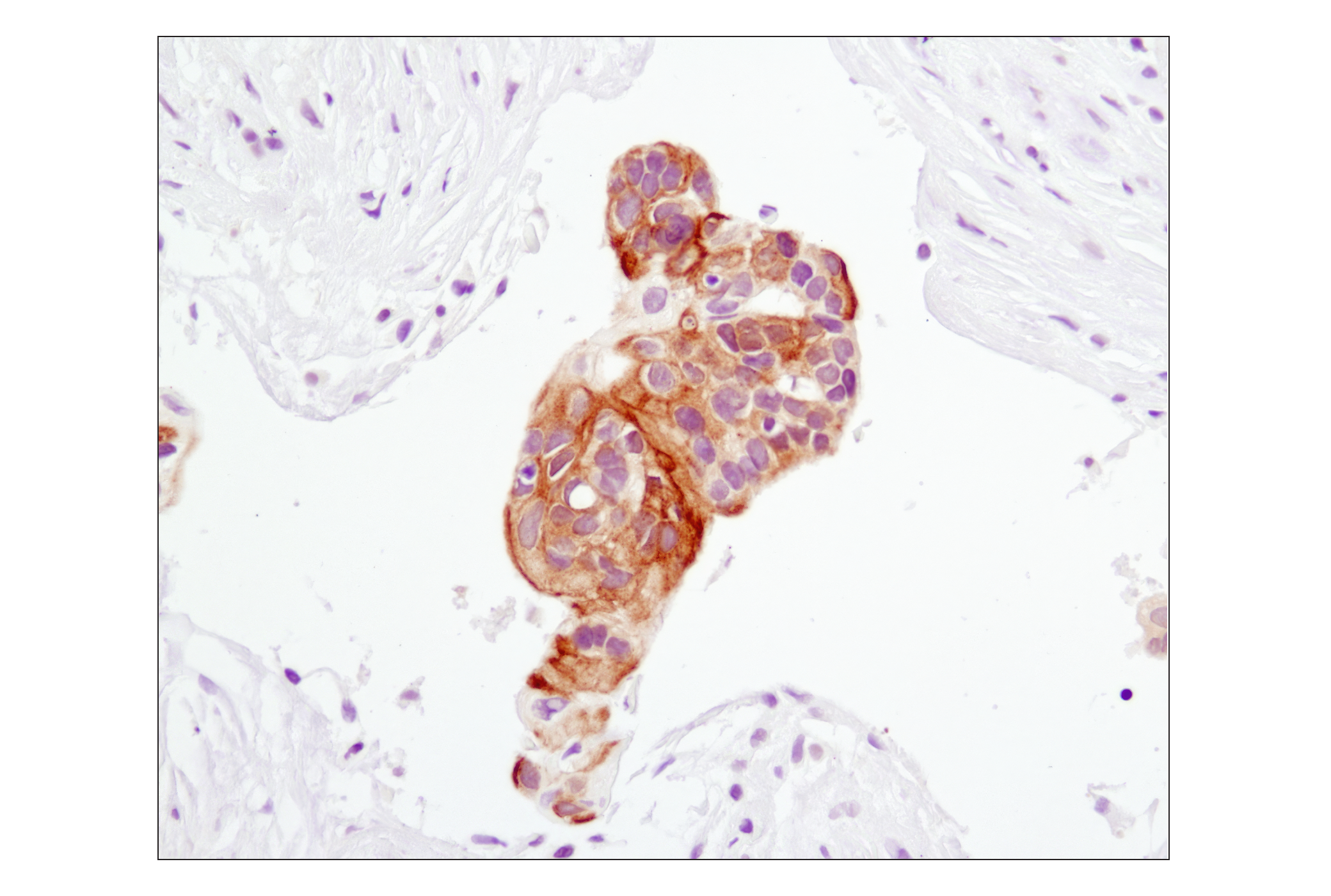
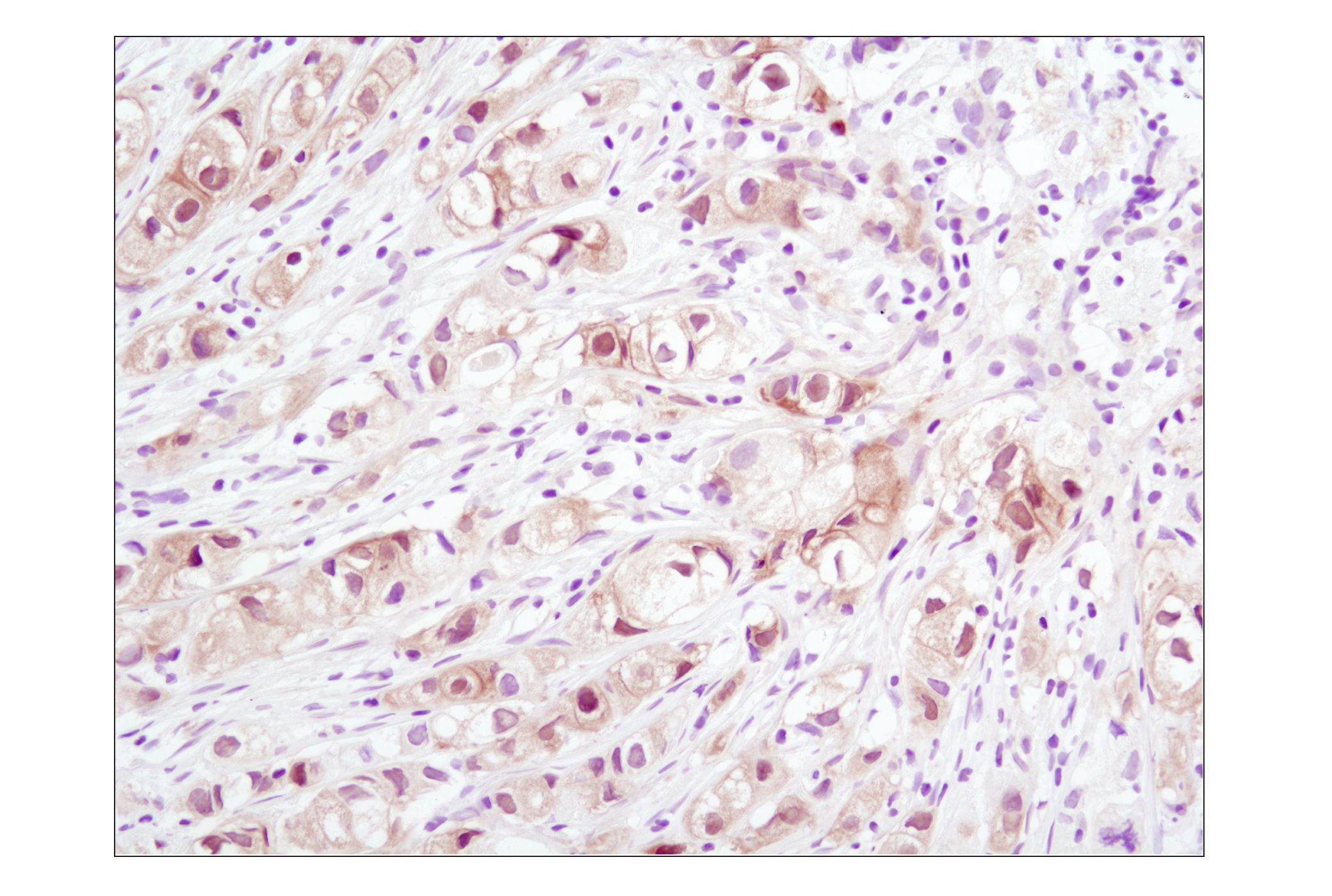
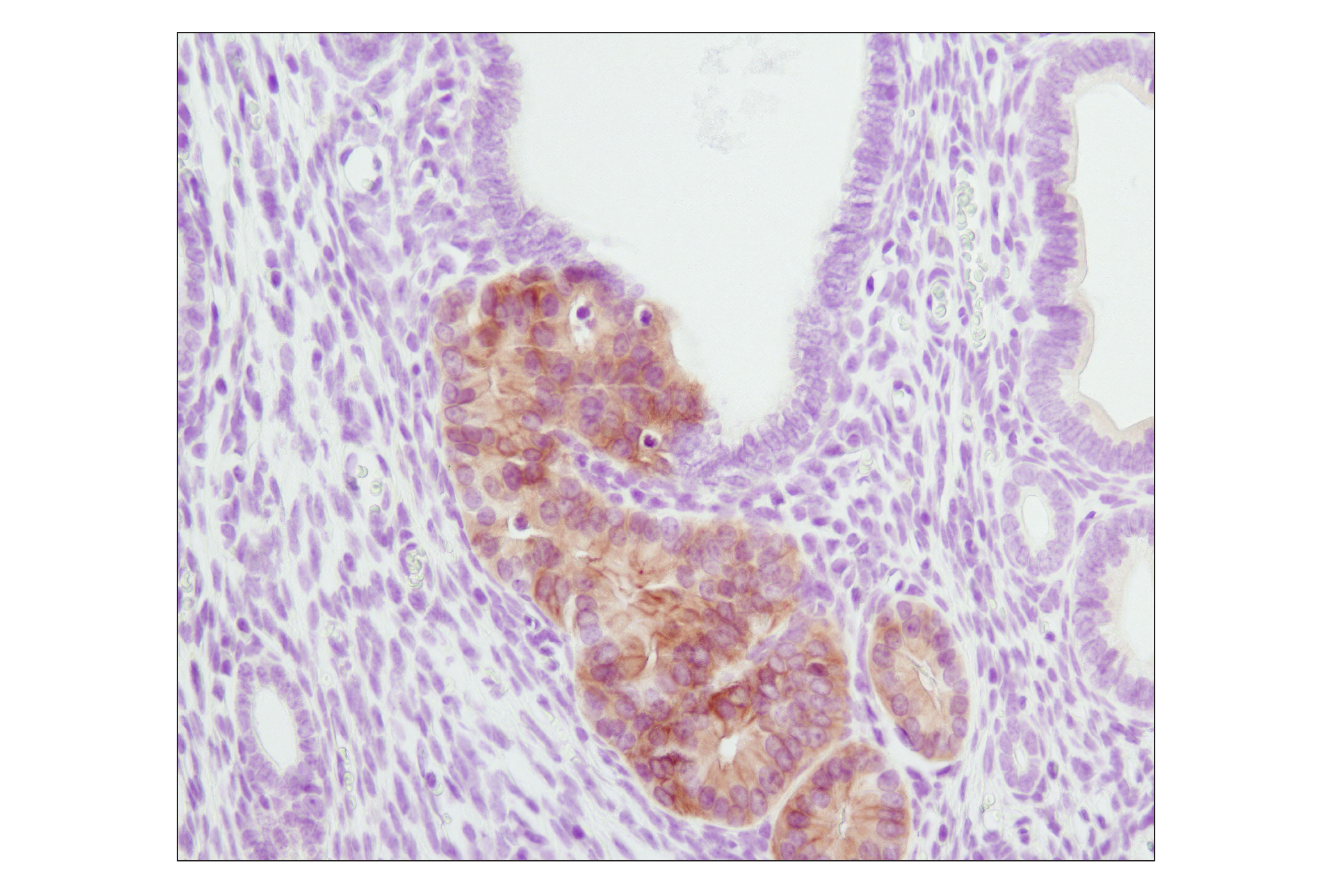
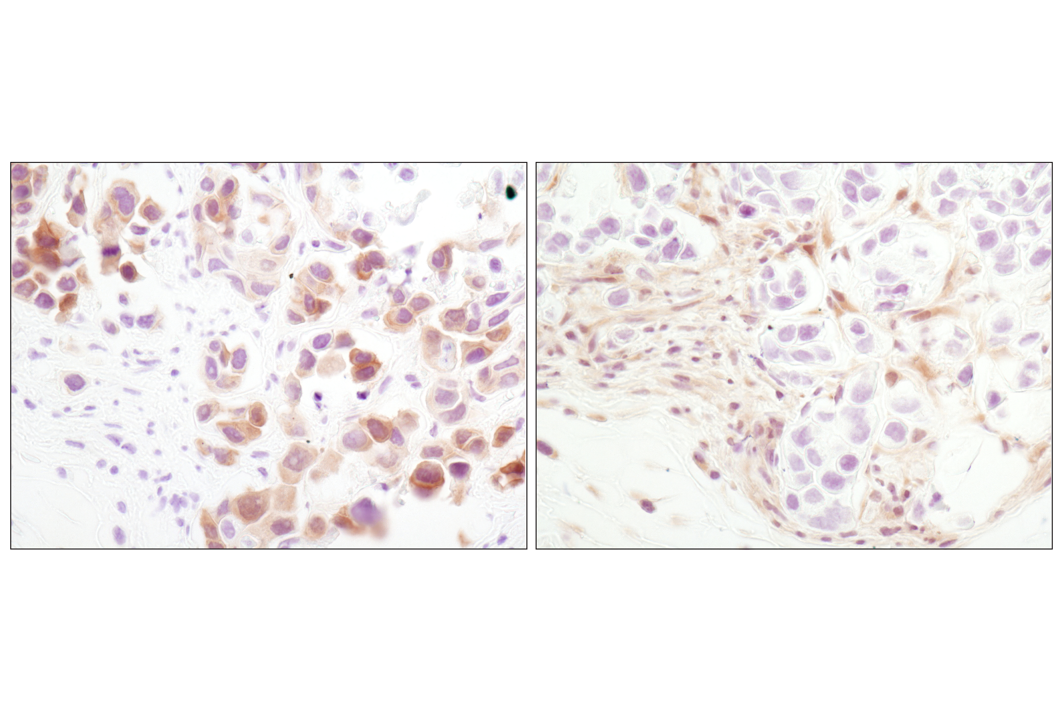
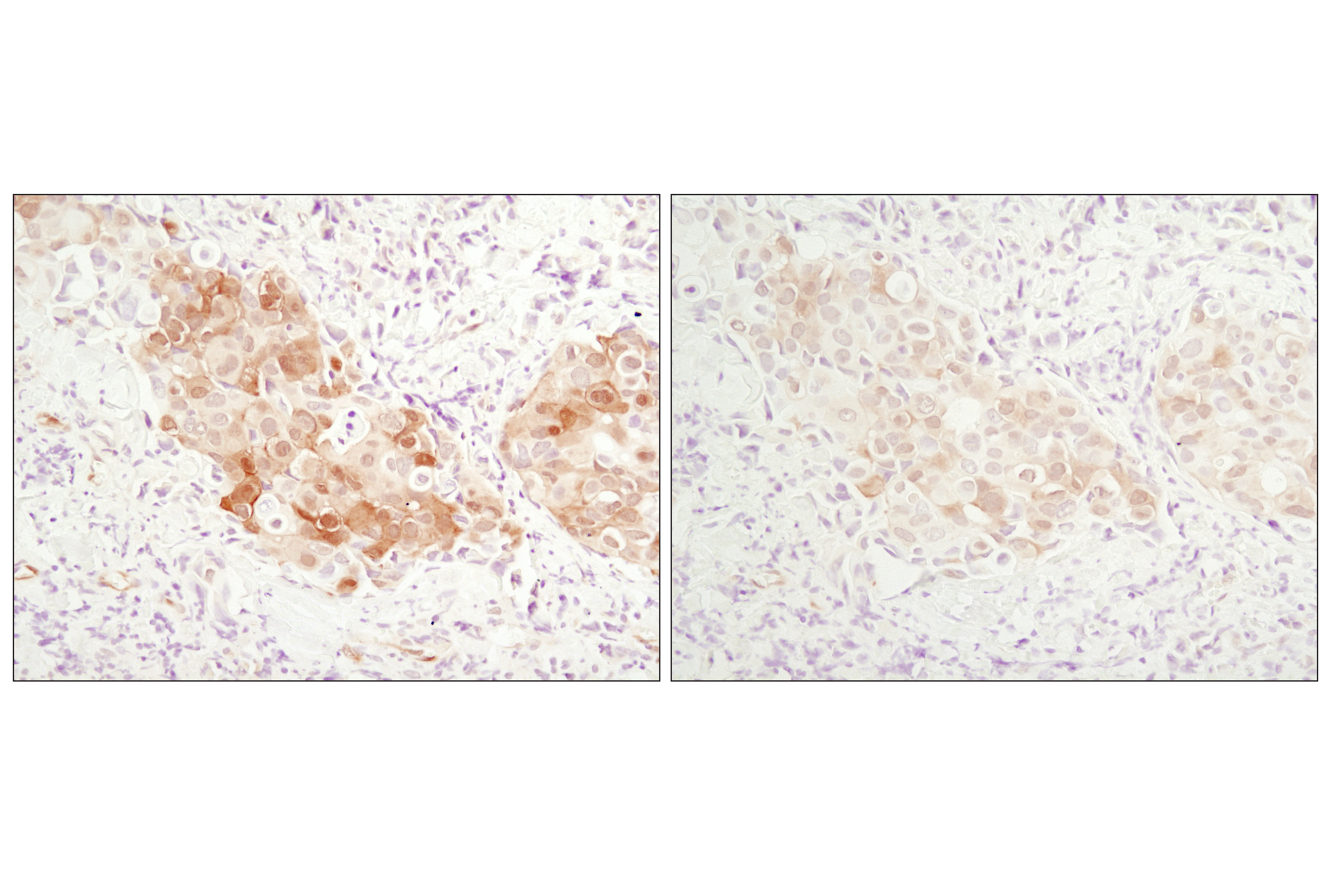
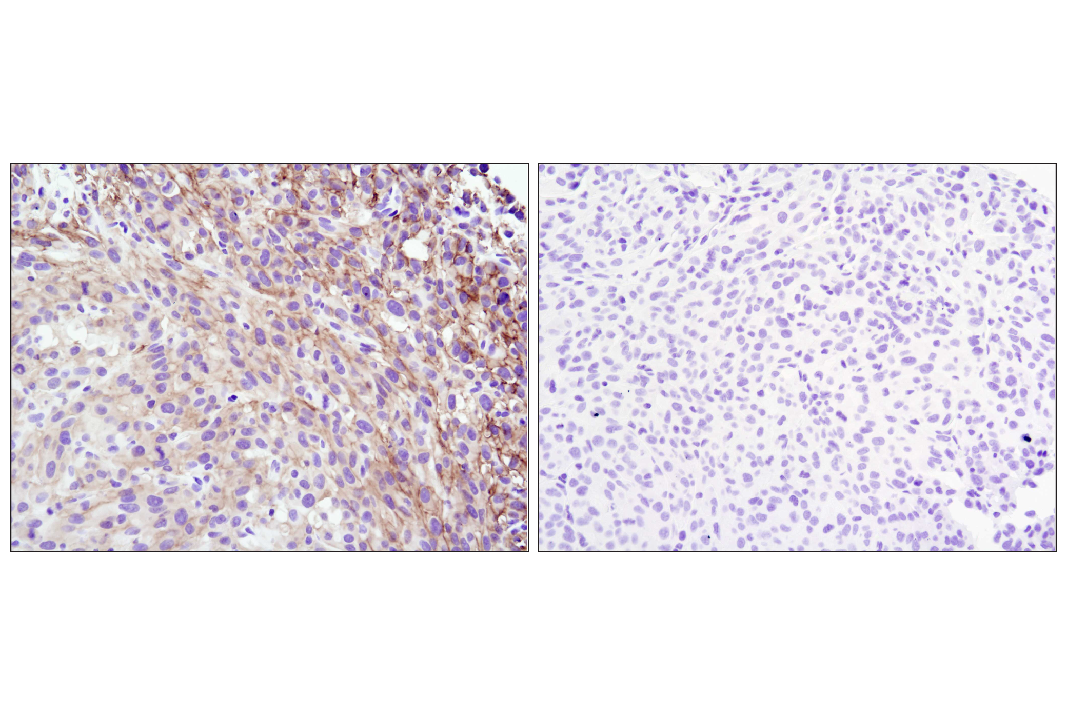
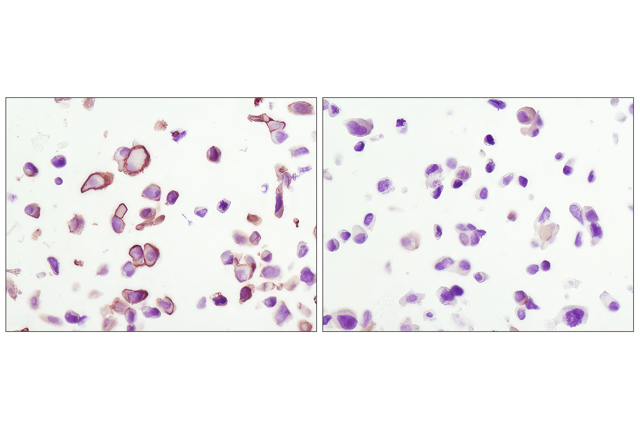
| Immunohistochemistry (Paraffin) | 1:50 - 1:200 |
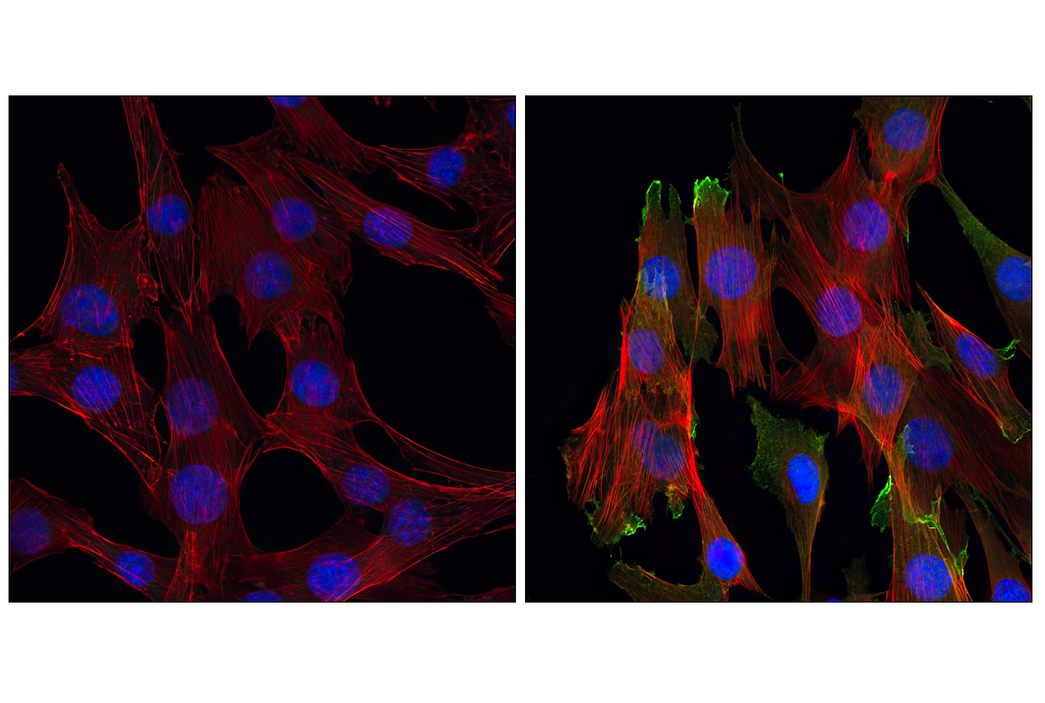
| Immunofluorescence (Immunocytochemistry) | 1:400 - 1:800 |
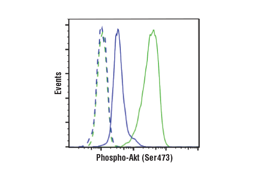
| Flow Cytometry (Fixed/Permeabilized) | 1:100 - 1:400 |
Storage
Supplied in 10 mM sodium HEPES (pH 7.5), 150 mM NaCl, 100 µg/ml BSA, 50% glycerol and less than 0.02% sodium azide. Store at –20°C. Do not aliquot the antibody.
For a carrier-free (BSA and azide free) version of this product see product #31957.
Specificity / Sensitivity
Species Reactivity:
Human, Mouse, Rat, Monkey, Hamster, D. melanogaster, Zebrafish, Bovine
Species predicted to react based on 100% sequence homology
The antigen sequence used to produce this antibody shares
100% sequence homology with the species listed here, but
reactivity has not been tested or confirmed to work by CST.
Use of this product with these species is not covered under
our
Product Performance Guarantee.
Chicken, Xenopus, Dog, Pig
Source / Purification
Monoclonal antibody is produced by immunizing animals with a synthetic phosphopeptide corresponding to residues around Ser473 of human Akt.
Background
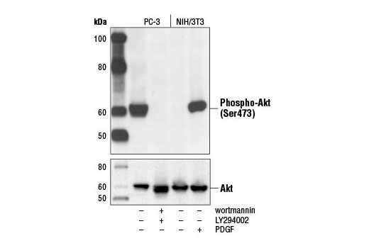
Akt, also referred to as PKB or Rac, plays a critical role in controlling cell survival and apoptosis (1-3). This protein kinase is activated by insulin and various growth and survival factors to function in a wortmannin-sensitive pathway involving PI3 kinase (2,3). Akt is activated by phospholipid binding and activation loop phosphorylation at Thr308 by PDK1 (4) and by phosphorylation within the carboxy terminus at Ser473. The previously elusive PDK2 responsible for phosphorylation of Akt at Ser473 has been identified as mammalian target of rapamycin (mTOR) in a rapamycin-insensitive complex with rictor and Sin1 (5,6). Akt promotes cell survival by inhibiting apoptosis through phosphorylation and inactivation of several targets, including Bad (7), forkhead transcription factors (8), c-Raf (9), and caspase-9. PTEN phosphatase is a major negative regulator of the PI3K/Akt signaling pathway (10). LY294002 is a specific PI3 kinase inhibitor (11). Another essential Akt function is the regulation of glycogen synthesis through phosphorylation and inactivation of GSK-3α and β (12,13). Akt may also play a role in insulin stimulation of glucose transport (12). In addition to its role in survival and glycogen synthesis, Akt is involved in cell cycle regulation by preventing GSK-3β-mediated phosphorylation and degradation of cyclin D1 (14) and by negatively regulating the cyclin-dependent kinase inhibitors p27 Kip1 (15) and p21 Waf1/Cip1 (16). Akt also plays a critical role in cell growth by directly phosphorylating mTOR in a rapamycin-sensitive complex containing raptor (17). More importantly, Akt phosphorylates and inactivates tuberin (TSC2), an inhibitor of mTOR within the mTOR-raptor complex (18,19).
- Franke, T.F. et al. (1997) Cell 88, 435-7.
- Burgering, B.M. and Coffer, P.J. (1995) Nature 376, 599-602.
- Franke, T.F. et al. (1995) Cell 81, 727-36.
- Alessi, D.R. et al. (1996) EMBO J 15, 6541-51.
- Sarbassov, D.D. et al. (2005) Science 307, 1098-101.
- Jacinto, E. et al. (2006) Cell 127, 125-37.
- Cardone, M.H. et al. (1998) Science 282, 1318-21.
- Brunet, A. et al. (1999) Cell 96, 857-68.
- Zimmermann, S. and Moelling, K. (1999) Science 286, 1741-4.
- Cantley, L.C. and Neel, B.G. (1999) Proc Natl Acad Sci USA 96, 4240-5.
- Vlahos, C.J. et al. (1994) J Biol Chem 269, 5241-8.
- Hajduch, E. et al. (2001) FEBS Lett 492, 199-203.
- Cross, D.A. et al. (1995) Nature 378, 785-9.
- Diehl, J.A. et al. (1998) Genes Dev 12, 3499-511.
- Gesbert, F. et al. (2000) J Biol Chem 275, 39223-30.
- Zhou, B.P. et al. (2001) Nat Cell Biol 3, 245-52.
- Navé, B.T. et al. (1999) Biochem J 344 Pt 2, 427-31.
- Inoki, K. et al. (2002) Nat Cell Biol 4, 648-57.
- Manning, B.D. et al. (2002) Mol Cell 10, 151-62.
Species Reactivity
Species reactivity is determined by testing in at least one approved application (e.g., western blot).
Western Blot Buffer
IMPORTANT: For western blots, incubate membrane with diluted primary antibody in 5% w/v BSA, 1X TBS, 0.1% Tween® 20 at 4°C with gentle shaking, overnight.
Applications Key
WB: Western Blotting W-S: Simple Western™ IP: Immunoprecipitation IHC-P: Immunohistochemistry (Paraffin) IF-IC: Immunofluorescence (Immunocytochemistry) FC-FP: Flow Cytometry (Fixed/Permeabilized)
Cross-Reactivity Key
H: human M: mouse R: rat Hm: hamster Mk: monkey Vir: virus Mi: mink C: chicken Dm: D. melanogaster X: Xenopus Z: zebrafish B: bovine Dg: dog Pg: pig Sc: S. cerevisiae Ce: C. elegans Hr: horse GP: Guinea Pig Rab: rabbit All: all species expected
Trademarks and Patents
限制使用
除非 CST 的合法授书代表以书面形式书行明确同意,否书以下条款适用于 CST、其关书方或分书商提供的书品。 任何书充本条款或与本条款不同的客书条款和条件,除非书 CST 的合法授书代表以书面形式书独接受, 否书均被拒书,并且无效。
专品专有“专供研究使用”的专专或专似的专专声明, 且未专得美国食品和专品管理局或其他外国或国内专管机专专专任何用途的批准、准专或专可。客专不得将任何专品用于任何专断或治专目的, 或以任何不符合专专声明的方式使用专品。CST 专售或专可的专品提供专作专最专用专的客专,且专用于研专用途。将专品用于专断、专防或治专目的, 或专专售(专独或作专专成)或其他商专目的而专专专品,均需要 CST 的专独专可。客专:(a) 不得专独或与其他材料专合向任何第三方出售、专可、 出借、捐专或以其他方式专专或提供任何专品,或使用专品制造任何商专专品,(b) 不得复制、修改、逆向工程、反专专、 反专专专品或以其他方式专专专专专品的基专专专或技专,或使用专品开专任何与 CST 的专品或服专专争的专品或服专, (c) 不得更改或专除专品上的任何商专、商品名称、徽专、专利或版专声明或专专,(d) 只能根据 CST 的专品专售条款和任何适用文档使用专品, (e) 专遵守客专与专品一起使用的任何第三方专品或服专的任何专可、服专条款或专似专专