| Cat. # | Size | Price | Inventory |
|---|---|---|---|
| 99991T | 1 Kit (4 x 20 microliters) |
| Product Includes | Quantity | Applications | Reactivity | MW(kDa) | Isotype |
|---|---|---|---|---|---|
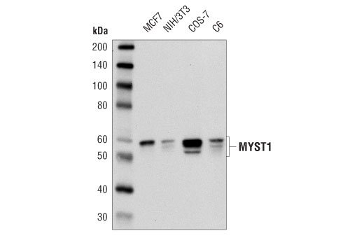
| MYST1 (D5T3R) Rabbit mAb 46862 | 20 µl |
|
H M R Mk | 53 | Rabbit IgG |
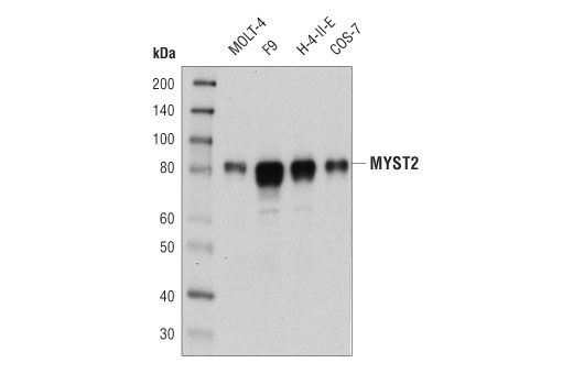
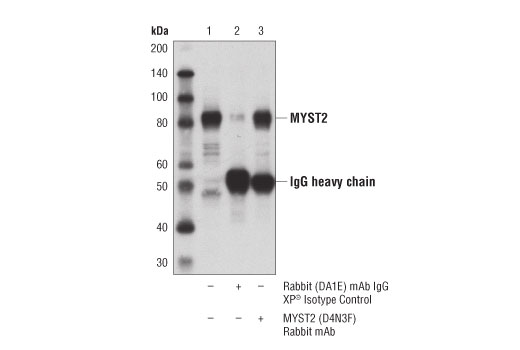
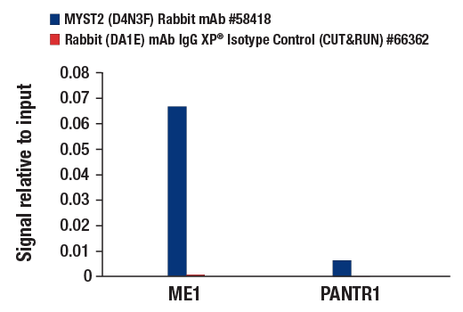
| MYST2 (D4N3F) Rabbit mAb 58418 | 20 µl |
|
H M R Mk | 80 | Rabbit IgG |
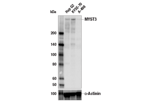
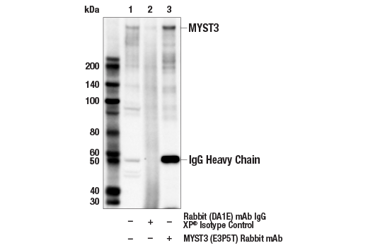
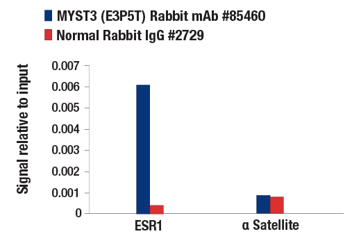
| MYST3 (E3P5T) Rabbit mAb 85460 | 20 µl |
|
H | 320 | Rabbit IgG |
| Anti-rabbit IgG, HRP-linked Antibody 7074 | 100 µl |
|
Rab | Goat |
Product Information
Monoclonal antibodies are produced by immunizing animals with recombinant proteins surrounding Val80 of human MYST1 protein and Val222 of human MYST2 protein; with a synthetic peptide corresponding to residues surrounding Pro927 of human MYST3 protein.
MYST1, also known as mammalian male absent on the first (MOF) and lysine acetyltransferase 8 (KAT8), is a member of the MYST (MOZ, YBF2, SAS2, and Tip60) family of histone acetyltransferases (1,2). As the catalytic subunit of two different histone acetyltransferase complexes, MSL and NSL, MYST1 is responsible for the majority of histone H4 lysine 16 acetylation in the cell. MYST1 also acetylates p53 on lysine 120 and is important for activation of pro-apoptotic genes (1,2). As a component of the MSL complex, MYST1 associates with MSL1, MSL2L1, and MSL3L1, and specifically acetylates histone H4 on lysine 16 (3-5). As part of the NSL complex, MYST1 associates with the MLL1 histone methyltransferase complex containing MLL1/KMT2A, ASH2L, HCFC1, WDR5, and RBBP5, and shows broader acetyltransferase activity for histone H4 on lysines 5, 8, and 16 (3-5). MYST1 plays a critical role in the regulation of transcription, DNA repair, autophagy, apoptosis, and emybryonic stem cell pluripotency and differentiation (1,2,6). Loss of MYST1 leads to a global reduction in histone H4 lysine 16 acetylation, a common hallmark found in many human cancers. A reduction of MYST1 protein levels and histone H4 lysine 16 acetylation is associated with poor prognosis in breast, renal, colorectal, gastric, and ovarian cancers (1).
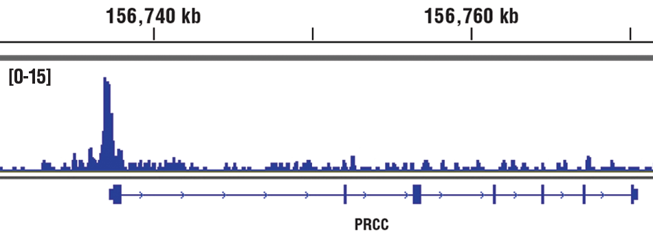
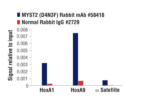
MYST2, also known as HBO1 and lysine acetyltransferase 7 (KAT7), is the catalytic subunit of the HBO1 acetyltransferase complex, which consists of MYST2, MYST/ESA1-associated factor 6 (MEAF6), inhibitor of growth protein 4 (ING4) or inhibitor of growth protein 5 (ING5), and one of two families of scaffold proteins (JADE-1/2/3 or BRPF1/2/3) (7,8). The substrate specificity of the HBO1 complex is determined by the associated scaffold protein. HBO1 complexes containing JADE scaffold proteins acetylate histone H4 on lysines 5, 8, and 12, while complexes containing BRPF scaffold proteins acetylate histone H3 on lysines 14 and 23 (8). In addition, the scaffold protein appears to regulate the function of the HBO1 complex. Complexes containing JADE scaffold proteins bind to origin recognition complex 1 (ORC1) and regulate licensing of DNA replication, while HBO1 complexes containing BRPF scaffold proteins regulate transcription (8-11). MYST2 is required for regulation of cell proliferation (1), adipogenesis (12), embryonic development (13), and survival of fetal liver erythroblasts (14). In addition, MYST2 is overexpressed in several human cancers, including cancers of the testis, ovary, breast, stomach, esophagus, and bladder (15). The MYST2 gene is amplified and protein is overexpressed in breast cancers, and overexpression of MYST2 increases anchorage-independent growth of several breast cancer cell lines (16).
MYST3, also known as monocytic leukemia zinc finger protein (MOZ) and lysine acetyltransferase 6A (KAT6A), was first discovered as a fusion partner of CREBBP in acute myeloid leukemia. MYST3 contributes to Hox gene expression and segment identity during development (17-20). MYST3 forms an evolutionarily conserved complex with ING5, EAF6, and BRD1 and has been shown to be a coactivator for many different transcription factors, including PU.1, NRF2, and Runx family members (21-23). MYST3 is critical in hematopoietic stem cell maintenance, where it acts synergistically with polycomb member BMI1 (24). Inhibitors of MYST3 are being investigated for therapeutic value as they can induce senescence and decrease tumor growth (25).
Except as otherwise expressly agreed in a writing signed by a legally authorized representative of CST, the following terms apply to Products provided by CST, its affiliates or its distributors. Any Customer's terms and conditions that are in addition to, or different from, those contained herein, unless separately accepted in writing by a legally authorized representative of CST, are rejected and are of no force or effect.
Products are labeled with For Research Use Only or a similar labeling statement and have not been approved, cleared, or licensed by the FDA or other regulatory foreign or domestic entity, for any purpose. Customer shall not use any Product for any diagnostic or therapeutic purpose, or otherwise in any manner that conflicts with its labeling statement. Products sold or licensed by CST are provided for Customer as the end-user and solely for research and development uses. Any use of Product for diagnostic, prophylactic or therapeutic purposes, or any purchase of Product for resale (alone or as a component) or other commercial purpose, requires a separate license from CST. Customer shall (a) not sell, license, loan, donate or otherwise transfer or make available any Product to any third party, whether alone or in combination with other materials, or use the Products to manufacture any commercial products, (b) not copy, modify, reverse engineer, decompile, disassemble or otherwise attempt to discover the underlying structure or technology of the Products, or use the Products for the purpose of developing any products or services that would compete with CST products or services, (c) not alter or remove from the Products any trademarks, trade names, logos, patent or copyright notices or markings, (d) use the Products solely in accordance with CST Product Terms of Sale and any applicable documentation, and (e) comply with any license, terms of service or similar agreement with respect to any third party products or services used by Customer in connection with the Products.