| Cat. # | Size | Price | Inventory |
|---|---|---|---|
| 84337S | 100 µl (50 tests) |
| REACTIVITY | H |
| SENSITIVITY | Endogenous |
| MW (kDa) | |
| Source/Isotype | Mouse IgG1 |
Product Information
| Application | Dilution |
|---|---|
| Flow Cytometry (Live) | 1:50 |
NOTE: Prepare solutions with reverse osmosis deionized (RODI) or equivalent grade water.
NOTE: When including fluorescent cellular dyes in your experiment (including viability dyes, DNA dyes, etc.), please refer to the dye product page for the recommended protocol. Visit www.cellsignal.com for a full listing of cellular dyes validated for use in flow cytometry.
NOTE: Count cells using a hemocytometer or alternative method.
NOTE: If using whole blood, lyse red blood cells and wash by centrifugation prior to immunostaining.
NOTE: Human Fc receptors cross-react with rabbit IgG. When cells of interest express high levels of Fc receptor protein (for example, macrophage/monocyte lineages), pre-incubate live cells with human Fc block prior to immunostaining with rabbit antibodies.
NOTE: Optimal centrifugation conditions will vary depending upon cell type and reagent volume. Generally, 150-300g for 1-5 minutes will be sufficient to pellet the cells.
posted June 2017
revised January 2022
Protocol Id: 1504
Human
Monoclonal antibody is produced by immunizing animals with cultured human B lymphoid cells treated with IFN-gamma.
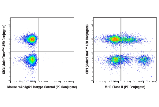
Major histocompatibility complex class II (MHC class II) molecules are heterodimeric, transmembrane glycoproteins expressed on the surface of antigen-presenting cells, such as macrophages, dendritic cells, and B cells. Expression can also be induced on other cell types through interferon-γ signaling (1). Prior to being displayed on the cell membrane, MHC class II molecules are loaded with exogenous peptide antigens approximately 15-24 amino acids in length that were derived from endocytosed extracellular proteins digested in the lysosome (2). Antigen-presentation through MHC class II is required for T cell activation during the immune response to extracellular pathogens (2). In humans, the MHC class II protein complex is encoded by the human leukocyte antigen gene complex (HLA). HLAs corresponding to MHC class II are HLA-DP, HLA-DM, HLA-DOA, HLA-DOB, HLA-DQ, and HLA-DR (3).
In the literature, this clone is reported to react with HLA-DR, HLA-DP, and HLA-DQ (4).
Except as otherwise expressly agreed in a writing signed by a legally authorized representative of CST, the following terms apply to Products provided by CST, its affiliates or its distributors. Any Customer's terms and conditions that are in addition to, or different from, those contained herein, unless separately accepted in writing by a legally authorized representative of CST, are rejected and are of no force or effect.
Products are labeled with For Research Use Only or a similar labeling statement and have not been approved, cleared, or licensed by the FDA or other regulatory foreign or domestic entity, for any purpose. Customer shall not use any Product for any diagnostic or therapeutic purpose, or otherwise in any manner that conflicts with its labeling statement. Products sold or licensed by CST are provided for Customer as the end-user and solely for research and development uses. Any use of Product for diagnostic, prophylactic or therapeutic purposes, or any purchase of Product for resale (alone or as a component) or other commercial purpose, requires a separate license from CST. Customer shall (a) not sell, license, loan, donate or otherwise transfer or make available any Product to any third party, whether alone or in combination with other materials, or use the Products to manufacture any commercial products, (b) not copy, modify, reverse engineer, decompile, disassemble or otherwise attempt to discover the underlying structure or technology of the Products, or use the Products for the purpose of developing any products or services that would compete with CST products or services, (c) not alter or remove from the Products any trademarks, trade names, logos, patent or copyright notices or markings, (d) use the Products solely in accordance with CST Product Terms of Sale and any applicable documentation, and (e) comply with any license, terms of service or similar agreement with respect to any third party products or services used by Customer in connection with the Products.