| Cat. # | Size | Price | Inventory |
|---|---|---|---|
| 26118T | 1 Kit (8 x 20 microliters) |
| Product Includes | Quantity | Applications | Reactivity | MW(kDa) | Isotype |
|---|---|---|---|---|---|
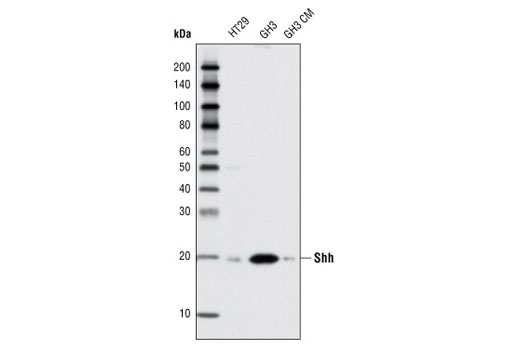
| Shh (C9C5) Rabbit mAb 2207 | 20 µl |
|
H R | 19, (45 precursor) | Rabbit IgG |
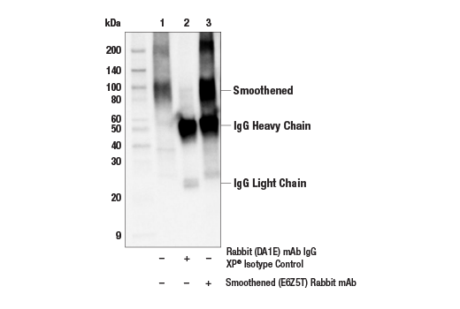
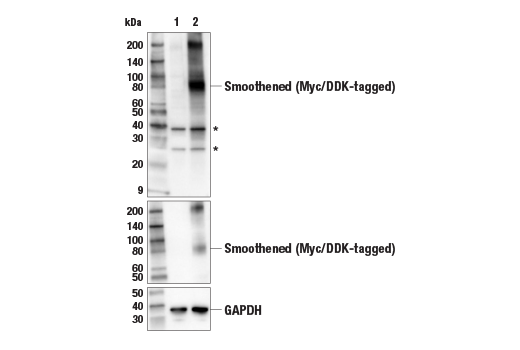
| Smoothened (E6Z5T) Rabbit mAb 92981 | 20 µl |
|
H | 85 | Rabbit IgG |
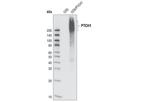
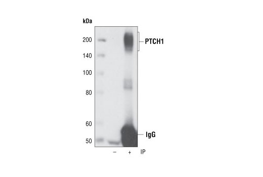
| PTCH1 (C53A3) Rabbit mAb 2468 | 20 µl |
|
H | 180-210 | Rabbit IgG |
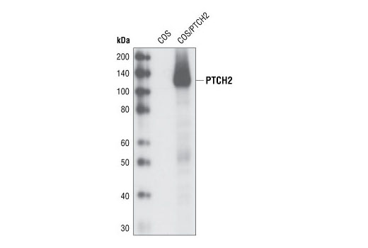
| PTCH2 (G1191) Antibody 2470 | 20 µl |
|
H | 130 | Rabbit |
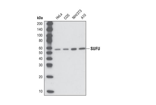
| SUFU (C54G2) Rabbit mAb 2520 | 20 µl |
|
H M R Mk | 54 | Rabbit IgG |
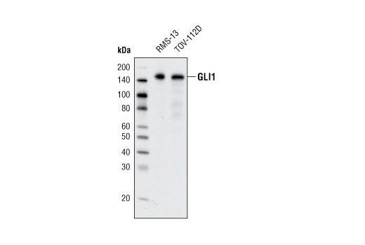
| GLI1 (C68H3) Rabbit mAb 3538 | 20 µl |
|
H | 160 | Rabbit IgG |
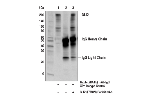
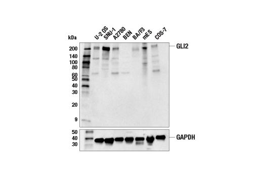
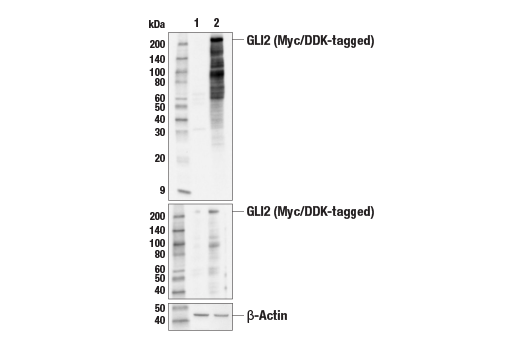
| GLI2 (E5V8N) Rabbit mAb 18773 | 20 µl |
|
H M Mk | 220 | Rabbit IgG |
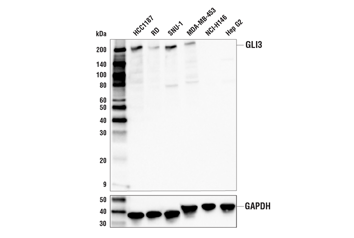
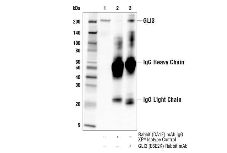
| GLI3 (E6E2K) Rabbit mAb 71107 | 20 µl |
|
H | 200 | Rabbit IgG |
| Anti-rabbit IgG, HRP-linked Antibody 7074 | 100 µl |
|
Goat |
Product Information
Polyclonal antibodies are produced by immunizing animals with a synthetic peptide corresponding to residues surrounding Gly1191 of human PTCH2. Polyclonal antibodies are purified by protein A and peptide affinity chromatography. Monoclonal antibodies are produced by immunizing animals with synthetic peptides corresponding to residues surrounding Glu53 of human Shh, Pro1307 of human PTCH1, Leu458 of human SUFU, Gly420 of human GLI1, and Pro1188 of human GLI2 protein, or recombinant proteins corresponding to human smoothened (SMO) protein, and the carboxy terminus of human GLI3.
The Hedgehog (Hh) signaling pathway plays critical roles in the regulation of cell fate, tissue patterning, and growth during embryonic development. It is downregulated during postnatal development, but can be reactivated to promote tissue repair and regeneration. Aberrant Hh signaling activity during prenatal development is associated with numerous birth defects (e.g., holoprosencephaly), while uncontrolled Hh pathway activation postnatally is linked to the development of several cancer types (1,2). There are three canonical Hh ligands: Sonic hedgehog (SHH), Indian hedgehog (IHH), and Desert hedgehog (DHH), all of which have distinct as well as overlapping roles and expression patterns (3-5). Patched1 and 2 (PTCH1 and PTCH2) are partially redundant 12-pass transmembrane proteins that function as receptors for Hh ligands (6-8). Smoothened (SMO) is a 7-pass transmembrane G protein-coupled receptor (GPCR) that functions as the key transducer of Hh signaling. In the absence of Hh ligands (off-state), PTCH proteins are localized to cilia, and function to suppress SMO activity (1,2). Suppressor of Fused (SUFU) simultaneously contributes to suppression of the pathway by sequestering the glioma-associated oncogene (GLI) family of transcription factors (9,10). Binding of Hh ligands to PTCH receptors results in derepression of SMO, in part by promoting its translocation to cilia; this leads to downregulation of SUFU, resulting in the stabilization and nuclear translocation of GLI transcription factors that regulate the transcription of genes involved in cell proliferation, migration, and survival (1).
Explore pathways related to this product.
STRING - Known and Predicted Protein-Protein Interactions.
Except as otherwise expressly agreed in a writing signed by a legally authorized representative of CST, the following terms apply to Products provided by CST, its affiliates or its distributors. Any Customer's terms and conditions that are in addition to, or different from, those contained herein, unless separately accepted in writing by a legally authorized representative of CST, are rejected and are of no force or effect.
Products are labeled with For Research Use Only or a similar labeling statement and have not been approved, cleared, or licensed by the FDA or other regulatory foreign or domestic entity, for any purpose. Customer shall not use any Product for any diagnostic or therapeutic purpose, or otherwise in any manner that conflicts with its labeling statement. Products sold or licensed by CST are provided for Customer as the end-user and solely for research and development uses. Any use of Product for diagnostic, prophylactic or therapeutic purposes, or any purchase of Product for resale (alone or as a component) or other commercial purpose, requires a separate license from CST. Customer shall (a) not sell, license, loan, donate or otherwise transfer or make available any Product to any third party, whether alone or in combination with other materials, or use the Products to manufacture any commercial products, (b) not copy, modify, reverse engineer, decompile, disassemble or otherwise attempt to discover the underlying structure or technology of the Products, or use the Products for the purpose of developing any products or services that would compete with CST products or services, (c) not alter or remove from the Products any trademarks, trade names, logos, patent or copyright notices or markings, (d) use the Products solely in accordance with CST Product Terms of Sale and any applicable documentation, and (e) comply with any license, terms of service or similar agreement with respect to any third party products or services used by Customer in connection with the Products.